. 2021 Dec 16;19(1):256.
doi: 10.1186/s12915-021-01190-2.

JNK signaling provides a novel therapeutic target for Rett syndrome

Affiliations

JNK signaling provides a novel therapeutic target for Rett syndrome

Clara Alice Musi et al. BMC Biol. .

Abstract

Background: Rett syndrome (RTT) is a monogenic X-linked neurodevelopmental disorder characterized by loss-of-function mutations in the MECP2 gene, which lead to structural and functional changes in synapse communication, and impairments of neural activity at the basis of cognitive deficits that progress from an early age. While the restoration of MECP2 in animal models has been shown to rescue some RTT symptoms, gene therapy intervention presents potential side effects, and with gene- and RNA-editing approaches still far from clinical application, strategies focusing on signaling pathways downstream of MeCP2 may provide alternatives for the development of more effective therapies in vivo. Here, we investigate the role of the c-Jun N-terminal kinase (JNK) stress pathway in the pathogenesis of RTT using different animal and cell models and evaluate JNK inhibition as a potential therapeutic approach.

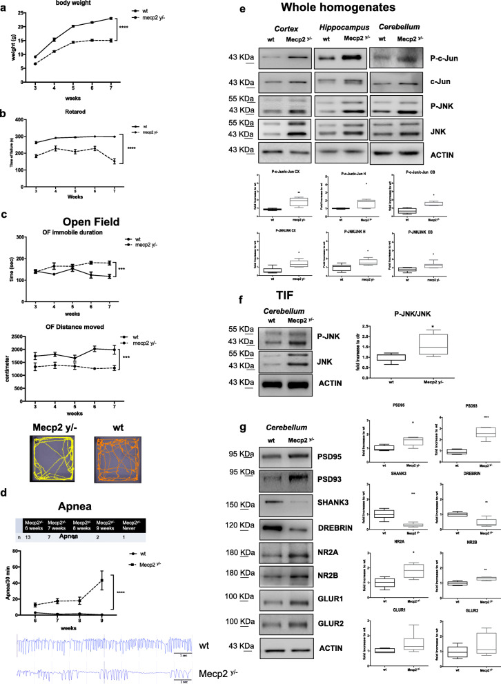
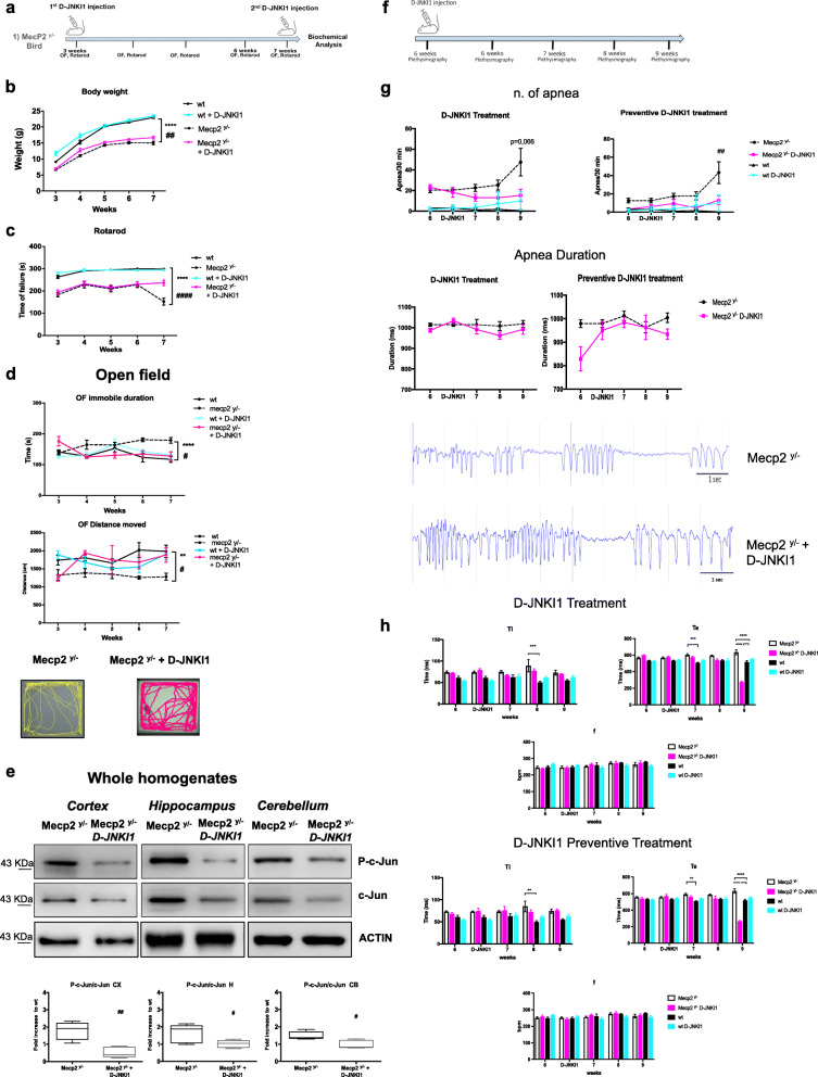
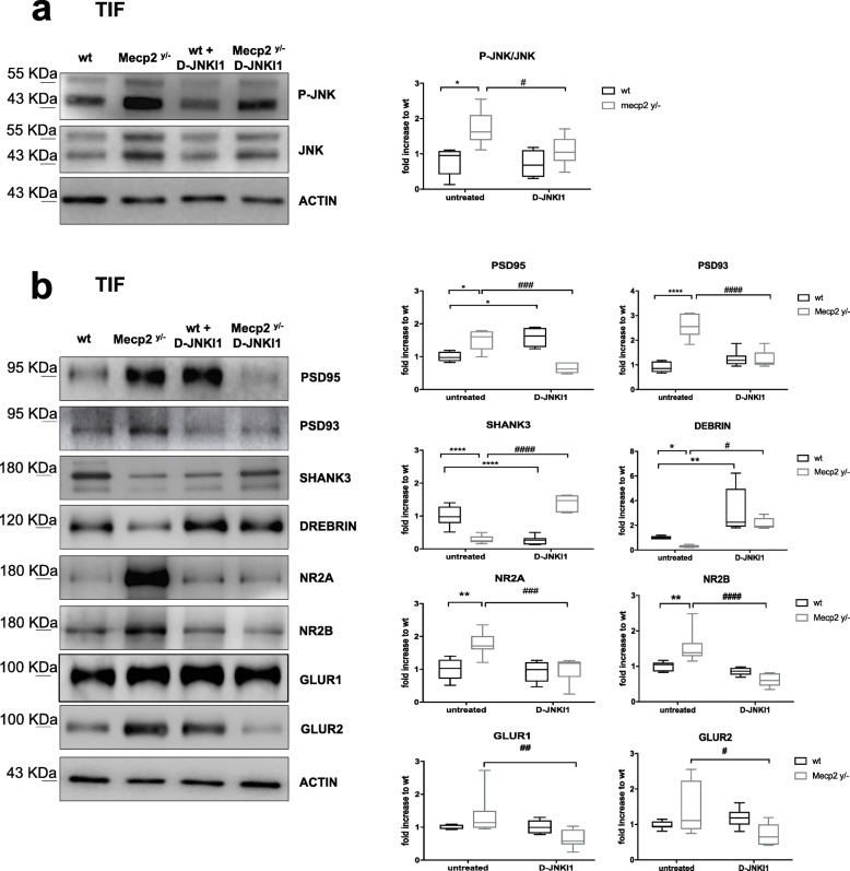
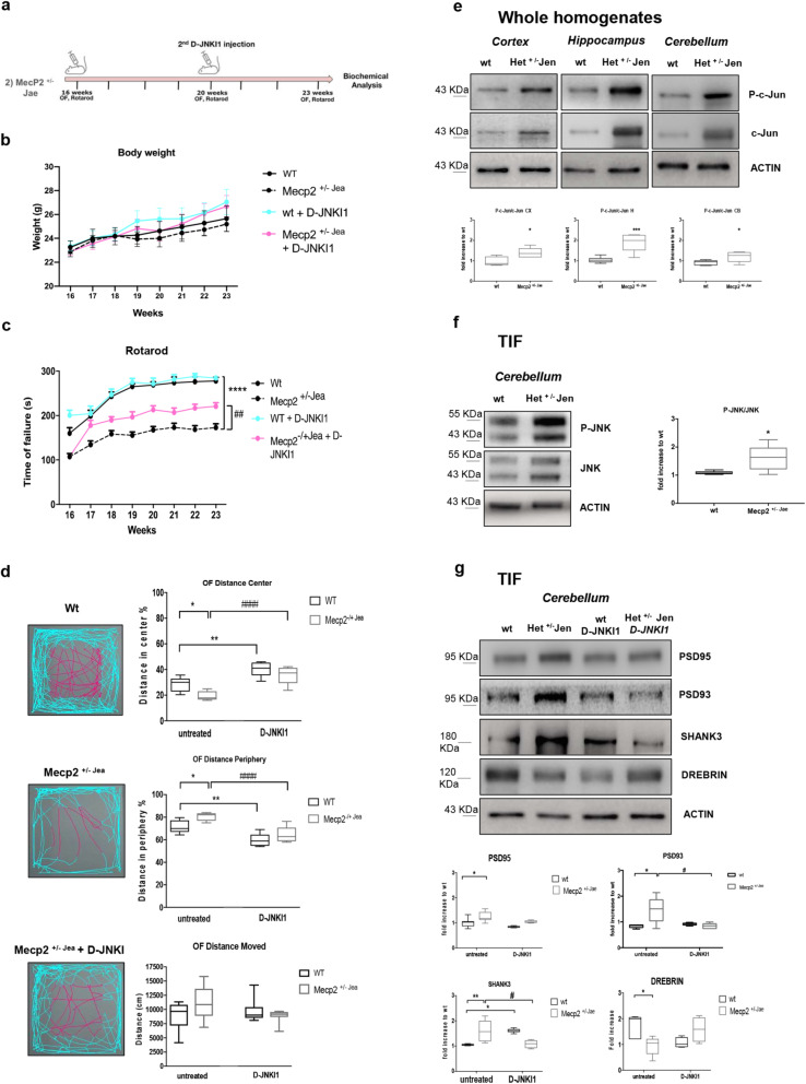
Results: We discovered that the c-Jun N-terminal kinase (JNK) stress pathway is activated in Mecp2-knockout, Mecp2-heterozygous mice, and in human MECP2-mutated iPSC neurons. The specific JNK inhibitor, D-JNKI1, promotes recovery of body weight and locomotor impairments in two mouse models of RTT and rescues their dendritic spine alterations. Mecp2-knockout presents intermittent crises of apnea/hypopnea, one of the most invalidating RTT pathological symptoms, and D-JNKI1 powerfully reduces this breathing dysfunction. Importantly, we discovered that also neurons derived from hiPSC-MECP2 mut show JNK activation, high-phosphorylated c-Jun levels, and cell death, which is not observed in the isogenic control wt allele hiPSCs. Treatment with D-JNKI1 inhibits neuronal death induced by MECP2 mutation in hiPSCs mut neurons.

Conclusions: As a summary, we found altered JNK signaling in models of RTT and suggest that D-JNKI1 treatment prevents clinical symptoms, with coherent results at the cellular, molecular, and functional levels. This is the first proof of concept that JNK plays a key role in RTT and its specific inhibition offers a new and potential therapeutic tool to tackle RTT.

Keywords: Apnea; D-JNKI1; MECP2; Neurodevelopmental disease; Neuroprotection; Synaptic dysfunction.


Conflict of interest statement

The authors declare that they have no competing interests.

Figures

Fig. 1
Fig. 1
JNK signaling activation in Mecp2y/- Bird male mice. a Growth curves of Mecp2y/- mice (n=25) compared to their wt littermates (n=25) from 3 to 7 weeks of age. b, c Behavioral analysis of Mecp2y/- mice (n=10) compared to their control wt (n=10) from 3 to 7 weeks of age in the Rotarod (b) and open field (c) tests (parameters shown: duration of immobility and distance moved). The distance moved by each experimental group was also presented in the arena plots under the open field graphs. d On-set and number of apnea in Mecp2y/- and wt mice at 6, 7, 8, and 9 weeks of ages in the table and the graphs, in the lower part, the representative plethysmographic traces of Mecp2y/− (n=15) vs wt (n=6) characterizing the respiratory patterns and breathing dysfunction. e JNK signaling pathway activation in the whole homogenate: Western blots and quantifications of P-c-Jun/c-Jun and P-JNK/JNK ratios in the cortex, hippocampus, and cerebellum of 7-week-old Mecp2y/- (n=10) and wt (n=10) mice. f Western blots and quantifications of TIF fraction (post-synaptic elements) showed the JNK activation in cerebellum of 7-week-old Mecp2y/- (n=10) compared to wt (n=10) mice. g Western blots and quantifications showed PSD alterations in Mecp2y/- (n=10) compared to wt (n=10) mice. Data were shown as mean ± SEM. Significance was calculated using two‐way ANOVA for repetitive measurements followed by Bonferroni post hoc test (panels a, b, c, and d) or Student’s t test followed by Tukey’s post hoc test (panels e, f, g). Statistical significance: *p<0.05, **p<0.01, ***p<0.001, and ****p<0.0001
Fig. 2
Fig. 2
D-JNKI1 rescues well-being conditions, locomotor impairments, and apnea numbers in Mecp2y/- male mice. a Timeline of D-JNKI1 treatment in Mecp2y/- male mice. b Growth curves of D-JNKI1-treated (blue sky) and untreated (black) wild type, and D-JNKI1-treated (fuchsia) and untreated (black-dotted) Mecp2y/- mice from 3 to 7 weeks of age (n=25 for each experimental group). c, d Behavioral analysis of D-JNKI1-treated vs untreated wt and Mecp2y/- mice (n=10 for each experimental group) from 3 to 7 weeks of age in the Rotarod (c) and open field (d) tests (parameters shown: time spent immobile and distance moved, with relative open field arena-plots). e Western blots and the quantification P-c-Jun/c-Jun ratio in the whole homogenate of the cortex, hippocampus, and cerebellum of 7-week-old D-JNKI1-treated and untreated Mecp2y/- mice. f Timeline of D-JNKI1 treatment and plethysmography analysis. g Number and duration of apnea in preventive and curative D-JNKI1 paradigm of treated (n=10) and untreated (n=6) wild type and Mecp2y/- mice (n=15) from 6 to 9 weeks of age. h Breathing analysis in preventive (lower part) and curative (upper part) D-JNKI1 paradigm of treated (fuchsia) and untreated Mecp2y/- (white) mice from 6 to 9 weeks of age and the treated (blue-sky) and untreated wild type (black) (parameters shown: Ti, Te, and f). Data were shown as mean ± SEM. Significance was calculated using two‐way ANOVA for repetitive measurements followed by Bonferroni post hoc test (panels a, b, c, e, f) or Student’s t test followed by Tukey’s post hoc test (panels d). Statistical significance relative to control **p<0.01, ***p<0.001, and ****p<0.0001; D-JNKI1-treated vs untreated Mecp2y/-: #p<0.05, ##p<0.01, ####p<0.0001
Fig. 3
Fig. 3
D-JNKI1 effect against dendritic spine alterations in Mecp2y/- male mice. a Western blots and quantifications in the cerebellum TIF (post-synaptic elements) of treated and untreated wt and Mecp2y/- mice to measure D-JNKI1 effect in vivo. D-JNKI1 significantly reduced JNK activation in Mecp2y/- mice, but not in control wt mice. b Western blots and quantifications of the post-synaptic elements in the cerebellum showed normalization of the PSD markers levels to control level in D-JNKI1-treated compared to untreated Mecp2y/- mice (n=10 for each experimental group). Data were shown as mean ± SEM. Significance was calculated using two‐way ANOVA followed by Bonferroni post hoc test. Significance relative to control *p<0.05, **p<0.01, and ****p<0.0001. D-JNKI1-treated vs untreated Mecp2y/− #p< 0.05, ##p< 0.01, ###p<0.001, and ####p<0.0001
Fig. 4
Fig. 4
Female Mecp2+/− Jaenisch neurological phenotype: JNK signaling activation and D-JNKI1 treatment. a Timeline of D-JNKI1 treatment in Female Mecp2+/− Jaenisch mice. b Growth curves of D-JNKI1-treated and untreated wt and Mecp2+/− Jae mice from 16 to 23 weeks of age. c Rotarod tests in D-JNKI1 Mecp2+/− Jae-treated mice (fuchsia line), Mecp2+/− Jae untreated (black dotted line), treated wt (blue-sky line), and untreated wt (black line). d Open field test. D-JNKI1 improved the behavioral performance of Mecp2+/− Jae (see plots for central (fuchsia) and peripheral (blue sky) movements of wt and Mecp2+/− Jae-treated and untreated mice). The last graph presented the distance moved: there were no genotypic differences. e Western blots and quantifications of c-Jun activation in the whole homogenate of the cortex, hippocampus, and cerebellum in 23-week-old wt and Mecp2+/− Jae mice. f Western blots and relative quantifications in the TIF cerebellum of 23-week-old wt and Mecp2+/− Jae mice confirmed JNK activation at the synaptic level in Mecp2+/− Jae mice. g Mecp2+/− Jae presented alterations of the PSD-region and D-JNKI1 treatment normalized the biochemical alterations in treated vs untreated Mecp2+/− Jae mice. Each experimental group: n=8. Data were shown as mean ± SEM. Significance was calculated using two‐way ANOVA for repetitive measurements followed by Bonferroni post hoc test (panels a, b, c), Student’s t test followed by Tukey’s post hoc test (panels d and e), and two‐way ANOVA followed by Bonferroni post hoc test (f). Significant differences from control *p<0.05, **p<0.01, ***p<0.001, and ****p<0.0001; D-JNKI1-treated vs untreated Mecp2+/−: #p<0.05, ##p<0.01, and #### p<0.0001
Fig. 5
Fig. 5
JNK signaling activation and D-JNKI1’s protective effects in Rett human iPSCs. a Neuronal differentiation of human iPSCs. The upper panel showed the neuronal differentiation protocol. The timing of critical steps was indicated in days from day 0 (d0). At the end of the differentiation (red arrow, d40), neurons were exposed to 2μM D-JNKI1 for 48 h before neurons were isolated for Western blot analysis. Immunofluorescence was used to define cell identity (lower panel): a1- OCT4/SSEA4 staining for iPSCs, a2- Nestin and SOX1 staining for telencephalic neural progenitors (d12/13), and a3- β3-Tubulin (TuJI) staining for neurons in terminally differentiated cultures and DAPI to stain nuclei. Scale bar: iPSCs and NPCs 100 μm and neurons 20 μm. b Western blots and quantifications of JNK activation in neurons differentiated from hiPSCs from RTT human patients; clones expressing either the wild type (hMecp2wt) or the mutated (hMecp2mut) MECP2 allele were differentiated in neurons. The hMecp2mut displayed higher P-JNK/JNK and P-c-Jun/c-Jun ratios than to hMecp2wt neurons. D-JNKI1 reduced hMecp2mut activation to hMecp2wt levels; D-JNKI1 in hMecp2wt neurons did not change P-JNK/JNK (n=4 and 5) and P-c-Jun/c-Jun ratios (n=5). c Cell death in hMecp2wt and hMecp2mut neurons: the hMecp2mut showed greater cell death than to hMecp2wt. D-JNKI1 reduced induced-cell death in the hMecp2mut neurons to the control level (hMecp2wt). Data were shown as mean ± SEM. Significance was calculated using two‐way ANOVA followed by Bonferroni post hoc test (panel b). Significance vs control *p<0.05, **p<0.01, and ****p<0.0001; D-JNKI1-treated vs untreated mutated neurons #p<0.05, ##p<0.01, and ###p<0.001. See additional file S3

References

    1. Percy AK. Rett syndrome: Current status and new vistas. Neurol Clin. 2002;20:1125–1141. - PubMed
    1. Amir RE, Van den Veyver IB, Wan M, Tran CQ, Francke U, Zoghbi HY. Rett syndrome is caused by mutations in X-linked MECP2, encoding methyl-CpG-binding protein 2. Nat Genet. 1999;23:185–188. - PubMed
    1. Jørgensen HF, Bird A. MeCP2 and other methyl-CpG binding proteins. Ment Retard Dev Disabil Res Rev. 2002;8:87–93. - PubMed
    1. Lewis JD, Meehan RR, Henzel WJ, Maurer-Fogy I, Jeppesen P, Klein F, et al. Purification, sequence, and cellular localization of a novel chromosomal protein that binds to methylated DNA. Cell. 1992;69:905–914. - PubMed
    1. Hagberg B. Clinical manifestations and stages of Rett syndrome. Ment Retard Dev Disabil Res Rev. 2002;8:61–65. - PubMed

Publication types