. 2021 Dec 15;40(1):395.
doi: 10.1186/s13046-021-02187-z.

WNT11/ROR2 signaling is associated with tumor invasion and poor survival in breast cancer

Affiliations

WNT11/ROR2 signaling is associated with tumor invasion and poor survival in breast cancer

Kerstin Menck et al. J Exp Clin Cancer Res. .

Abstract

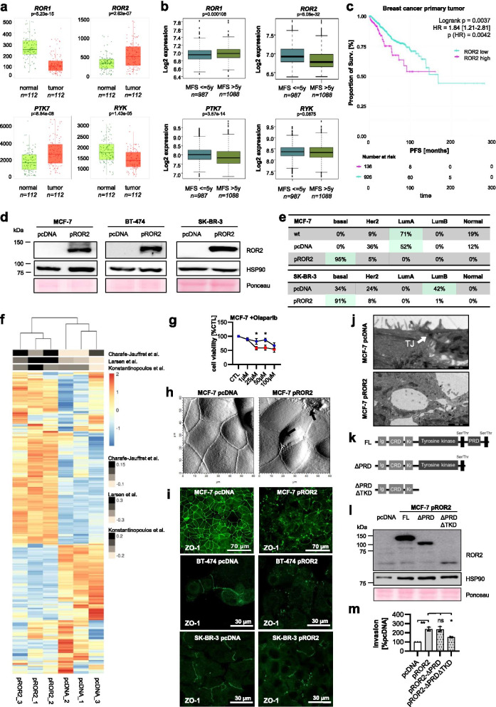
Background: Breast cancer has been associated with activation of the WNT signaling pathway, although no driver mutations in WNT genes have been found yet. Instead, a high expression of the alternative WNT receptor ROR2 was observed, in particular in breast cancer brain metastases. However, its respective ligand and downstream signaling in this context remained unknown.

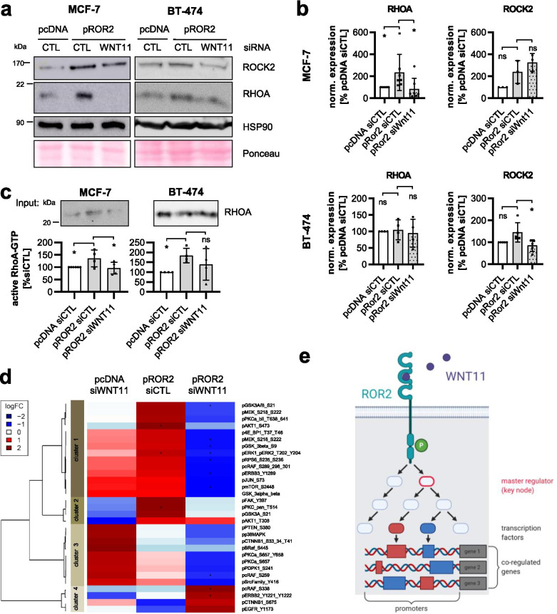
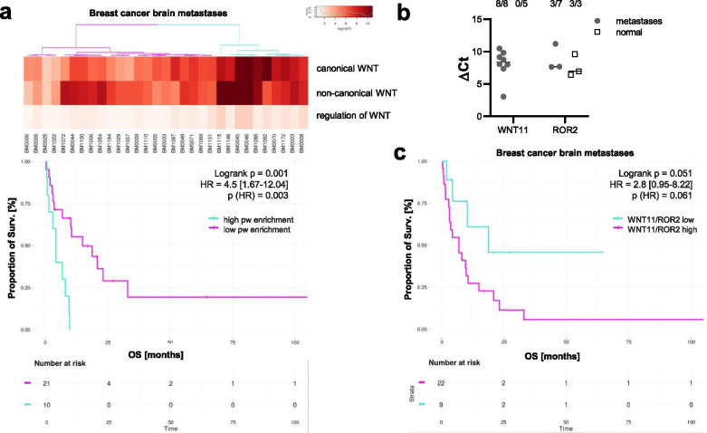
Methods: We modulated the expression of ROR2 in human breast cancer cells and characterized their gene and protein expression by RNA-Seq, qRT-PCR, immunoblots and reverse phase protein array (RPPA) combined with network analyses to understand the molecular basis of ROR2 signaling in breast cancer. Using co-immunoprecipitations, we verified the interaction of ROR2 with the identified ligand, WNT11. The functional consequences of WNT11/ROR2 signaling for tumor cell aggressiveness were assessed by microscopy, impedance sensing as well as viability and invasion assays. To evaluate the translational significance of our findings, we performed gene set enrichment, expression and survival analyses on human breast cancer brain metastases.

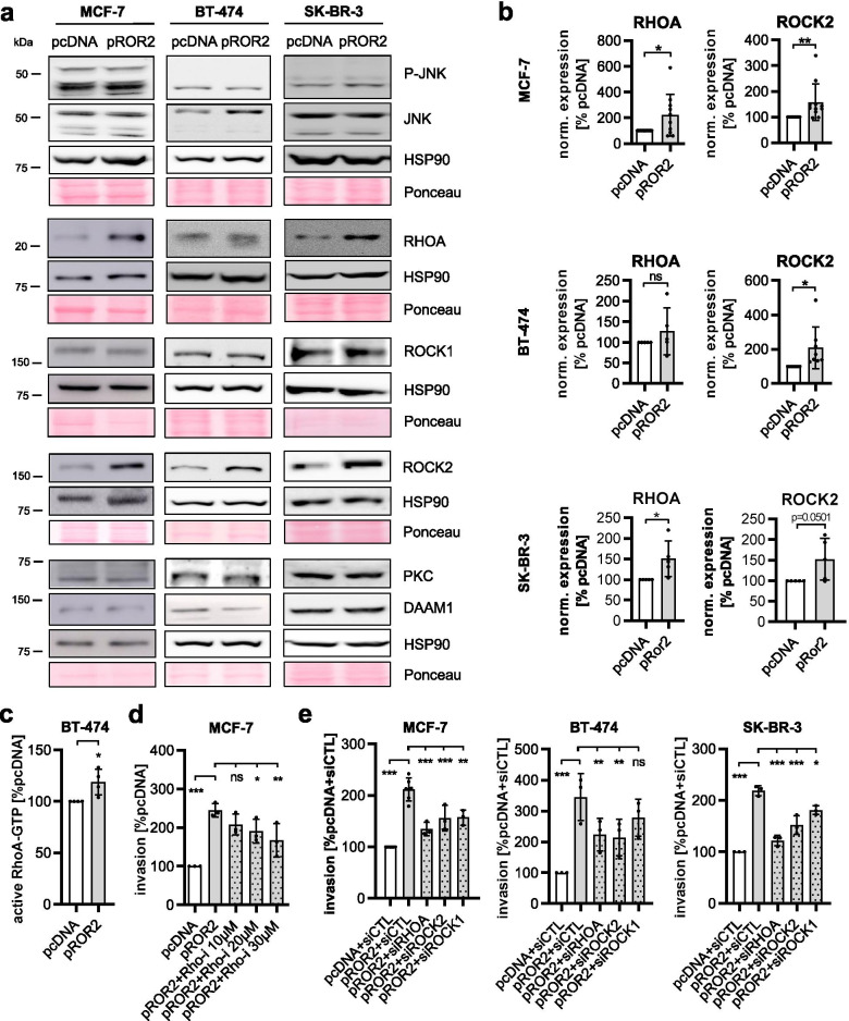
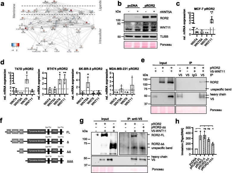
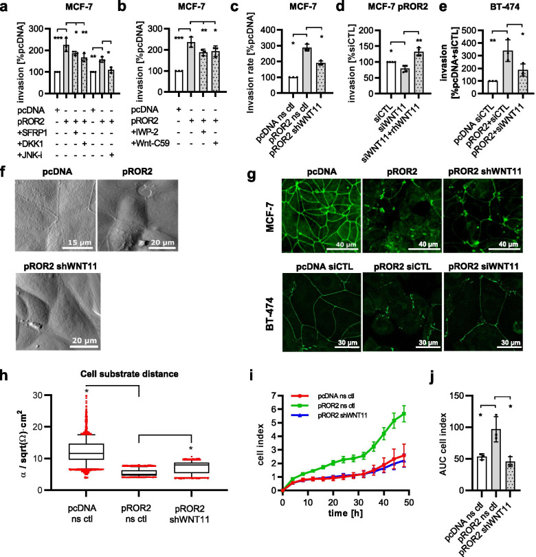
Results: We found ROR2 to be highly expressed in aggressive breast tumors and associated with worse metastasis-free survival. ROR2 overexpression induced a BRCAness-like phenotype in a cell-context specific manner and rendered cells resistant to PARP inhibition. High levels of ROR2 were furthermore associated with defects in cell morphology and cell-cell-contacts leading to increased tumor invasiveness. On a molecular level, ROR2 overexpression upregulated several non-canonical WNT ligands, in particular WNT11. Co-immunoprecipitation confirmed that WNT11 indeed interacts with the cysteine-rich domain of ROR2 and triggers its invasion-promoting signaling via RHO/ROCK. Knockdown of WNT11 reversed the pro-invasive phenotype and the cellular changes in ROR2-overexpressing cells.

Conclusions: Taken together, our study revealed a novel auto-stimulatory loop in which ROR2 triggers the expression of its own ligand, WNT11, resulting in enhanced tumor invasion associated with breast cancer metastasis.

Keywords: BRCAness; Breast cancer; Metastasis; Network analysis; ROR2; WNT11.


Conflict of interest statement

The authors declare that they have no competing interests.

Figures

Fig. 1
Fig. 1
Expression of ROR2 induces a highly aggressive phenotype in breast cancer cells. a RNA-Seq: Gene expression of the four non-canonical WNT co-receptors ROR1, ROR2, PTK7 and RYK in normal breast (green) and matched breast cancer tissue (red) from the TNMplot database. Significance was calculated with a paired Wilcoxon statistical test. b Microarray gene expression data from 2075 breast cancer primary tumors were correlated with either poor (≤ 5 years) or better (> 5 years) metastasis-free survival (MFS). Significance was calculated with a t-test. Boxes represent the 25-75th percentiles with the line at the median. Outliers are marked as small dots. c Progression-free survival (PFS) of 1067 breast cancer patients from TCGA stratified by ROR2 mRNA expression. d MCF-7, BT-474 and SK-BR-3 human breast cancer cells were transfected either with an empty vector (pcDNA) or with a hROR2 overexpression plasmid (pROR2). Transfection efficiency was confirmed by western blot. e Determination of the molecular breast cancer subtype based on RNA-Seq data from the indicated cell lines using the PAM50 gene signature. f RNA-Seq of MCF-7 cells: GSVA analysis for three independently published BRCAness gene expression signatures. g MTT assay: Cells were treated for 96 h with the indicated concentrations of olaparib (mean ± SD, n = 3, *p < 0.05). h Atomic force microscopy (AFM) of MCF-7 pcDNA and pROR2 cells. i Immunofluorescence staining of the tight junction marker ZO-1. j Electron microscopy of a representative tight junction (TJ) in MCF-7 pcDNA cells. An example for a defective cell-cell-junction is shown for pROR2 cells (scale bar: 1 μm). k Schematic overview of the pROR2 C-terminal deletion constructs. l+m C-terminal deletion constructs were overexpressed in MCF-7. Expression was confirmed by western blot (l) and the invasion rate measured in Boyden chambers (m) (mean ± SD, n = 3, *p < 0.001, **p < 0.0001, ns = not significant). Significance was calculated with a one-way ANOVA with Dunnett’s multiple comparison test
Fig. 2
Fig. 2
RHOA/ROCK mediate ROR2-induced tumor invasion. a pcDNA and pROR2 cells were characterized for the expression of non-canonical WNT signaling proteins by western blot. b Densitometric quantification of RHOA and ROCK2 in pcDNA and pROR2 cells normalized on HSP90 expression (mean ± SD, *p < 0.05, **p < 0.01, ns = not significant). c Active RHOA-GTP was measured in BT-474 cells by ELISA (mean ± SD, n = 4, *p < 0.05). d Invasion assay: MCF-7 pROR2 cells were treated for 96 h with the indicated concentration of Rhosin, a RHO inhibitor (mean ± SD, n = 3, *p < 0.05, **p < 0.01, ***p < 0.0001, ns = not significant). Significance was calculated with a one-way ANOVA with Dunnett’s multiple comparison test. e Cells were transfected with the indicated siRNAs (10 nM) and cell invasion was measured in Boyden chambers (mean ± SD, n = 3, *p < 0.05, **p < 0.01, ***p < 0.001, ns = not significant). Significance was calculated with a one-way ANOVA with Dunnett’s multiple comparison test
Fig. 3
Fig. 3
WNT11 is a novel ligand for ROR2 in humans. a RNA-Seq of MCF-7 pROR2 cells: Network of differentially expressed genes associated with non-canonical WNT signaling grouped according to their cellular localization. b MCF-7 cells were stimulated for 24 h with rWNT5A (100 ng/ml) and WNT11 expression was analyzed by western Blot. c+d Expression of the non-canonical WNT ligands was measured by qRT-PCR in MCF-7 (c) or the indicated ROR2-overexpressing human breast cancer cell lines (d) (mean ± SD, n = 3–9, *p < 0.05, p < 0.01, n.e. = not expressed). Expression values were calculated relative to the empty vector control cells. e Co-immunoprecipitation (Co-IP) of V5-WNT11 in MCF-7 pROR2 cells detects ROR2 by western blot. f Schematic representation of the ROR2 N-terminal deletion constructs. g Co-IP of V5-Wnt11 in MCF-7 expressing either pROR2-FL or pROR2-ΔΔ. h Cell invasion assays of MCF-7 expressing N-terminal ROR2 deletion constructs (mean ± SD, n = 3, *p < 0.01, **p = 0.0001, ns = not significant). Significance was calculated with a one-way ANOVA with Dunnett’s multiple comparison test
Fig. 4
Fig. 4
WNT11 mediates the pro-tumoral effects of ROR2. a+b Invasion assay: MCF-7 pcDNA or pROR2 cells were treated with WNT inhibitors (a) or Porcupine inhibitors (b) (mean ± SD, n = 3, *p < 0.05, **p < 0.01, ***p < 0.0001). Significance was calculated with a one-way ANOVA with Dunnett’s multiple comparison test. c Invasion assay of MCF-7 pROR2 cells stably expressing a non-sense control (ns ctl) or WNT11 (shWNT11) shRNA (mean ± SD, n = 3, *p < 0.001). Significance was calculated with a one-way ANOVA with Dunnett’s multiple comparison test. d Invasion of MCF-7 pROR2 cells transfected with control (siCTL) or WNT11 siRNA (siWNT11) +/− rhWNT11 (100 ng/ml) was assessed in Boyden chambers (mean ± SD, n = 3, *p < 0.05, **p < 0.001). Significance was calculated with a one-way ANOVA with Dunnett’s multiple comparison test. e Invasion assay of BT-474 pcDNA and pROR2 cells transfected with either siCTL or siWNT11 (mean ± SD, n = 3, *p < 0.05, **p < 0.001). Significance was calculated with a one-way ANOVA with Dunnett’s multiple comparison test. f AFM of cell-cell-junctions in the indicated cell lines. g Immunofluorescence for the tight junction protein ZO-1. h ECIS measurements of MCF-7 cells (box: 25-75th percentile, line at median, *p < 0.0001). Significance was calculated with a one-way ANOVA with Dunnett’s multiple comparison test. i+j xCELLigence measurements of MCF-7 cells (mean ± SD). Shown is one representative example (i) and a corresponding Area Under the Curve (AUC) analysis for all three independent experiments (mean ± SD, *p < 0.01). Significance was calculated with a one-way ANOVA with Dunnett’s multiple comparison test
Fig. 5
Fig. 5
WNT11 mediates ROR2 signaling. a+b Western blot: RHOA and ROCK2 in MCF-7 and BT-474 pROR2 cells treated with control siRNA (siCTL) or siRNA against WNT11 (siWNT11) (a) with corresponding densitometric quantification normalized on HSP90 expression (b) (mean ± SD, *p < 0.05, ns = not significant). Significance was calculated with a one-way ANOVA with Dunnett’s multiple comparison test. c Levels of active RHOA-GTP in the indicated cells were assessed by ELISA (mean ± SD, *p < 0.05, ns = not significant). Significance was calculated with a one-way ANOVA with Dunnett’s multiple comparison test. c MCF-7 pcDNA and pROR2 siCTL/siWNT11 cells were characterized by RPPA for phospho-proteins associated with the WNT signaling pathway (n = 3, *p < 0.05). d Schematic representation of the master regulator analysis of ROR2/WNT11 signaling. The illustration was created with BioRender.com
Fig. 6
Fig. 6
ROR2/WNT11 are expressed in metastatic breast cancer and associated with poor survival. a Pathway (pw) enrichment for RNA-Seq data from 31 patients with brain metastases given for different WNT subpathways. Significance was calculated with a log rank test. b qRT-PCR: Expression of ROR2 and WNT11 in samples of human brain metastases (line at median). The upper numbers indicate the number of samples with positive signals out of all investigated samples. c Kaplan-Meier survival curves showing the OS of metastatic patients based on their averaged WNT11 and ROR2 expression. The separation high/low was computed based on an optimal cutoff using the maxstat method. Significance was calculated with a log rank test

Similar articles

Cited by

References

    1. Bray F, Ferlay J, Soerjomataram I, Siegel RL, Torre LA, Jemal A. Global cancer statistics 2018: GLOBOCAN estimates of incidence and mortality worldwide for 36 cancers in 185 countries. CA Cancer J Clin. 2018;68:394–424. - PubMed
    1. Niehrs C. The complex world of WNT receptor signalling. Nat Rev Mol Cell Biol. 2012;13:767–779. - PubMed
    1. De A. Wnt/Ca2+ signaling pathway: a brief overview. Acta Biochim Biophys Sin. 2011;43:745–756. - PubMed
    1. Habas R, Kato Y, He X. Wnt/Frizzled activation of Rho regulates vertebrate gastrulation and requires a novel Formin homology protein Daam1. Cell. 2001;107:843–854. - PubMed
    1. Habas R, Dawid IB, He X. Coactivation of Rac and Rho by Wnt/Frizzled signaling is required for vertebrate gastrulation. Genes Dev. 2003;17:295–309. - PMC - PubMed