. 2021 Dec 21;118(51):e2112625118.
doi: 10.1073/pnas.2112625118.

MEKK3-TGFβ crosstalk regulates inward arterial remodeling

Affiliations

MEKK3-TGFβ crosstalk regulates inward arterial remodeling

Hanqiang Deng et al. Proc Natl Acad Sci U S A. .

Abstract

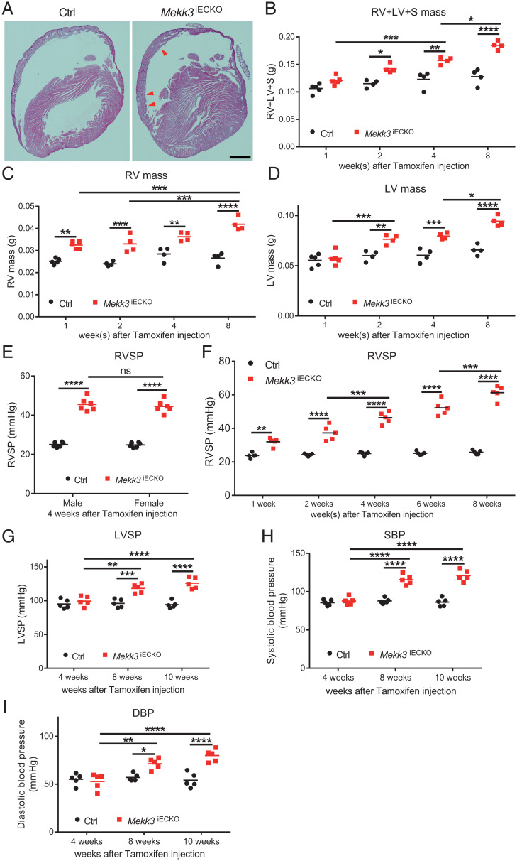
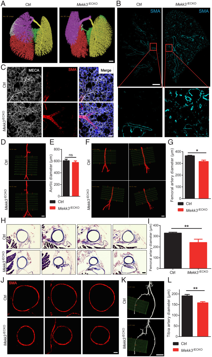
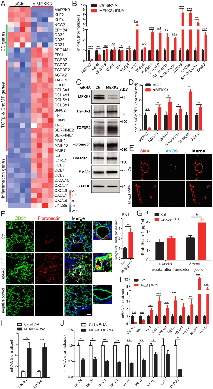
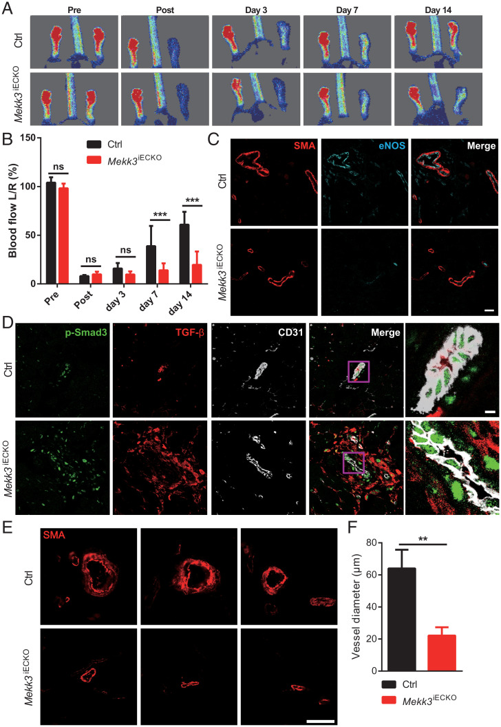
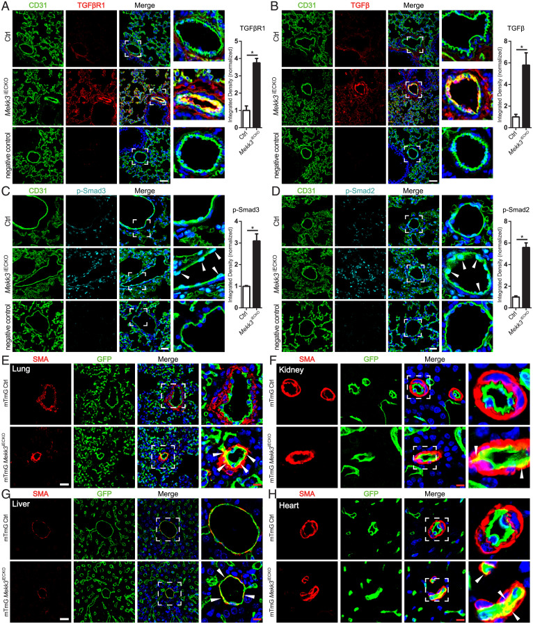
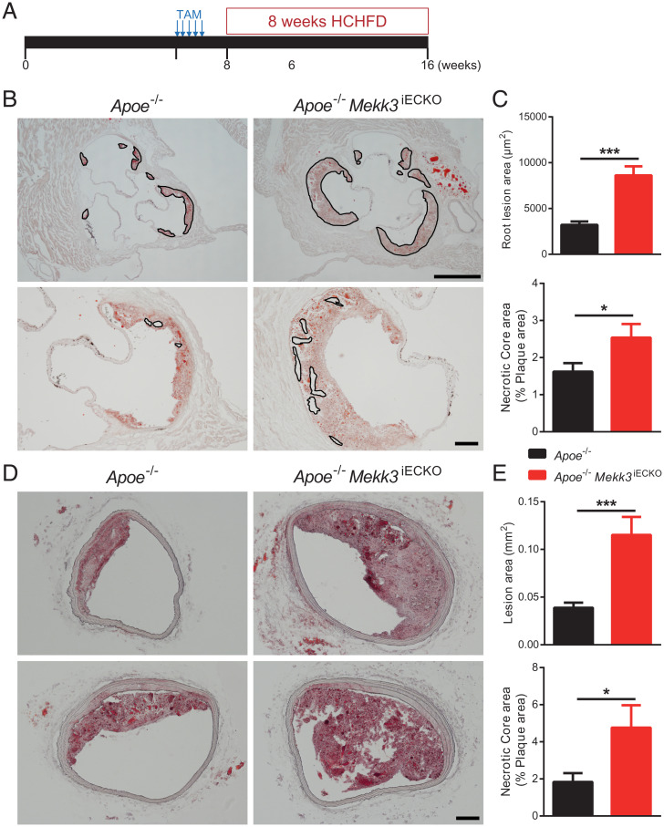
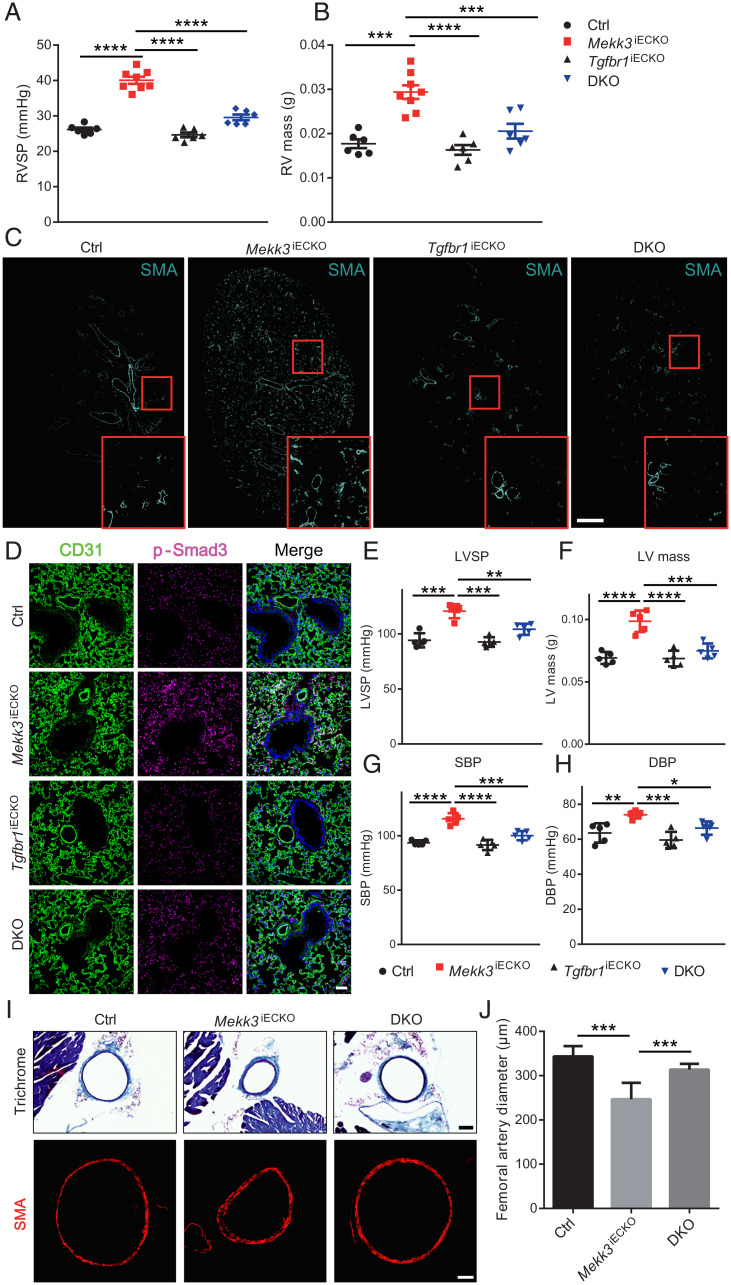
Arterial remodeling is an important adaptive mechanism that maintains normal fluid shear stress in a variety of physiologic and pathologic conditions. Inward remodeling, a process that leads to reduction in arterial diameter, plays a critical role in progression of such common diseases as hypertension and atherosclerosis. Yet, despite its pathogenic importance, molecular mechanisms controlling inward remodeling remain undefined. Mitogen-activated protein kinases (MAPKs) perform a number of functions ranging from control of proliferation to migration and cell-fate transitions. While the MAPK ERK1/2 signaling pathway has been extensively examined in the endothelium, less is known about the role of the MEKK3/ERK5 pathway in vascular remodeling. To better define the role played by this signaling cascade, we studied the effect of endothelial-specific deletion of its key upstream MAP3K, MEKK3, in adult mice. The gene's deletion resulted in a gradual inward remodeling of both pulmonary and systematic arteries, leading to spontaneous hypertension in both vascular circuits and accelerated progression of atherosclerosis in hyperlipidemic mice. Molecular analysis revealed activation of TGFβ-signaling both in vitro and in vivo. Endothelial-specific TGFβR1 knockout prevented inward arterial remodeling in MEKK3 endothelial knockout mice. These data point to the unexpected participation of endothelial MEKK3 in regulation of TGFβR1-Smad2/3 signaling and inward arterial remodeling in artery diseases.

Keywords: MEKK3; atherosclerosis; inward arterial remodeling; pulmonary arterial hypertension; systemic hypertension.


Conflict of interest statement

The authors declare no competing interest.

Figures

Fig. 1.
Fig. 1.
Deletion of endothelial MEKK3 in adult mice induces pulmonary and systemic hypertension. (A) Representative Hematoxylin and Eosin (H&E) staining of Ctrl and Mekk3iECKO mice hearts at 4 wk after tamoxifen injection. Arrowheads point to the right ventricle hypertrophy. (Scale bar, 1,000 μm.) (BD) Time-course analysis of RV, LV, and septum (S) mass for Ctrl and Mekk3iECKO mice at indicated time points, n = 5 mice at 1 wk and n = 4 mice at 2, with 4 and 8 wk per time point and per group; all mice are male. (E) RVSP of male and female Ctrl and Mekk3iECKO mice at 4 wk after tamoxifen injection, showing no difference between male and female. n = 6 mice for per genotype. (F) Time-course analysis of RVSP for Ctrl and Mekk3iECKO mice at indicated time points, n = 5 mice (3 male and 2 female) per time point and per group. (GI) Time-course analysis of LV systolic pressure (LVSP), Systolic blood pressure (SBP), and Diastolic blood pressure (DBP) from Ctrl and Mekk3iECKO mice at indicated time points, n = 5 male mice per time point and per group. Bars represent the mean. *P < 0.05, **P < 0.01, ***P < 0.001, ****P < 0.0001, and ns: not significant, calculated by two-way ANOVA with Tukey’s multiple comparison tests.
Fig. 2.
Fig. 2.
Endothelial MEKK3 deletion leads to inward vascular remolding. (A) Representative micro-CT images of Ctrl and Mekk3iECKO entire lungs at 6 wk after tamoxifen injection. Each color represents one lung lobe. (Scale bar, 1,000 μm.) (B) Representative SMA immunostaining in Ctrl and Mekk3iECKO entire lung sections. (Scale bar, 1 mm.) (C) Representative higher-magnification images of SMA staining of Ctrl and Mekk3iECKO lungs. (Scale bar, 50 μm.) (D) Representative micro-CT images of Ctrl and Mekk3iECKO aorta. (Scale bar: 1,000 μm.) (E) Quantification of aortic diameter (n = 3 male mice). (F) Representative micro-CT images of Ctrl and Mekk3iECKO femoral artery. (Scale bar, 1,000 μm.) (G) Quantification of femoral artery diameter for Ctrl (n = 6 male mice) and Mekk3iECKO mice (n = 5 male mice) at 8 wk after tamoxifen injection. (H) Masson Trichrome staining of Ctrl and Mekk3iECKO femoral artery sections. (Scale bar, 100 μm.) (I) Quantification of femoral artery diameter. n = 6 male mice per group. Data represent mean ± SD. (J) SMA immunostaining in Ctrl and Mekk3iECKO femoral artery sections. (Scale bar, 50 μm.) (K) Representative micro-CT images of Ctrl and Mekk3iECKO tibia artery. (Scale bar, 1,000 μm.) (L) Quantification of tibia artery diameter for Ctrl (n = 6 male mice) and Mekk3iECKO mice (n = 5 male mice) at 8 wk after tamoxifen injection. ns: not significant, *P < 0.05, and **P < 0.01, calculated by Mann–Whitney U test.
Fig. 3.
Fig. 3.
Loss of MEKK3 in ECs induces TGFβ-signaling. (A) Bulk RNA-seq analysis of HUVECs treated with Ctrl or MEKK3 siRNA. Heatmap shows EC, TGFβ, EndMT genes, and inflammatory genes. n = 4 samples for each group. (B) qpcr analysis of TGFβ pathway and EndMT markers expression in HUVECs treated with Ctrl or MEKK3 siRNA (n = 3). Data represent mean ± SD. (C and D) Representative WB and densitometric quantification of MEKK3 (n = 3), TGFβR1 (n = 4), TGFβR2 (n = 3), fibronectin (n = 3), collagen (n = 3), and SM22α (n = 3) expression in HUVECs treated with Ctrl or MEKK3 siRNA. Data represent mean ± SEM. (E) Immunostaining of eNOS and SMA in Ctrl and Mekk3iECKO lungs. (Scale bar, 20 μm.) (F) Immunostaining and quantification of Fibronectin in Ctrl and Mekk3iECKO lungs. Negative control means no primary antibody control. (Scale bar, 25 μm,) Data represent mean ± SEM. (G) Circulating endothelin-1 concentration in plasma from Ctrl and Mekk3iECKO mice at 4 and 8 wk after tamoxifen injection. n = 4 male mice per group. (H) qpcr analysis of EndMT markers in lung ECs isolated from Ctrl and Mekk3iECKO mice. Data represent mean ± SD. n = 4 mice per group. (I) qpcr analysis of LIN28a and LIN28b (n = 4) expression in HUVECs treated with Ctrl or MEKK3 siRNA. Data represent mean ± SD. (J) qpcr analysis of let-7 miRNA family (n = 3) expression in HUVECs treated with Ctrl or MEKK3 siRNA. ns: not significant, *P < 0.05, **P < 0.01, and ***P < 0.001, calculated by unpaired t test (B, D, F, H, I, and J) and two-way ANOVA with Sidak’s multiple comparison tests (G).
Fig. 4.
Fig. 4.
Loss of MEKK3 in ECs impairs blood-flow recovery and increases TGFβ-signaling in hindlimb ischemia (HLI) model. (A) Representative images of blood-flow recovery from Ctrl and Mekk3iECKO mice at indicated days. (B) Quantification of blood-flow recovery for Ctrl and Mekk3iECKO mice. Data represent mean ± SD. n = 6 male mice per group. (C and D) Immunostaining of eNOS and SMA (C) and TGFβ and p-Smad3 (D) in gracilis muscle blood vessels from Ctrl and Mekk3iECKO mice at day 7. (Scale bar, 20 μm.) (E and F) SMA immunostaining and quantification of blood vessels diameter for Ctrl and Mekk3iECKO mice. Data represent mean ± SEM. n = 6 male mice per group. (Scale bar, 50 μm.) **P < 0.01, ***P < 0.001, and ns: not significant, calculated by unpaired t test (F) and two-way ANOVA with Sidak’s multiple comparison tests (B).
Fig. 5.
Fig. 5.
Loss of MEKK3 in the endothelium induces EndMT. (AD) Representative immunostaining and quantification of TGFβR1 (A), TGFβ (B), p-Smad3 Ser423/425 (C), and p-Smad2 Ser465/467 (D) in lung sections from Ctrl and Mekk3iECKO mice at 4 wk after tamoxifen injection. n = 4 mice per group. Negative control means no primary antibody control. Arrowheads show p-Smad2 and 3 staining in ECs. (Scale bar, 25 μm,) Data represent mean ± SEM. *P < 0.05, calculated by Mann–Whitney U test. (EG) Representative GFP, SMA, and DAPI staining in lung (E), kidney (F), liver (G), and heart (H) from mTmG Ctrl and mTmG Mekk3iECKO mice at 4 wk after tamoxifen injection. (Scale bars, 25 μm [white] and 8 μm [red].) Arrowheads point to ECs expressing SMA.
Fig. 6.
Fig. 6.
Endothelial MEKK3 knockout increases atherosclerotic plaque growth. (A) Experiment timeline: at 6 wk, Apoe−/− mice and Apoe−/− Mekk3iECKO mice were intraperitoneally injected tamoxifen for 5 consecutive d. From 8 wk, mice were fed HCHFD for 8 wk, then were euthanized for atherosclerosis analysis. (B) Representative Oil-Red-O (ORO) staining of aortic root. Outlines in the upper images indicate plaque lesion area and in the lower images indicate necrotic core area. (Scale bars, 500 μm [Upper] and 100 μm [Lower].) (C) Quantification of aortic root lesion area and necrotic core area for Apoe−/− mice (n = 10 male mice) and Apoe−/− Mekk3iECKO mice (n = 7 male mice). (D) Representative ORO staining of brachiocephalic artery. (Scale bar, 100 μm.) (E) Quantification of brachiocephalic artery lesion area and necrotic core area for Apoe−/− mice (n = 10 male mice) and Apoe−/− Mekk3iECKO mice (n = 7 male mice). Data represent mean ± SEM. *P < 0.05 and ***P < 0.001, calculated by unpaired t test.
Fig. 7.
Fig. 7.
Genetic deletion of endothelial TGFβR1 attenuates MEKK3 deficiency-induced hypertension. (A and B) Right ventricle systolic pressure (RVSP) and RV mass of Ctrl (n = 6, 3 male and 3 female), Mekk3iECKO (n = 8, 4 male and 4 female), Tgfbr1iECKO (n = 6, 3 male and 3 female), and DKO mice (n = 6, 3 male and 3 female) at 3 wk after tamoxifen injection. (C) Representative immunostaining of SMA of Ctrl and Mekk3iECKO, Tgfbr1iECKO, and DKO entire lungs. (Scale bar, 1 mm.) (D) Immunostaining of p-Smad3 in Ctrl and Mekk3iECKO, Tgfbr1iECKO, and DKO lungs. (Scale bar, 50 μm.) (EH) Left ventricle systolic pressure (LVSP), LV mass, Systolic blood pressure (SBP), and Diastolic blood pressure (DBP) of Ctrl, Mekk3iECKO, Tgfbr1iECKO, and DKO mice at 8 wk after tamoxifen injection. (I) Representative Masson Trichrome staining (up) and SMA staining (down) of Ctrl, Mekk3iECKO, and DKO mice femoral artery sections. (Scale bar, 100 μm [Upper] and 50 μm [Lower].) (J) Quantification of femoral artery diameter from Trichrome-stained sections (n = 30 sections from three pairs of mice per group). (EH) n = 5 mice (3 male and 2 female) for each group. Data represent mean ± SEM. *P < 0.05, **P < 0.01, ***P < 0.001, and ****P < 0.0001, calculated by one-way ANOVA with Tukey’s multiple comparison tests.

References

    1. Baeyens N., Bandyopadhyay C., Coon B. G., Yun S., Schwartz M. A., Endothelial fluid shear stress sensing in vascular health and disease. J. Clin. Invest. 126, 821–828 (2016). - PMC - PubMed
    1. Renna N. F., de Las Heras N., Miatello R. M., Pathophysiology of vascular remodeling in hypertension. Int. J. Hypertens. 2013, 808353 (2013). - PMC - PubMed
    1. Castorena-Gonzalez J. A., Staiculescu M. C., Foote C., Martinez-Lemus L. A., Mechanisms of the inward remodeling process in resistance vessels: Is the actin cytoskeleton involved? Microcirculation 21, 219–229 (2014). - PMC - PubMed
    1. Ward M. R., et al. , Low blood flow after angioplasty augments mechanisms of restenosis: Inward vessel remodeling, cell migration, and activity of genes regulating migration. Arterioscler. Thromb. Vasc. Biol. 21, 208–213 (2001). - PubMed
    1. Peti W., Page R., Molecular basis of MAP kinase regulation. Protein Sci. 22, 1698–1710 (2013). - PMC - PubMed

Publication types

MeSH terms

Substances