. 2021 Dec 15;11(1):24066.
doi: 10.1038/s41598-021-02174-6.

Eruca sativa seed napin structural insights and thorough functional characterization

Affiliations

Eruca sativa seed napin structural insights and thorough functional characterization

Binish Khaliq et al. Sci Rep. .

Abstract

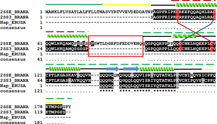
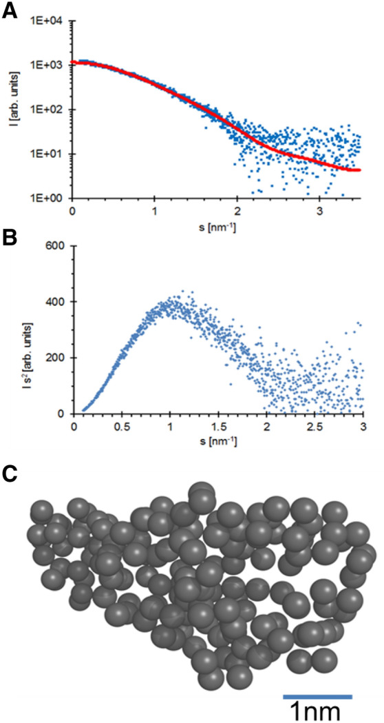
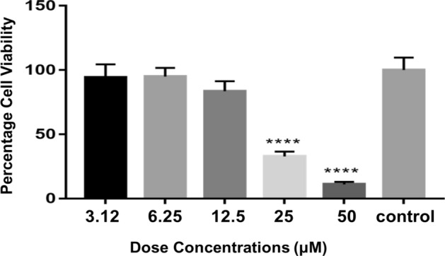
A potent napin protein has been thoroughly characterized from seeds of rocket salad (Eruca sativa). Eruca sativa napin (EsNap) was purified by ammonium sulfate precipitation (70%) and size-exclusion chromatography. Single intact 16 kDa EsNap band was reduced to 11 and 5 kDa bands respectively on SDS-PAGE. Nano LC-MS/MS yielded two fragments comprising of 26 residues which showed 100% sequence identity with napin-3 of Brassica napus. CD spectroscopy indicated a dominant α-helical structure of EsNap. Monodispersity of EsNap was verified by dynamic light scattering, which also confirmed the monomeric status with a corresponding hydrodynamic radius of 2.4 ± 0.2 nm. An elongated ab initio shape of EsNap was calculated based on SAXS data, with an Rg of 1.96 ± 0.1 nm. The ab initio model calculated by DAMMIF with P1 symmetry and a volume of approx. 31,100 nm3, which corresponded to a molecular weight of approximately 15.5 kDa. The comparison of the SAXS and ab initio modeling showed a minimized χ2-value of 1.87, confirming a similar molecular structure. A homology model was predicted using the coordinate information of Brassica napus rproBnIb (PDB ID: 1SM7). EsNap exhibited strong antifungal activity by significantly inhibiting the growth of Fusarium graminearum. EsNap also showed cytotoxicity against the hepatic cell line Huh7 and the obtained IC50 value was 20.49 µM. Further, strong entomotoxic activity was experienced against different life stages of stored grain insect pest T. castaneum. The result of this study shows insights that can be used in developing potential antifungal, anti-cancerous and insect resistance agents in the future using EsNap from E. sativa.


Conflict of interest statement

The authors declare no competing interests.

Figures

Figure 1
Figure 1
Purification and molecular weight determination of E. sativa seed napin (EsNap). (A) Partial purification of EsNap from crude extract, lane 1; crude extract, lane 2 and 3; 50% ammonium sulfate saturation of crude extract supernatant, lane 4; re-dissolved and dialyzed 70% ammonium sulfate saturation pellet, M; Protein Ladder (Catalog, 22,610). (B) Purification of EsNap by calibrated size exclusion chromatography (SEC, HiLoad 16/60 Superdex 75) subsequent to ammonium sulfate precipitation (70% saturated solution). Chromatogram showed the P1 and P2 with retention volume of 48 and 80.7 ml respectively. (C) SDS-PAGE showed that P1 has cruciferin and EsNap was found in P2 of the chromatogram while lane 1 is the 70% solubilized pellet before SEC. (D) SDS-PAGE analysis of napin under non-reducing condition (lane 1) and in the presence of 20 mM DTT (lane 2) which splitted the intact band into approximately 11 and 5 kDa respectively.
Figure 2
Figure 2
Multiple sequence alignment of EsNap with other closely related plant napins 2SS3_BRANA (Napin-3: Brassica napus), 2SSE_BRANA (Napin embryo-specific: Brassica napus) and Nap_ ERUSA (Napin: Eruca sativa). Secondary structure elements (α-helices and β-sheets) of EsNap are indicated at the top. Signal peptide, N/C terminus and turns are indicated with green, yellow and black bars respectively. Identically conserved residues are labeled by asterisks (*), while semi-conserved substitutions are labeled by single dots (.) and cysteine involved in disulfide bonds between two chains are shown in red color. The highly flexible region is highlighted with a red box. Smaller and larger chains are shown in dot line (–) upper on the secondary structure elements in green and red color respectively Multiple sequences alignment was performed by using ClustalW in the default set up and BoxShade server.
Figure 3
Figure 3
CD spectroscopy of EsNap. Far-UV CD spectrum of EsNap is indicating predominantly α-helical secondary structure (38%).
Figure 4
Figure 4
The particle size distribution obtained by dynamic light scattering reveals the monodispersity of the EsNap solution (blue line) with a hydrodynamic radius of 2.4 ± 0.2 nm. A broadening and shift in hydrodynamic radius (1.8 ± 0.2 nm; red line) was observed after the addition of 20 mM DTT to EsNap solution which confirmed the splitted banding pattern as already observed on SDS-PAGE under reduced conditions. Obtained results indicated that EsNap is a monomeric globular protein.
Figure 5
Figure 5
(A) Small-angle X-ray scattering intensity plot of pure EsNap and the corresponding calculated fit curve (red) resembling a single ab initio model as displayed in panel C and calculated by GASBOR. The plot shows the dependency of the scattering intensity I on the momentum transfers. (B) Kratky plot of the scattering intensity distribution indicating a compact and relatively rigid protein structure (C) Ab initio model possessing P1 symmetry; the scale bar is 1 nm in length.
Figure 6
Figure 6
Overall structure of EsNap in-silico model and structural alignments. (1A) The 3D structure was predicted by homology modelling and is shown as ribbon diagram. The 3D model of EsNap consisted of four helices and a hypervariable loop comprising of two antiparallel β-sheets (yellow color) marked by red circle. (2A)The smaller chain composed of N-terminal (green) to H1 helix (purple) and longer composed of H2, H3, H4 and two shortβ-sheets (red). The disulfide bridges are represented by yellow spheres (B) Ab initio model of EsNap with P1 symmetry (grey spheres), Lys 9, Arg 11, Lys 12 (blue) and Lys 105 (red) form flexible N and C-terminal of ab initio model and 3D model of EsNap have been superimposed; involved in antifungal and anticancer activity. (C) Structural alignment of E. sativa napin (Cyan, EsNap) with BnIb from Brassica napus (Green, pdb code: 1SM7) and 2S albumin from Moringa oleifera (Purple, pdb code: 5DOM). Hypervariable loops and connecting loops between two polypeptide domains are colored in yellow and red, respectively.
Figure 7
Figure 7
Inhibitory activities of EsNap towards growth of Fusarium graminearum mycelia. (A) All the three EsNap concentrations (30, 50 and 100 µg) in medium inhibited the fungal growth, while regular and optimal fungal growth was observed in medium mixed with buffer and 30–100 µg BSA as well as only in buffer as negative control. No fungal growth was observed in the medium with fungicide TOPSIN as positive control. (B) Inhibition of mycelia growth of Fusarium graminearum in gene frame chamber. [a: BSA (100 µg); b: phosphate buffer; c: Concentrated EsNap protein (100 µg)].
Figure 8
Figure 8
Effect of different doses of EsNap on Huh-7 cells visualized by plotting the protein concentration (µM) against the cell viability (%). Asterisks are indicating significant mycelia inhibition at concentrations of 25 and 50 µM; however, calculated IC50 value of EsNap was 20.49 µM.

Similar articles

Cited by

References

    1. Deice Raasch-Fernandes L, et al. Induction of phytoalexins and proteins related to pathogenesis in plants treated with extracts of cutaneous secretions of southern Amazonian Bufonidae amphibians. PLoS ONE. 2019;14:e0211020. - PMC - PubMed
    1. Deveau A, et al. Bacterial–fungal interactions: Ecology, mechanisms and challenges. FEMS Microbiol. Rev. 2018;42:335–352. - PubMed
    1. Edqvist J, Blomqvist K, Nieuwland J, Salminen TA. Plant lipid transfer proteins: Are we finally closing in on the roles of these enigmatic proteins? J. Lipid Res. 2018;59:1374–1382. - PMC - PubMed
    1. Tam JP, Wang S, Wong KH, Tan WL. Antimicrobial peptides from plants. Pharmaceuticals. 2015;8:711–757. - PMC - PubMed
    1. Ishaq N, Bilal M, Iqbal H. Medicinal potentialities of plant defensins: A review with applied perspectives. Medicines. 2019;6:29. - PMC - PubMed

Publication types

MeSH terms