. 2022 Apr;126(6):865-873.
doi: 10.1038/s41416-021-01619-z. Epub 2021 Dec 15.

Molecular pathways in post-colonoscopy versus detected colorectal cancers: results from a nested case-control study

Affiliations

Molecular pathways in post-colonoscopy versus detected colorectal cancers: results from a nested case-control study

Roel M M Bogie et al. Br J Cancer. 2022 Apr.

Abstract

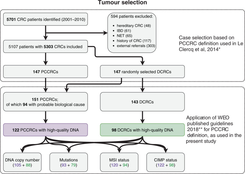
Background: Post-colonoscopy colorectal cancers (PCCRCs) pose challenges in clinical practice. PCCRCs occur due to a combination of procedural and biological causes. In a nested case-control study, we compared clinical and molecular features of PCCRCs and detected CRCs (DCRCs).

Methods: Whole-genome chromosomal copy number changes and mutation status of genes commonly affected in CRC were examined by low-coverage WGS and targeted sequencing, respectively. MSI and CIMP status was also determined.

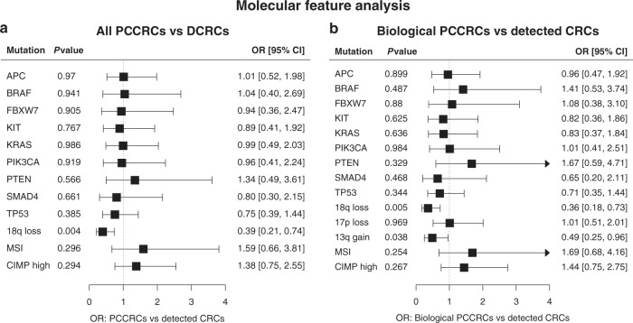
Results: In total, 122 PCCRCs and 98 DCRCs with high-quality DNA were examined. PCCRCs were more often located proximally (P < 0.001), non-polypoid appearing (P = 0.004), early stage (P = 0.009) and poorly differentiated (P = 0.006). PCCRCs showed significantly less 18q loss (FDR < 0.2), compared to DCRCs. No significant differences in mutations were observed. PCCRCs were more commonly CIMP high (P = 0.014) and MSI (P = 0.029). After correction for tumour location, only less 18q loss remained significant (P = 0.005).

Conclusion: Molecular features associated with the sessile serrated lesions (SSLs) and non-polypoid colorectal neoplasms (CRNs) are more commonly seen in PCCRCs than in DCRCs. These together with the clinical features observed support the hypothesis that SSLs and non-polypoid CRNs are contributors to the development of PCCRCs. The future focus should be directed at improving the detection and endoscopic removal of these non-polypoid CRN and SSLs.

Clinical trial registration: NTR3093 in the Dutch trial register ( www.trialregister.nl ).


Conflict of interest statement

AM received funding from the Dutch Cancer Society, from the Dutch Organization for Health Research and Development, from Pentax Europe GmBH. AM has given scientific advice to Kyowa Kirin, Bayer, and Takeda. BC has several patents pending. The remaining authors declare no competing interests.

Figures

Fig. 1
Fig. 1. Flowchart of the process of selection of CRC cases for molecular analysis.
PCCRCs post-colonoscopy CRCs, DCRCs detected CRCs. *le Clercq et al. [5]. **Rutter et al. [20].
Fig. 2
Fig. 2. Comparison of molecular features between post-colonoscopy colorectal cancers (PCCRCs) and detected colorectal cancers (DCRCs).
Forest plots showing the associations of several molecular features of (a) PCCRCs compared to DCRCs, and (b) PCCRCs with plausible biological aetiology compared to DCRCs, after multiple imputation of missing values and correction for age, gender and tumour location. OR odds ratio, CI confidence interval.
Fig. 3
Fig. 3. Unsupervised hierarchal cluster analysis based on the molecular features of all CRCs analysed.
In the cluster analysis results of the nine genes most commonly mutated in this study (APC, TP53, KRAS, KIT, PIK3CA, BRAF, FBXW7, SMAD4 and PTEN), the significant chromosomal alterations (loss of 17p, loss of 18q and gain of 13q), CIMP status and MSI, were included. a Heatmap displaying the distribution of all clinical and molecular features of all CRCs analysed in this study. Orange indicates presence, while blue indicates the absence of these features. The first three columns represent CRC type (Biological PCCRCs: red, Procedural PCCRCs: blue, DCRCs: green), CRC location (proximal: yellow, distal: dark blue) and CRC morphology (polypoid: purple, non-polypoid: light blue). After clustering of the CRCs, three large branches can be detected (blue, green, and red). b Overview of hallmark features of each branch from clustering analysis. Red: PCCRC, green: DCRC, yellow: proximal location, dark blue: distal location, light blue: non-polypoid, purple: polypoid, orange: MSI/high CIMP present, blue: MSI/high CIMP absent.

References

    1. Singh S, Singh PP, Murad MH, Singh H, Samadder NJ. Prevalence, risk factors, and outcomes of interval colorectal cancers: a systematic review and meta-analysis. Am J Gastroenterol. 2014;109:1375–89. doi: 10.1038/ajg.2014.171. - DOI - PubMed
    1. Rex DK. Preventing colorectal cancer and cancer mortality with colonoscopy: what we know and what we don’t know. Endoscopy. 2010;42:320–3. doi: 10.1055/s-0029-1244067. - DOI - PubMed
    1. le Clercq CM, Sanduleanu S. Interval colorectal cancers: what and why. Curr Gastroenterol Rep. 2014;16:375. doi: 10.1007/s11894-014-0375-3. - DOI - PubMed
    1. Pabby A, Schoen RE, Weissfeld JL, Burt R, Kikendall JW, Lance P, et al. Analysis of colorectal cancer occurrence during surveillance colonoscopy in the dietary Polyp Prevention Trial. Gastrointest Endosc. 2005;61:385–91. doi: 10.1016/S0016-5107(04)02765-8. - DOI - PubMed
    1. le Clercq CM, Bouwens MW, Rondagh EJ, Bakker CM, Keulen ET, de Ridder RJ, et al. Postcolonoscopy colorectal cancers are preventable: a population-based study. Gut. 2014;63:957–63. doi: 10.1136/gutjnl-2013-304880. - DOI - PubMed

Publication types