Review
. 2021 Nov 29:12:778913.
doi: 10.3389/fimmu.2021.778913. eCollection 2021.

Corona Virus Disease 2019 (COVID-19) as a System-Level Infectious Disease With Distinct Sex Disparities

Affiliations
Review

Corona Virus Disease 2019 (COVID-19) as a System-Level Infectious Disease With Distinct Sex Disparities

Modjtaba Emadi-Baygi et al. Front Immunol. .

Abstract

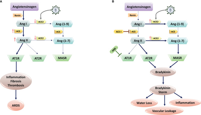
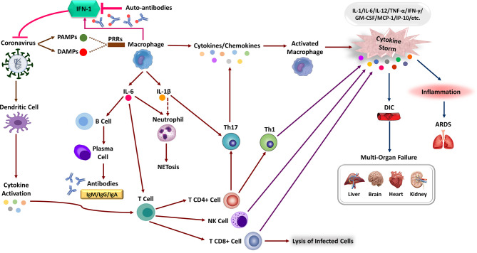
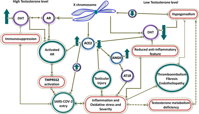
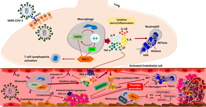
The current global pandemic of the Severe Acute Respiratory Syndrome CoronaVirus 2 (SARS-CoV-2) causing COVID-19, has infected millions of people and continues to pose a threat to many more. Angiotensin-Converting Enzyme 2 (ACE2) is an important player of the Renin-Angiotensin System (RAS) expressed on the surface of the lung, heart, kidney, neurons, and endothelial cells, which mediates SARS-CoV-2 entry into the host cells. The cytokine storms of COVID-19 arise from the large recruitment of immune cells because of the dis-synchronized hyperactive immune system, lead to many abnormalities including hyper-inflammation, endotheliopathy, and hypercoagulability that produce multi-organ dysfunction and increased the risk of arterial and venous thrombosis resulting in more severe illness and mortality. We discuss the aberrated interconnectedness and forthcoming crosstalks between immunity, the endothelium, and coagulation, as well as how sex disparities affect the severity and outcome of COVID-19 and harm men especially. Further, our conceptual framework may help to explain why persistent symptoms, such as reduced physical fitness and fatigue during long COVID, may be rooted in the clotting system.

Keywords: COVID-19; coagulation; immunity; interconnectedness; thromboembolism.


Conflict of interest statement

MN was employed by Erythrogen Medical Genetics Lab. The remaining authors declare that the research was conducted in the absence of any commercial or financial relationships that could be construed as a potential conflict of interest.

Figures

Figure 1
Figure 1
SARS-CoV-2 and Renin-Angiotensin System (RAS) interactions. (A) SARS-CoV-2 infection is mediated via the human Angiotensin-Converting Enzyme 2 (ACE2), which would cause ACE2 downregulation. Subsequent block of Ang II/Ang–(1-7) metabolism lead to the high levels of Ang II production through ACE upregulation. The imbalanced Ang II/Ang 1–7 causes the suppression of AT2 and Mas receptors with anti-inflammatory properties. In contrast, Ang II and its receptor AT1R activations induce pro-inflammatory effects, fibrosis, and thrombosis, leading to Acute Respiratory Distress Syndrome (ARDS) in lungs. (B) In patients with comorbidities, including hypertension, administration of RAS blockers (e.g., ACE-I, and ARBs) suppresses the ACE pathway. In these patients, SARS-CoV-2 infection increase ACE2 expression. Upregulated ACE-2 converts Ang I to Ang–(1-9) and Ang–(1-7), activating the bradykinin pathway via MAS and AT2R receptors. Upregulated bradykinin (bradykinin storm) in the lungs of COVID-19 patients would increase SARS-CoV-2 virulence by inducing hypotension, vascular permeability, water loss, and inflammation.
Figure 2
Figure 2
The immune response corporation in covid-19 disease. Type 1 interferon (IFN-I) immune response plays a pivotal role in effective immunity against SARS-CoV-2 infection and rapid viral clearance. Following SARS-CoV-2 infection, the IFN-1 initiates via recognizing PAMPs/DAMPs by PRRs of the human immune cells and releasing inflammatory cytokines (e.g., IL-1 beta, IL-6). The expression of numerous inflammatory cytokines leads to activation of neutrophils (NET formation) and attraction of different immune cells toward the site of infection. Macrophages activation, Dendritic Cell (DC) maturation and inflammatory cytokines stimulate the adaptive immune response to join the fight against SARS-CoV-2. Activation of T-cells via cytokines promotes multiple T-cell differentiation (i.e., CD4 +, CD8 +, NK cell, Th1) that directly kills virus-infected cells. Activated B-cells produce virus-specific antibodies that would participate in a successive immune response. In patients with severe forms of COVID-19, the delayed IFN-I pathways or neutralizing auto-Abs will lead to immune system overreaction and the generation of high inflammatory cytokine levels. The aberrant induction of the immune system and the production of various pro-inflammatory cytokines (e.g., IL-1, IL-6, MCP-1, TNF-α, and etc.), the so-called “cytokine storm,” leads to severe COVID-19 immunopathology. These can cause severe local damage to the lungs (e.g., ARDS) and other organs (e.g., DIC), and in the worst case, can lead to Multi-Organ Failures (MOF) and even death.
Figure 3
Figure 3
High and low testosterone impact on COVID-19 severity. Controlled by androgen, both TMPRSS2 and ACE2 are regulated by genes on the X chromosome. At high testosterone levels, both receptors are up-regulated, resulting in SARS-CoV-2 entry. Testosterone has an immunosuppressive feature, which leads to more virus entry and disease severity. In contrast, it has linked low testosterone level caused by hypogonadism in men to increased infection of COVID-19. Therefore, in men with hypogonadism, caused by various factors such as reduction of ACE2, testicular damage, and thromboembolism through hyperinflammation caused by the viral infection, the level of testosterone is reduced and the anti-inflammatory effect of testosterone is blocked, resulting in disease severity. Low testosterone results in downregulation of ACE2 that through activation of AR1T by ANG II leads to inflammation and disease severity.
Figure 4
Figure 4
Interactions between the components of the immune system and coagulation cascades that induce thrombosis in COVID-19. In brief, the SARS-CoV-2 infection triggers an inflammatory response in the alveolar lumen by macrophages and neutrophils. Overexpression of PAI-1 and its interaction with TLR4 induce the IL-6 expression and STAT3 activation in the lung of COVID-19 patients. Subsequent production of inflammatory cytokines such as IL-1β, IL-8, IL-6, and TNF-α causes neutrophils to be recruited and release Neutrophil Extracellular Traps (NETs). NETs directly activate factor XII, thereby activating the contact-dependent clotting pathway. The PAR-1 receptor mediates cytokine-dependent Tissue Factor (TF) activation, which stimulates the STAT3/PAI-1 complex in blood’s monocytes and enhances inflammatory cytokine production leading to extrinsic coagulation cascade activation and thrombosis formation. Moreover, inflammatory cytokine in combination with the direct binding of SARS-CoV-2 to the endothelial cells triggers Von Willebrand Factor (VWF) secretion cleaved by ADAMTS13 to the regular size. Activated TF/VWF/FVIII complex in cooperation with NETs recruits platelets to adhere to endothelial surfaces and provoke intrinsic coagulation cascades. Altogether these events ultimately driving thrombin formation from circulating prothrombin, which cleaves fibrinogen to fibrin and stimulates thrombosis formation. Besides, suppression of endothelial enzymes and Plasminogen Activators (tPA/uPA) would aggravate the coagulopathy state by preventing effective fibrinolysis. Both tPA and uPA participate in the normal coagulation-plasmin-fibrin pathway, can be inhibited by PAI-1/HIF-1α in severe COVID-19 patients with ARDS.

Similar articles

Cited by

References

    1. Tay MZ, Poh CM, Rénia L, MacAry PA, Ng LFP. The Trinity of COVID-19: Immunity, Inflammation and Intervention. Nat Rev Immunol (2020) 20(6):363–74. doi: 10.1038/s41577-020-0311-8 - DOI - PMC - PubMed
    1. Giovanetti M, Benedetti F, Campisi G, Ciccozzi A, Fabris S, Ceccarelli G, et al. . Evolution Patterns of SARS-CoV-2: Snapshot on Its Genome Variants. Biochem Biophys Res Commun (2021) 538:88–91. doi: 10.1016/j.bbrc.2020.10.102 - DOI - PMC - PubMed
    1. Haney S, Bardwell L, Nie Q. Ultrasensitive Responses and Specificity in Cell Signaling. BMC Syst Biol (2010) 4:119. doi: 10.1186/1752-0509-4-119 - DOI - PMC - PubMed
    1. Rowland MA, Greenbaum JM, Deeds EJ. Crosstalk and the Evolvability of Intracellular Communication. Nat Commun (2017) 8(1):1–8. doi: 10.1038/ncomms16009 - DOI - PMC - PubMed
    1. Lin L, Lu L, Cao W, Li T. Hypothesis for Potential Pathogenesis of SARS-CoV-2 Infection–A Review of Immune Changes in Patients With Viral Pneumonia. Emerg Microbes Infect (2020) 9(1):727–32. doi: 10.1080/22221751.2020.1746199 - DOI - PMC - PubMed

Publication types

MeSH terms