. 2021 Dec 6:2021:8381115.
doi: 10.1155/2021/8381115. eCollection 2021.

Bevacizumab Increases Endothelin-1 Production via Forkhead Box Protein O1 in Human Glomerular Microvascular Endothelial Cells In Vitro

Affiliations

Bevacizumab Increases Endothelin-1 Production via Forkhead Box Protein O1 in Human Glomerular Microvascular Endothelial Cells In Vitro

Satoru Nihei et al. Int J Nephrol. .

Abstract

Molecular mechanisms underlying the nephrotoxicity associated with bevacizumab are unclear. Endothelin-1 (ET-1) is involved in podocyte injury and proteinuria, and its level increases in most cases of kidney disorders. Forkhead box protein O1 (FoxO1), a transcription factor, is a major determinant of ET-1 promoter activation and is regulated by protein kinase B (Akt) phosphorylation-dependent nuclear exclusion. We evaluated the effect of bevacizumab on ET-1 production in human glomerular microvascular endothelial cells (hGECs). We analyzed the changes in the mRNA and protein levels of ET-1 in hGECs treated with bevacizumab using real-time reverse transcription-polymerase chain reaction and enzyme-linked immunosorbent assay. Changes in the protein levels and phosphorylation status of Akt and FoxO1 in hGECs treated with bevacizumab were analyzed by western blotting. After cell lysis, FoxO1 protein was isolated from the cytoplasmic and nuclear fractions. We also investigated the effects of AS1842856 (a FoxO1 inhibitor) on bevacizumab-induced ET-1 production. Bevacizumab significantly and dose-dependently increased the mRNA and protein levels of ET-1 in hGECs (p < 0.05). Bevacizumab treatment also led to a decrease in phosphorylated Akt protein levels. Inhibition of Akt activity by LY294002 promoted ET-1 production. Bevacizumab also induced an increase in FoxO1 protein levels in the nucleus. Inhibition of FoxO1 activity by AS1842856 resulted in decreased ET-1 levels in bevacizumab-treated hGECs. ET-1 axis activation, Akt inactivation, and FoxO1 nuclear localization are the molecular mechanisms underlying bevacizumab-induced nephrotoxicity. Therefore, inhibition of renal ET-1 production could be a promising approach to protect against or treat bevacizumab-induced nephrotoxicity.


Conflict of interest statement

The authors declare that they have no conflicts of interest.

Figures

Figure 1
Figure 1
Bevacizumab induced ET-1 production in cultured human glomerular microvascular endothelial cells (hGECs). hGECs were plated in 24-well plates (1 × 105 cells/well) for ELISA or 60 mm culture dishes (1 × 106 cells/dish) for RT-PCR. hGECs that were 90% confluent were serum-starved for 24 h before the experiments were performed. hGECs were treated with 0.1 or 1 µM bevacizumab for 8 h, RNA was extracted from the cells, and ET-1 mRNA level was examined by real-time RT-PCR (a). The medium was collected and ET-1 protein level was examined by the ELISA (b). Data shown are means ± SD (n = 3; p < 0.05). The experiments were repeated at least three times, with reproducible results.
Figure 2
Figure 2
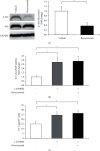
The Akt pathway was inactivated in human glomerular microvascular endothelial cells (hGECs) treated with bevacizumab. hGECs were plated in 24-well plates (1 × 105 cells/well) for ELISA or 60 mm culture dishes (1 × 106 cells/dish) for RT-PCR and western blot. hGECs that were 90% confluent were serum-starved for 24 h before the experiments were performed. hGECs were treated with 1 µM bevacizumab or 10 µM LY294002 (PI3K/Akt inhibitor) for 8 h, and western blots were used to evaluate the changes in Akt phosphorylation after bevacizumab treatment (a). The levels of ET-1 mRNA (b) and ET-1 protein (c) were measured using real-time RT-PCR and the ELISA after LY294002 treatment. Data shown are means ± SD (n = 3; p < 0.05). The experiments were repeated at least three times, with reproducible results.
Figure 3
Figure 3
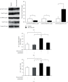
The nuclear localization of FoxO1 was increased in human glomerular microvascular endothelial cells (hGECs) treated with bevacizumab. hGECs were plated in 24-well plates (1 × 105 cells/well) for ELISA or 60 mm culture dishes (1 × 106 cells/dish) for RT-PCR and western blot. hGECs that were 90% confluent were serum-starved for 24 h before the experiments were performed. hGECs were treated with 1 µM bevacizumab and 0.1 µM AS1842856 (FoxO1 inhibitor) for 8 h, and western blots were used to evaluate the changes in FoxO1 protein level in cytosolic (cyto) and nuclear (nu) fractions after bevacizumab treatment (a). The levels of ET-1 mRNA (b) and ET-1 protein (c) were measured using real-time RT-PCR and the ELISA after bevacizumab and AS1842856 treatment. Data shown are means ± SD (n = 3; p < 0.05). The experiments were repeated at least three times, with reproducible results.
Figure 4
Figure 4
Vascular endothelial growth factor A (VEGFA) is associated with the suppression of ET-1 production in cultured human glomerular microvascular endothelial cells (hGECs). hGECs were plated in 24-well plates (1 × 105 cells/well) for ELISA or 60 mm culture dishes (1 × 106 cells/dish) for RT-PCR. hGECs that were 90% confluent were serum-starved for 24 h before the experiments were performed. hGECs were treated with 0, 10, and 40 ng/mL of VEGFA or with a combination of 40 ng/mL VEGFA and 1 µM bevacizumab for 8 h. The levels of ET-1 mRNA (a) and ET-1 protein (b) were measured using real-time RT-PCR and the ELISA after VEGFA and bevacizumab treatment. Data shown are means ± SD (n = 3; p < 0.05). The experiments were repeated at least three times, with reproducible results.
Figure 5
Figure 5
A schematic showing signaling pathways that increase ET-1 production on FoxO1 nuclear localization induced by bevacizumab in human glomerular microvascular endothelial cells (hGECs). Abbreviations: VEGFA, vascular endothelial growth factor A; Akt, protein kinase B; FoxO1, forkhead box protein O1; ET-1, endothelin-1.

References

    1. Kabbinavar F., Hurwitz H. I., Fehrenbacher L., et al. Phase II, randomized trial comparing bevacizumab plus fluorouracil (FU)/leucovorin (LV) with FU/LV alone in patients with metastatic colorectal cancer. Journal of Clinical Oncology . 2003;21(1):60–65. doi: 10.1200/jco.2003.10.066. - DOI - PubMed
    1. Sandler A., Gray R., Perry M. C., et al. Paclitaxel-carboplatin alone or with bevacizumab for non-small-cell lung cancer. New England Journal of Medicine . 2006;355(24):2542–2550. doi: 10.1056/nejmoa061884. - DOI - PubMed
    1. Rini B. I., Halabi S., Rosenberg J. E., et al. Phase III trial of bevacizumab plus interferon alfa versus interferon alfa monotherapy in patients with metastatic renal cell carcinoma: final results of CALGB 90206. Journal of Clinical Oncology . 2010;28(13):2137–2143. doi: 10.1200/jco.2009.26.5561. - DOI - PMC - PubMed
    1. Ferrara N., Hillan K. J., Gerber H.-P., Novotny W. Discovery and development of bevacizumab, an anti-VEGF antibody for treating cancer. Nature Reviews Drug Discovery . 2004;3(5):391–400. doi: 10.1038/nrd1381. - DOI - PubMed
    1. Ferrara N., Hillan K. J., Novotny W. Bevacizumab (Avastin), a humanized anti-VEGF monoclonal antibody for cancer therapy. Biochemical and Biophysical Research Communications . 2005;333(2):328–335. doi: 10.1016/j.bbrc.2005.05.132. - DOI - PubMed