

[Clinical application of superior lateral genicular artery perforator propeller flap in repair of soft-tissue defects around knee joint]
- PMID: 34913318
- PMCID: PMC8669197
- DOI: 10.7507/1002-1892.202106024
[Clinical application of superior lateral genicular artery perforator propeller flap in repair of soft-tissue defects around knee joint]
Abstract
Objective: To explore the feasibility and effectiveness of using the superior lateral genicular artery (SLGA) perforator propeller flap to reconstruct soft-tissue defects around the knee joint.
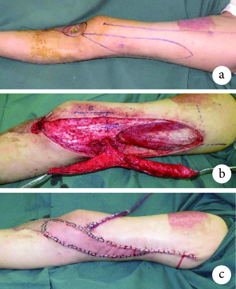
Methods: Between October 2013 and May 2019, 10 patients underwent repairing of soft-tissue defects around the knee joint using the SLGA perforator propeller flap. There were 6 males and 4 males, with a median age of 34.5 years (range, 6-66 years). Etiologies included radical tumor resection in 4 cases, post-burn scar contracture in 3 cases, post-burn hypertrophic scar in 2 cases, and prothesis exposure after knee arthroplasty in 1 case. Defects located on the lateral knee in 6 cases, proximal lateral leg in 2 cases, popliteal fossa in 1 cases, and infrapatellar region in 1 case. The size of soft-tissue defects was from 6 cm×4 cm to 14 cm×8 cm. The extraction range of the flap was from 10.0 cm×5.5 cm to 23.0 cm×7.0 cm; the length of the perforator pedicle was 2.5-5.0 cm, with an average of 3.65 cm; the flaps were rotated 180°, the large paddle of the propeller flap was used to repair the defect, and the small paddle was used to assist the closure of donor site.
Results: Blister was observed in the distal 3-cm of one flap and the flap survived after conservative management. All the flaps survived, and the wounds in the donor and recipient areas healed by first intention. There was no vascular crisis, incision dehiscence, infection, or other complications. All 10 patients were followed up 4 to 48 months, with an average of 12.6 months. The color and texture of the flap were similar to those of the recipient area, and there was no need for secondary operation for degreasing and thinning. Scar contracture was corrected; no tumor recurrence was found in tumor patients; the artificial knee joint was preserved, the knee joint flexion and extension activities were good, and all the patient and family members were satisfied with the appearance and function of the lower limbs after operation.
Conclusion: The SLGA perforator propeller flap surgery is relatively simple without the need of microvascular anastomosis, has the minimal donor-site morbidities, and can provide a compound flap for the repairing of a complex wound. The SLGA perforator propeller flap is one of the optional methods to repair soft-tissue defects around the knee joint.
目的: 探讨应用膝上外侧动脉(superior lateral genicular artery,SLGA)穿支螺旋桨皮瓣修复膝关节周围软组织缺损的可行性及疗效。.
方法: 2013年10月—2019年5月,应用SLGA穿支螺旋桨皮瓣修复膝关节周围软组织缺损10例。其中男6例,女4例;年龄6~66岁,中位年龄34.5岁。病因:肿瘤根治性切除4例,烧伤后瘢痕挛缩3例,烧伤后瘢痕增生2例,人工膝关节置换术后组织坏死假体外露1例。部位:膝关节外侧6例,小腿近端外侧2例,腘窝1例,髌骨下方1例。软组织缺损范围 6 cm×4 cm~14 cm×8 cm。皮瓣切取范围为10.0 cm×5.5 cm~23.0 cm×7.0 cm;穿支血管蒂长度为2.5~5.0 cm,平均3.65 cm;皮瓣均旋转180°。螺旋桨皮瓣的大桨修复缺损,小桨辅助供区关闭。.
结果: 术后1例患者皮瓣远端3 cm处出现水疱,经常规换药后愈合。所有皮瓣全部成活,供受区创面均Ⅰ期愈合,无血管危象及切口裂开、感染等并发症发生。10例患者均获随访,随访时间4~48个月,平均12.6个月。所有皮瓣颜色、质地和厚度与受区相似,无需二次手术去脂修薄。瘢痕挛缩得到矫正;肿瘤患者未见肿瘤复发;人工膝关节得以保留,膝关节屈伸活动度良好。患者及家属对术后下肢外观及功能满意。.
结论: SLGA穿支螺旋桨皮瓣切取相对简单,无需显微血管吻合,供区损伤小,可以切取复合组织瓣,是修复膝关节周围皮肤软组织缺损可选择的方法之一。.
Keywords: Knee joint; perforator propeller flap; soft-tissue defect; superior lateral genicular artery; wound repair.
Conflict of interest statement
利益冲突:所有作者声明,在课题研究和文章撰写过程中不存在利益冲突。
Figures


References
-
- Demirseren ME, Efendioglu K, Demiralp CO, et al Clinical experience with a reverse-flow anterolateral thigh perforator flap for the reconstruction of soft-tissue defects of the knee and proximal lower leg. J Plast Reconstr Aesthet Surg. 2011;64(12):1613–1620. doi: 10.1016/j.bjps.2011.06.047. - DOI - PubMed
MeSH terms
LinkOut - more resources
Full Text Sources
Medical