. 2021 Dec 16;16(12):e0261127.
doi: 10.1371/journal.pone.0261127. eCollection 2021.

Metformin attenuates osteoclast-mediated abnormal subchondral bone remodeling and alleviates osteoarthritis via AMPK/NF-κB/ERK signaling pathway

Affiliations

Metformin attenuates osteoclast-mediated abnormal subchondral bone remodeling and alleviates osteoarthritis via AMPK/NF-κB/ERK signaling pathway

Haohui Guo et al. PLoS One. .

Retraction in

Abstract

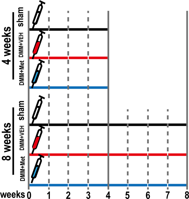
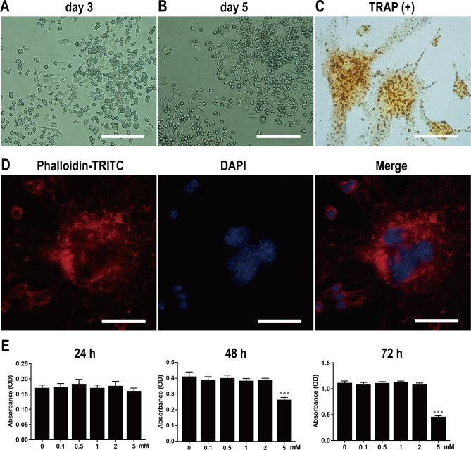
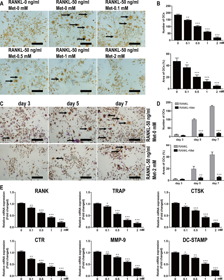
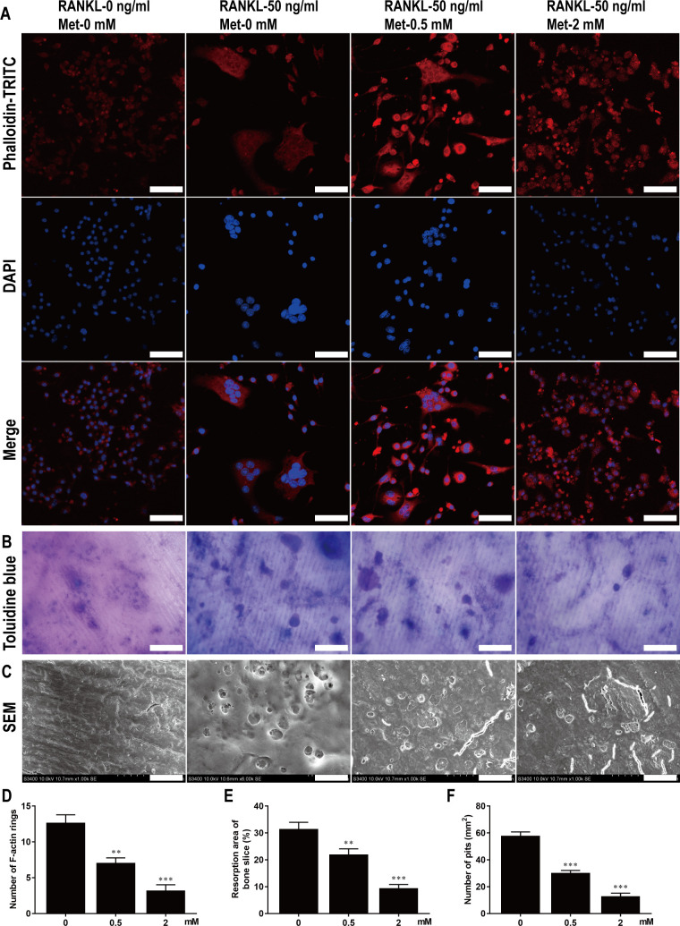
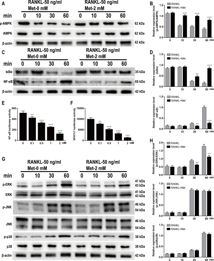
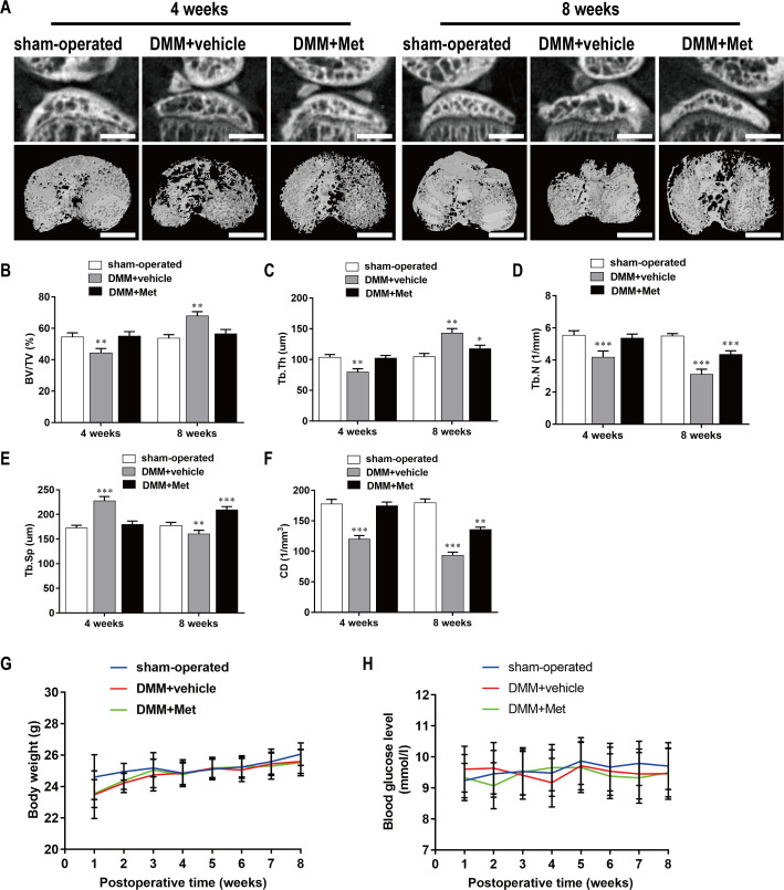
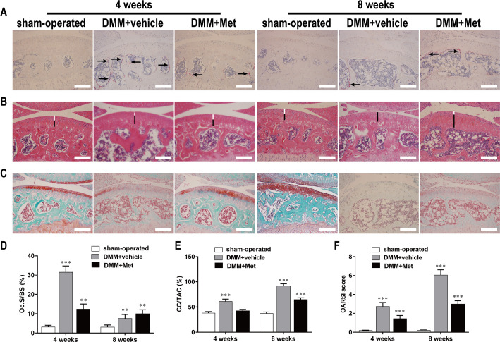
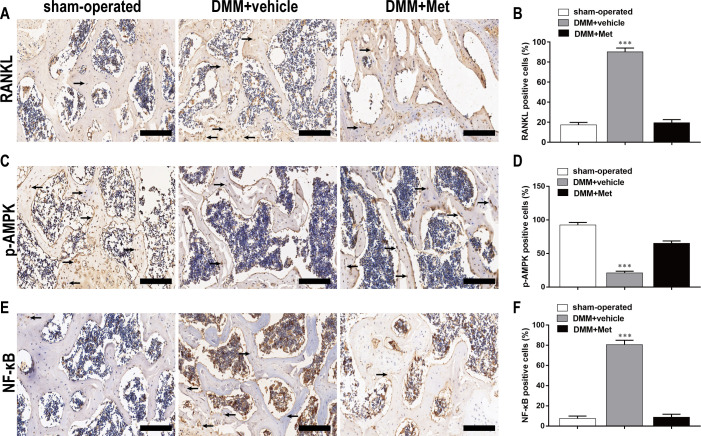
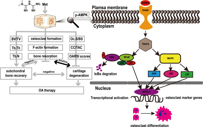
This study explored the mechanism by which metformin (Met) inhibits osteoclast activation and determined its effects on osteoarthritis (OA) mice. Bone marrow-derived macrophages were isolated. Osteoclastogenesis was detected using tartrate-resistant acid phosphatase (TRAP) staining. Cell proliferation was evaluated using CCK-8, F-actin rings were detected by immunofluorescence staining, and bone resorption was detected using bone slices. Nuclear factor kappa-B (NF-κB) and nuclear factor of activated T-cell cytoplasmic 1 (NFATc1) were detected using luciferase assays, and the adenosine monophosphate-activated protein kinase (AMPK), NF-κB, and mitogen-activated protein kinase (MAPK) signaling pathways were detected using western blotting. Finally, expression of genes involved in osteoclastogenesis was measured using quantitative polymerase chain reaction. A knee OA mouse model was established by destabilization of the medial meniscus (DMM). Male C57BL/6J mice were assigned to sham-operated, DMM+vehicle, and DMM+Met groups. Met (100 mg/kg/d) or vehicle was administered from the first day postoperative until sacrifice. At 4- and 8-week post OA induction, micro-computed tomography was performed to analyze microstructural changes in the subchondral bone, hematoxylin and eosin staining and Safranin-O/Fast Green staining were performed to evaluate the degenerated cartilage, TRAP-stained osteoclasts were enumerated, and receptor activator of nuclear factor κB ligand (RANKL), AMPK, and NF-κB were detected using immunohistochemistry. BMM proliferation was not affected by Met treatment below 2 mM. Met inhibited osteoclast formation and bone resorption in a dose-dependent manner in vitro. Met suppressed RANKL-induced activation of p-AMPK, NF-κB, phosphorylated extracellular regulated protein kinases (p-ERK) and up-regulation of genes involved in osteoclastogenesis. Met reversed decreases in BV/TV, Tb.Th, Tb.N, and CD, and an increase in Tb.Sp at 4 weeks postoperatively. The number of osteoclasts and OARSI score were decreased by Met without effect on body weight or blood glucose levels. Met inhibited RANKL, p-AMPK, and NF-κB expression in early OA. The mechanism by which Met inhibits osteoclast activation may be associated with AMPK/NF-κB/ERK signaling pathway, indicating a novel strategy for OA treatment.


Conflict of interest statement

The authors have declared that no competing interests exist.

Figures

Fig 1
Fig 1. Schematic of animal experiment design.
A total of 30 male C57BL/6J mice were assigned into 3 groups containing 10 mice: sham-operated group, DMM+vehicle group, and DMM+Met group. Mice in each group respectively received VEH, VEH, and Met (100 mg/kg/d) for 4 and 8 weeks post-operated.
Fig 2
Fig 2. Isolation and culture of primary BMMs, and osteoclast differentiation, identification, and proliferation.
Light microscopy of primary BMMs on 3rd (A) and 5th d (B). Scale bar, 200 μm. Light microscopy of TRAP-positive osteoclasts with multiple nuclei (C). The F-actin ring structure stained with phalloidin-TRITC and DAPI was observed under LSCM (D). Scale bar, 40 μm. BMMs were stimulated by various concentrations of Met for 24, 48 and 72 hours, and the absorbance value was detected by CCK-8 assay at 450nm (E). n = 3 per group. ***P < 0.001, compared with control group.
Fig 3
Fig 3. Met inhibits the differentiation of BMMs into osteoclasts in vitro.
(A) BMMs were stimulated with 30 ng/ml M-CSF, 50 ng/ml RANKL, and various concentrations of Met for 7 d and then subjected to TRAP staining. Scale bar, 200 μm. (B) Quantitative analysis of the numbers and areas of osteoclasts from panel A. (C) BMMs were stimulated with 30 ng/ml M-CSF and 50 ng/ml RANKL in the presence or absence of 2 mM DHA for 3, 5, and 7 d and then subjected to TRAP staining. Scale bar, 200 μm. (D) Quantitative analysis of the numbers and areas of osteoclasts from panel C. (E) BMMs were stimulated with 30 ng/ml M-CSF, 50 ng/ml RANKL, and various concentrations of Met for 7 d and then genes related to osteoclastogenesis were detected using RT-qPCR. n = 3 per group. *P < 0.05, **P < 0.01 and ***P < 0.001, compared with control group.
Fig 4
Fig 4. Met restrains F-actin rings formation and bone resorption of osteoclast in vitro.
(A) BMMs were stimulated with 30 ng/ml M-CSF, 50 ng/ml RANKL, and various concentrations of Met for 7 d and then stained with TRITC-conjugated phalloidin and DAPI to show F-actin rings and nucleus. Scale bar, 50 μm. (B) BMMs were seeded on bone slices and stimulated with 30 ng/ml M-CSF, 50 ng/ml RANKL, and various concentrations of Met for 9 d, bone resorption areas stained with toluidine blue were examined. Scale bar, 50 μm. (C) Bone resorption pits were shown by SEM. Scale bar, 20 μm. Quantitative analysis of (D) F-actin rings, (E) resorption areas and (F) resorption pits. n = 3 per group. **P < 0.01 and ***P < 0.001, compared with control group.
Fig 5
Fig 5. Met inhibits RANKL-induced AMPK, NF-κB and ERK activation during osteoclastogenesis.
BMMs were pretreated with or without Met (2 mM) for 2 h and then stimulated with 30 ng/ml M-CSF and 50 ng/ml RANKL for indicated time period (0, 10, 20, 30, 60 min), and the cell lysates were quantitatively analyzed using western blot for AMPK (A and B), NF-κB (C and D) and MAPK (G and H) signaling pathways. RAW264.7 cells transfected with NF-κB and NFATc1 were pretreated with various concentrations of Met for 2 h, and then incubated with α-MEM containing 30 ng/ml M-CSF and 50 ng/mL RANKL for 6 h to activate NF-κB and 24 h to activate NFATc1. Luciferase activities of (E) NF-κB and (F) NFATc1 were quantitatively analyzed. n = 3 per group. **P < 0.01 and ***P < 0.001, compared with control group.
Fig 6
Fig 6. Met attenuates bone loss during the early stage of DMM-induced OA.
(A) The DMM-induced OA mice were treated with or without Met for 4 and 8 weeks, and then microarchitecture in tibial subchondral bone was examined by μCT. Scale bar, 1,000 μm. (B, C, D, E, and F) Quantitative μCT analyses of microarchitecture in tibial subchondral bone: (B) BV/TV (%), (C) Tb.Th, (D) Tb.N, (E)Tb.Sp and (F) CD. (G and H) Quantitative analyses of body weight and blood glucose: (G) body weight and (H) blood glucose. n = 5 per group/time point. *P < 0.05, **P < 0.01 and ***P < 0.001 compared with the sham-operated group.
Fig 7
Fig 7. Met suppresses cartilage degeneration by inhibiting osteoclastogenesis in the early stage of DMM-induced OA.
The DMM-induced OA mice were treated with or without Met for 4 and 8 weeks, and histological analysis of osteoclasts in subchondral and cartilage were stained with (A) TRAP, (B) HE and (C) Safranin O-Fast Green respectively. Scale bar, 100 μm. Quantitative analysis of (D) Oc.S/BS, (E) CC/TAC, and (F) OARSI scores. n = 5 per group/time point. **P < 0.01 and ***P < 0.001, compared with the sham-operated group.
Fig 8
Fig 8. Met inhibits RANKL, NF-κB expression and promoted p-AMPK expression in the early stage of DMM-induced OA.
The DMM-induced OA mice were treated with or without DHA for 4 weeks, and expression of (A) RANKL, (C) p-AMPK, and (E) NF-κB was shown by immunohistochemistry staining. Scale bar, 100 μm. Quantitative analysis of (B) RANKL, (D) p-AMPK, and (F) NF-κB. n = 5 per group/time point. ***P < 0.001, compared with the sham-operated group.
Fig 9
Fig 9. Proposed mechanism of Met-induced attenuation of OA via the inhibition of osteoclast formation.
RANKL binds to RANK and recruits TRAF6 to activate NF-κB and MAPK pathways. The signal is then transmitted to NFATc1 and c-Fos. Sequentially the stimulated NFATc1 translocates into nucleus and initiates the expression of marker genes related to osteoclastogenesis and resorption, including RANK, TRAP, CTSK, CTR, MMP-9, and DC-STAMP. While Met can block the above activities by inhibiting the phosphorylation of AMPK.

References

    1. Martel-Pelletier J, Barr AJ, Cicuttini FM, Conaghan PG, Cooper C, Goldring MB, et al. Osteoarthritis. Nature reviews. Disease primers. 2016;2:16072. doi: 10.1038/nrdp.2016.72 - DOI - PubMed
    1. Hunter DJ, March L, Chew M. Osteoarthritis in 2020 and beyond: a Lancet Commission. Lancet (London, England). 2020;396(10264):1711–2. doi: 10.1016/S0140-6736(20)32230-3 - DOI - PubMed
    1. Biver E, Berenbaum F, Valdes AM, Araujo De Carvalho I, Bindels LB, Brandi ML, et al. Gut microbiota and osteoarthritis management: An expert consensus of the European society for clinical and economic aspects of osteoporosis, osteoarthritis and musculoskeletal diseases (ESCEO). AGEING RES REV. 2019;55:100946. doi: 10.1016/j.arr.2019.100946 - DOI - PubMed
    1. Goldring SR, Goldring MB. Changes in the osteochondral unit during osteoarthritis: structure, function and cartilage-bone crosstalk. Nature reviews. Rheumatology. 2016;12(11):632–44. doi: 10.1038/nrrheum.2016.148 - DOI - PubMed
    1. Casper-Taylor ME, Barr AJ, Williams S, Wilcox RK, Conaghan PG. Initiating factors for the onset of OA: A systematic review of animal bone and cartilage pathology in OA. Journal of orthopaedic research: official publication of the Orthopaedic Research Society. 2020;38(8):1810–8. - PMC - PubMed

Publication types

MeSH terms