. 2022 Jan 1;36(1-2):70-83.
doi: 10.1101/gad.349072.121. Epub 2021 Dec 16.

Guide RNA acrobatics: positioning consecutive uridines for pseudouridylation by H/ACA pseudouridylation loops with dual guide capacity

Affiliations

Guide RNA acrobatics: positioning consecutive uridines for pseudouridylation by H/ACA pseudouridylation loops with dual guide capacity

Beáta E Jády et al. Genes Dev. .

Abstract

Site-specific pseudouridylation of human ribosomal and spliceosomal RNAs is directed by H/ACA guide RNAs composed of two hairpins carrying internal pseudouridylation guide loops. The distal "antisense" sequences of the pseudouridylation loop base-pair with the target RNA to position two unpaired target nucleotides 5'-UN-3', including the 5' substrate U, under the base of the distal stem topping the guide loop. Therefore, each pseudouridylation loop is expected to direct synthesis of a single pseudouridine (Ψ) in the target sequence. However, in this study, genetic depletion and restoration and RNA mutational analyses demonstrate that at least four human H/ACA RNAs (SNORA53, SNORA57, SCARNA8, and SCARNA1) carry pseudouridylation loops supporting efficient and specific synthesis of two consecutive pseudouridines (ΨΨ or ΨNΨ) in the 28S (Ψ3747/Ψ3749), 18S (Ψ1045/Ψ1046), and U2 (Ψ43/Ψ44 and Ψ89/Ψ91) RNAs, respectively. In order to position two substrate Us for pseudouridylation, the dual guide loops form alternative base-pairing interactions with their target RNAs. This remarkable structural flexibility of dual pseudouridylation loops provides an unexpected versatility for RNA-directed pseudouridylation without compromising its efficiency and accuracy. Besides supporting synthesis of at least 6% of human ribosomal and spliceosomal Ψs, evidence indicates that dual pseudouridylation loops also participate in pseudouridylation of yeast and archaeal rRNAs.

Keywords: RNA-guided RNA modification; RNA–RNA interaction; box H/ACA RNAs; guide RNA acrobatics; pseudouridine; pseudouridylation.


Figures

Figure 1.
Figure 1.
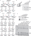
Selection of substrate uridines for pseudouridylation by box H/ACA guide RNAs. (A) Schematic structure of eukaryotic bipartite box H/ACA pseudouridylation guide RNAs. The consensus sequences of the conserved H and ACA box motifs and the internal pseudouridylation guide loops (Ψ-loop) are shown. (B) Schematic consensus structure of the 5′ and 3′ hairpins of H/ACA pseudouridylation guide RNPs. The antisense sequences (blue dots) and the selected target sequences (gray dots) with the target U (Ψ) positioned for pseudouridylation are shown. The proximal and distal stems (PS and DS) together with the upstream and downstream antisense target stems (UATS and DATS) are indicated. The four H/ACA core proteins (dyskerin, Nop10, Nhp2, and Gar1) are shown. Archaeal ACA RNAs contain K-turns or K-loops. (C) Potential base-pairing interactions of the proposed 5′-terminal pseudouridylation guide loop of human SNORA53 with 28S rRNA positioning U3747 or U3749 for pseudouridylation.
Figure 2.
Figure 2.
Human SNORA53 is required for synthesis of both Ψ3747 and Ψ3749 in the 28S rRNA. (A) A strategy for CRISPR/Cas9-mediated deletion of human SNORA53 gene in HAP1 cells. Schematic structure of a fragment of the human SLC25A3 gene encompassing exons 4–6 (E4–E6) and the intronic SNORA53 gene (open arrow) is shown. Positions of the forward (F1 and F2) and reverse (R1 and R2) primers used for genomic and RT-PCR analysis are indicated. The genomic sequences targeted by sgRNAs are presented in sense orientation. The PAM sequences are in italics. (B) PCR analysis of genomic DNAs extracted from SNORA53 knockout (SNORA53-KO) and parental HAP1 cells. The amplified DNA fragments were analyzed on 1% agarose gel. (Lane M) DNA size markers in base pairs (bp). Schematic structures and expected lengths of the amplified fragments are shown at the right. (C) RT-PCR analysis of the accumulation of spliced SLC25A3 mRNA in SNORA53-KO and HAP1 cells. Structure of the amplified cDNA fragment is shown. (D) SNORA53 accumulation. RNAs isolated from HAP1 and SNORA53-KO cells were annealed with internally labeled SNORA53- or U85-specific antisense RNA probes and digested with a mixture of RNase A and T1. The protected RNA fragments were analyzed on a 6% sequencing gel. (Lane M) Single-stranded DNA size markers in nucleotides (nt). (E) Mapping of 28S rRNA pseudouridylation in HAP1 and SNORA53-KO cells before and after restoration of SNORA53 expression (SNORA53-RST). Ψs covalently modified by CMCT were detected by primer extension analysis using a terminally labeled 28S-specific primer and AMV reverse transcriptase. (Lanes A,G,C,U) Dideoxy sequencing reactions performed on 28S ribosomal DNA with the same 28S-specific primer. (Lanes NT) Primer extension reactions performed on nontreated control RNAs. Ψs corresponding to the detected RT stop signals are indicated at the right. (F) Restoration of SNORA53 accumulation in SNORA53-KO cells. Schematic structure of the pIRESpuro2/GL/SNORA53 expression construct is shown. The coding region of the human SNORA53 gene (open arrow) was inserted into the second intron of the truncated β-globin gene (E1–E3) placed under the control of the cytomegalovirus (CMV) promoter and the globin polyadenylation signal (PA). The resulting CMV-globin-SNORA53 expression unit was inserted into the pIRESpuro2 expression plasmid and transfected into SNORA53-KO cells. Puromycin-resistant cell colonies were isolated, and expression of SNORA53 in SNORA53-RST cells was verified by RNase A/T1 mapping.
Figure 3.
Figure 3.
The 5′-terminal pseudouridylation loop of SNORA53 directs pseudouridylation of U3747 and U3749 in the 28S rRNA. (A) A computer-predicted secondary structure of the 5′ hairpin of human SNORA53 based on minimum free energy calculation (Zuker 2003). The evolutionarily invariant nucleotides predicted to base-pair with 28S rRNA are in blue. Other conserved nucleotides are in red. Arrows indicate internal deletions d1 and d2. (B) Expression of mutant SNORA53 RNAs in SNORA53-KO cells monitored by RNase A/T1 protection with sequence-specific antisense RNA probes. As controls, accumulation of endogenous SNORA53 was also tested in HAP1 and SNORA53-KO cells. (Lane M) Single-stranded DNA size markers. (C) Primer extension mapping of 28S rRNA pseudouridylation in HAP1 and SNORA53-KO cells either lacking or expressing mutant SNORA53 RNAs as indicated above the lanes. For details, see the legend for Figure 2E. (D) Proposed structural rearrangements of the three-way helical junctions formed by the 5′-terminal pseudouridylation loop of human SNORA53 and 28S rRNA positioning either U3747 or U3749 for pseudouridylation. The proposed alternative base-pairing interactions leading to “active” configuration A or B are indicated by blue and red lines, respectively. The common base-pairing interactions present in both configurations are shaded. The deleted or altered nucleotides in mutant SNORA53-CUU133del and SNORA53-CU132GA RNAs are boxed.
Figure 4.
Figure 4.
Structural requirements of the dual pseudouridylation guide activity of the 5′ hairpin of SNORA53. (A) In vivo pseudouridylation of 28S rRNA with a mutant SNORA53 RNA. (Top left panel) A predicted interaction of endogenous 28S rRNA with an ectopically expressed mutant SNORA53 RNA carrying an extra A (A+, in red) inserted between U13 and A14. The 28S-U3752 residue is underlined. Please note that 28S-U3751 could also bulge out in configuration A, but the upstream antisense element of SNORA53-U13+A is able to base-pair with both U3751 and U3752. Accumulation of SNORA53-U13+A in SNORA53-KO cells was verified by RNase mapping. As controls, accumulation of endogenous SNORA53 and U85 RNAs was also tested. (Bottom panel) Pseudouridylation of 28S rRNA was monitored by primer extension mapping in HAP1 and SNORA53-KO cells lacking or expressing SNORA53-U13+A. For other details, see the legend for Figure 2E. (B) In vivo pseudouridylation of transiently expressed mutant 28S rRNA (28S-U3752del) sequences lacking U3752 in mouse 3T3 cells. (Top panel) A predicted interaction of endogenous mouse SNORA53 with 28S-U3752del rRNA and schematic structure of the mouse pW(Xb/Xh) expression construct are shown. The mouse RNA polymerase I promoter (Pol I prom) and terminator (term), the 5′- and 3′-terminal regions of the 5′ and 3′ external transcribed sequences (ETS; gray boxes), and a fragment of the chloramphenicol acetyltransferase (CAT) gene are indicated (Hadjiolova et al. 1994). The nucleotide sequences of wild-type (28S-WT) and mutant (28S-U3752del) 28S rRNA fragments with the known ribosomal Ψs are shown. (Bottom panel) Pseudouridylation of the expressed 28S-WT and 28S-U3752del ribosomal minigene transcripts was monitored by primer extension analysis. (C) Correct positioning of the H box relative to the selected substrate Us is essential for the dual guide activity of the 5′ hairpin of SNORA53. (Left panel) Interaction of the descending strand of the 5′ hairpin of SNORA53 with 28S rRNA positioning U3747 and U3749 for pseudouridylation. A putative H-box-like motif is highlighted in the dashed box. The A149 residue was deleted in SNORA53-A149del and replaced for C in SNORA53-A149C. (Middle panel) Accumulation of ectopically expressed SNORA53-A149C and SNORA53-A149del RNAs and endogenous U85 scaRNA in SNORA53-KO cells was measured by RNase mapping. (Right panel) Primer extension mapping of 28S rRNA pseudouridylation in SNORA53-KO cells lacking or expressing SNORA53-A149C or SNORA53-A149del RNAs.
Figure 5.
Figure 5.
Human pseudouridylation guide loops with possible dual guide capacities. (A) Closely located human spliceosomal and ribosomal Ψs are listed together with the corresponding pseudouridylation guide RNA hairpins (5′hp and 3′hp) connected to their synthesis. Hairpins newly implicated in dual pseudouridylation reactions and their predicted target Ψs are highlighted in red. Participation of the shaded H/ACA hairpins in the synthesis of their predicted target Ψs has been experimentally validated in this study. (B) Potential alternative base-pairing interactions of H/ACA pseudouridylation guide loops with their target RNAs positioning consecutive ribosomal and spliceosomal Us for pseudouridylation. Direction of the synthesis of Ψ7, Ψ44, and Ψ89 in the U2 snRNA by human SCARNA14, SCARNA8, and SCARNA1, respectively, has been proposed earlier (Darzacq et al. 2002; Kiss et al. 2004; Schattner et al. 2006). The newly predicted target Ψs are highlighted in red. The functional interactions shaded in light gray have been experimentally validated in this work. Base-pairing interactions common in the alternative functional configurations are shaded in dark gray. For the interaction of SNORA53 and 28S rRNA, see Figure 3D.
Figure 6.
Figure 6.
The 3′-terminal pseudouridylation loops of human SCARNA1, SNORA57, and SCARNA8 possess dual guide capacities. (A) Expression of SCARNA1, SNORA57, SCARNA8, and SCARNA14 measured by RNase mappings. After deletion of the SCARNA1, SNORA57, SCARNA8, and SCARNA14 genes in HAP1 cells (Supplemental Fig. S3), each KO cell line was transformed by pIRESpuro/GL plasmids expressing SCARNA1, SCARNA8, or SNORA57 (RST cells). (B) Primer extension mapping of U2 snRNA and 18S rRNA pseudouridylation in the absence and presence of SCARNA1, SCARNA8, SNORA57, and SCARNA14 as indicated above the lanes. For other details, see the legend for Figure 2E.
Figure 7.
Figure 7.
Mutational analyses of SNORA57 and SCARNA1. (A) Predicted base-pairing interactions of wild-type (WT) and mutant SNORA57 RNAs with 28S rRNA in configurations A and B directing synthesis of Ψ1045 and Ψ1046, respectively. (B) Accumulation of mutant SNORA57 RNAs in SNORA57-KO cells and pseudouridylation of endogenous 28S rRNA. (Left panel) Accumulation of SNORA57-UUC126del and SNORA57-U125A RNAs was detected by RNase protection with sequence-specific probes. (Right panel) The pseudouridylation state of 18S rRNA in wild-type HAP1 and SNORA57-KO cells lacking or expressing the indicated mutant SNORA57 RNAs was measured by primer extension. (C) Predicted interactions of wild-type and mutant SCARNA1 RNAs with U2 snRNA directing pseudouridylation of U89 (configuration A) or U91 (configurations B). The nucleotide alterations in the mutant SCARNA1 RNAs are highlighted in red. (D) In vivo pseudouridylation activity of mutant SCARNA1 RNAs. (Top panel) Expression of mutant SCARNA1 RNAs in SCARNA1-KO cells was monitored by RNase protection. The lower band in the SCARNA1-G145U lane likely represents a degradation product of SCARNA1-G145U. (Bottom panel) Primer extension mapping of U2 snRNA pseudouridylation at U89 and U91 in the absence (SCARNA1-KO) or presence of wild-type (HAP1) and mutant SCARNA1 RNAs as indicated above the lanes. For other details, see the legend for Figure 2E.

Comment in

References

    1. Adachi H, De Zoysa MD, Yu YT. 2019. Post-transcriptional pseudouridylation in mRNA as well as in some major types of noncoding RNAs. Biochim Biophys Acta Gene Regul Mech 1862: 230–239. 10.1016/j.bbagrm.2018.11.002 - DOI - PMC - PubMed
    1. Arnez JG, Steitz TA. 1994. Crystal structure of unmodified tRNAGln complexed with glutaminyl-tRNA synthetase and ATP suggests a possible role for pseudo-uridines in stabilization of RNA structure. Biochemistry 33: 7560–7567. 10.1021/bi00190a008 - DOI - PubMed
    1. Baker DL, Youssef OA, Chastkofsky MI, Dy DA, Terns RM, Terns MP. 2005. RNA-guided RNA modification: functional organization of the archaeal H/ACA RNP. Genes Dev 19: 1238–1248. 10.1101/gad.1309605 - DOI - PMC - PubMed
    1. Bakin AV, Ofengand J. 1998. Mapping of pseudouridine residues in RNA to nucleotide resolution. Methods Mol Biol 77: 297–309. - PubMed
    1. Balakin AG, Smith L, Fournier MJ. 1996. The RNA world of the nucleolus: two major families of small RNAs defined by different box elements with related functions. Cell 86: 823–834. 10.1016/S0092-8674(00)80156-7 - DOI - PubMed

LinkOut - more resources