. 2021 Dec 16;6(1):153.
doi: 10.1038/s41541-021-00420-6.

Development of multivalent mRNA vaccine candidates for seasonal or pandemic influenza

Affiliations

Development of multivalent mRNA vaccine candidates for seasonal or pandemic influenza

Sudha Chivukula et al. NPJ Vaccines. .

Abstract

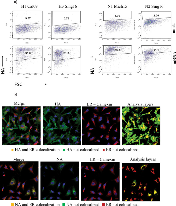
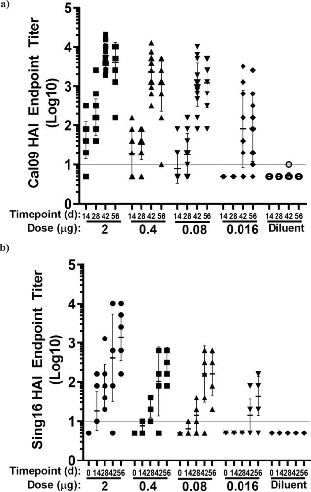
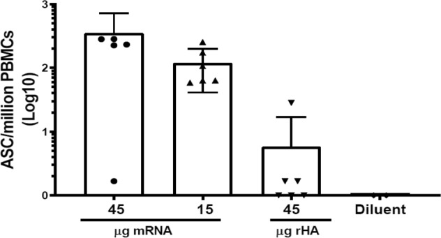
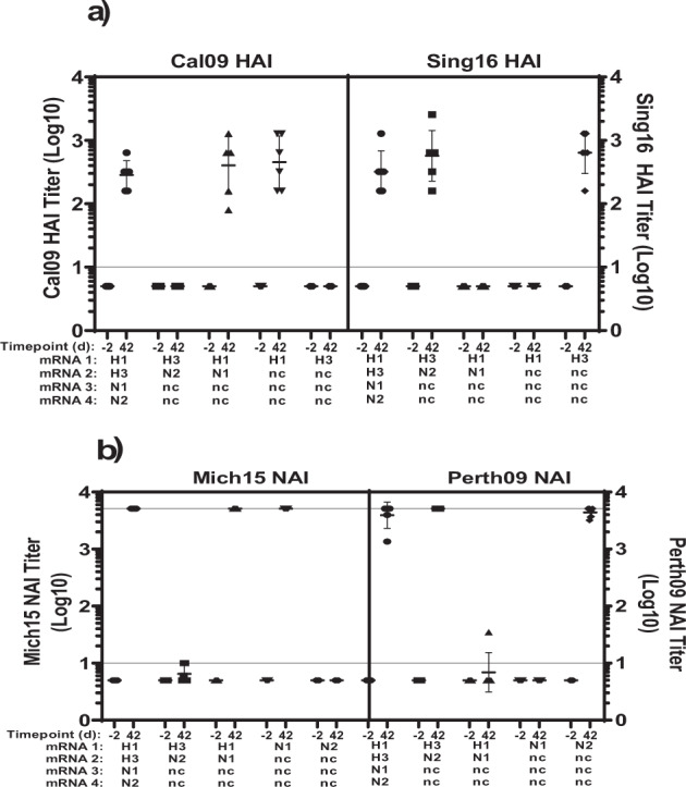
Recent approval of mRNA vaccines for emergency use against COVID-19 is likely to promote rapid development of mRNA-based vaccines targeting a wide range of infectious diseases. Compared to conventional approaches, this vaccine modality promises comparable potency while substantially accelerating the pace of development and deployment of vaccine doses. Already demonstrated successfully for single antigen vaccines such as for COVID-19, this technology could be optimized for complex multi-antigen vaccines. Herein, utilizing multiple influenza antigens, we demonstrated the suitability of the mRNA therapeutic (MRT) platform for such applications. Seasonal influenza vaccines have three or four hemagglutinin (HA) antigens of different viral subtypes. In addition, influenza neuraminidase (NA), a tetrameric membrane protein, is identified as an antigen that has been linked to protective immunity against severe viral disease. We detail the efforts in optimizing formulations of influenza candidates that use unmodified mRNA encoding full-length HA or full-length NA encapsulated in lipid nanoparticles (LNPs). HA and NA mRNA-LNP formulations, either as monovalent or as multivalent vaccines, induced strong functional antibody and cellular responses in non-human primates and such antigen-specific antibody responses were associated with protective efficacy against viral challenge in mice.


Conflict of interest statement

The research is funded by Translate Bio, a Sanofi company and Sanofi Pasteur Inc. Sudha Chivukula, Timothy Tibbitts, Donghui Zhang, Natalie Anosova, Lu Li, Younghoon Kim, Heesik Yoon, Charles Lai, Sophia T Mundle, Yanhua Yan, Gregory Ulinski, Peter Piepenhagen, Victoria Landolfi, Joshua DiNapoli, Danilo Casimiro, Frank DeRosa, Ron Swearingen, Shrirang Karve, Rebecca Goldman, Ashish Sarode, Anusha Dias, Hardip Gopani, Asad Khanmohammed and Dustin Cooper are employees of Sanofi Pasteur or Translate Bio and may hold stock in the company.

Figures

Fig. 1
Fig. 1. In vitro expression and localization of HA and NA in HskMC and HeLa cells transformed with mRNA.
a Frequency of HA and NA expressing human skeletal muscle cells were evaluated by flow cytometry. HskMC cells were transfected with mRNA coding for Cal09 HA, Sing16 HA, Mich15 NA and Sing16 NA. Corresponding antigen expression was confirmed by intracellular staining with each antigen specific mouse or rabbit antibodies followed by fluorochrome conjugated goat anti-mouse IgG or goat anti-rabbit IgG secondary antibody. The numbers represent the frequency of antigen expression as determined by comparison with mock transfected HskMCs. For gating strategy see Supplementary Fig. 2. b Intracellular localization of expressed HA and NA in HeLa cells. HA and NA protein expression for a bivalent H3N2-mRNA LNP analyzed along with Calnexin an endoplasmic reticulum marker. Image panels show merged image of HA and ER (upper) or NA and ER (lower) antibody staining. Middle two images display separate HA and NA antibody staining (green) and ER marker Calnexin (red). Images in the analysis layer column display results of image analysis to measure colocalization of HA or NA with ER marker. Images contain image analysis mask layers to denote regions of HA or NA colocalization with ER (yellow), HA or NA not colocalized with the ER (green), and ER not containing any HA or NA expression (red).
Fig. 2
Fig. 2. Serological Evaluation of HA mRNA-LNP vaccine in mice.
BALB/c mice (n = 8 per group) were immunized twice IM, 4 weeks apart with 2, 0.4, 0.08 and 0.016 μg of either Cal09 HA mRNA-LNP or Sing16 HA mRNA-LNP. Log10 HAI titers recorded against (a) A/California/07/2009 H1N1 influenza virus (b) A/Singapore/INFIMH160019/2016 H3N2 influenza virus shown. Each dot represents an individual animal, and the line represents the geometric mean for the group. Lower horizontal line in each panel represents the lower limit of assay read out. See Supplementary Fig. 4 for ELISA titers.
Fig. 3
Fig. 3. Serological Evaluation of NA mRNA-LNP vaccine in mice.
BALB/c mice (n = 8 per group) were immunized twice IM 4 weeks apart with 2, 0.4, 0.08, and 0.016 μg of either Mich15 NA mRNA-LNP or Sing16 NA mRNA-LNP. Log10 NAI (ELLA) titers recorded for sera against (a) A/Michigan/45/2015 (N1): A/Mallard/Sweden/2002 (H6) chimeric influenza virus, b A/Singapore/INFIMH160019/2016 (N2): A/Mallard/Sweden/2002 (H6) chimeric virus are depicted. Each dot represents an individual animal, and the line represents the geometric mean for the group. Lower horizontal line in each panel represents the lower limit of assay read out. See Supplementary Fig. 5 for ELISA titers.
Fig. 4
Fig. 4. Protective efficacy of Cal09 HA mRNA-LNP vaccine in mice after lethal A/Belgium/2009 H1N1 virus challenge.
Mice (n = 8) received two IM doses of Cal09 HA mRNA-LNP (0.4 μg each) on day 0 and day 28. Control animals received two IM doses of diluent on day 0 and day 28. a HAI titers are reported as Log10 for serum samples taken at study days -7, 14, 28, 42, 56, 92, 107 are reported. Each dot represents an individual animal, and the line represents the geometric mean for the group. Lower horizontal line represents the lower limit of assay read out. b Daily weights after intranasal challenge with 4LD50 of A/Belgium/2009 H1N1 strain on day 93 are reported. Weights are presented as the percentage of weight lost from the day of challenge. Individual lines represent each animal. c Immunization and challenge schedules are provided.
Fig. 5
Fig. 5. Protective efficacy single dose of unmodified Mich15 NA mRNA-LNP in mice after lethal A/Belgium/2009 H1N1 virus challenge.
Mice (n = 16) were injected by the IM route with 0.4 μg or 0.016 μg of A/Michigan/45/2015 NA mRNA-LNP. Half of the mice only received one injection (1 dose) on study day 0, while the other half (2 doses) received two injections given at study day 0 and day 28. Control animals received two IM doses of diluent or hEPO mRNA-LNP (0.6 μg) on day 0 and day 28. a NAI titers are reported as Log10 for serum samples taken at study days -1, 14, 28, 42, 88, 102 for one dose group; -1, 14, 28, 42, 114 for 2 dose group and -1, 14, 28, 42 for control group. Each dot represents an individual animal, and the line represents the geometric mean for the group. Lower horizontal line represents the lower limit of assay read out, b daily weight change after intranasal challenge on day 89 for single dose group and day 117 (89 days after second dose) for two dose group with 4LD50 of Belgium09 H1N1 are reported. Weights are presented as the percentage of weight lost from the day of challenge. Individual lines represent each animal. c Immunization and challenge schedules are provided.
Fig. 6
Fig. 6. Serological Evaluation of HA Sing16 HA mRNA-LNP vaccine in NHP.
Cynomolgus macaques (n = 6 per group) were injected twice, 4 weeks apart by IM route, with 15, 45, 135, or 250 μg of Sing16 HA mRNA-LNP. Serum samples were collected at days -6, 14, 28, 42, and 56. (a) Log 10 HAI titers against A/Singapore/INFIMH160019/2016 virus and (b) Log 10 micro neutralization (MN) titers against A/Singapore/INFIMH160019/2016 virus are shown. Each dot represents an individual animal, and the line represents the geometric mean for the group. Lower horizontal line represents the lower limit of assay read out. See Supplementary Fig. 6 for ELISA titers against recombinant Sing16 HA protein and Supplementary Table 2 for statistical analysis.
Fig. 7
Fig. 7. T cell responses in NHP vaccinated with Sing16 HA mRNA-LNP vaccine.
Cynomolgus macaques (n = 6 per group) were injected twice, 4 weeks apart by IM route, with 45, 135, or 250 μg of Sing16 HA mRNA-LNP. T cells were determined by ELISPOT on day 42 in PBMC stimulated in vitro with peptide pools to represent the entire HA open reading frame. The responses of PBMC secreting IFNɣ (a) or IL-13 (b) calculated as spots forming cells (SFC) per million PBMC are shown. Each symbol represents an individual sample, and the bar represents the mean with standard deviation for the group.
Fig. 8
Fig. 8. Secretion of A/Singapore/16/H3-specific IgG by memory B cells on day 180 in NHP vaccinated with Sing16 HA mRNA-LNP vaccine.
Cynomolgus macaques (n = 6 per group) were injected twice, 4 weeks apart by IM route, with 15 or 45 μg of Sing16 HA mRNA-LNP. The Human IgG Single-Color memory B cell ELISPOT kit (CAT# NC1911372, CTL) was used to measure A/Singapore/16/H3-specific and total IgG+ antibody-secreting cells (ASCs). Differentiation of MBCs into ASCs was performed in PBMC collected at day 180 by using a stimulation cocktail provided by the kit. The number of IgG+ and number of A/Singapore/16/H3-specific ASCs was calculated per million of PBMCs for each animal and the frequency of antigen-specific ASCs is shown. Each symbol represents an individual sample, and the bar represents the mean with standard deviation for the group. The percentage of antigen-specific ASCs to the total IgG+ ASCs is shown in Supplementary Table 3.
Fig. 9
Fig. 9. Delivery of bivalent combinations of influenza vaccine in mice.
BALB/c mice (n = 8 per group) were immunized twice IM, 4 weeks apart with a total 0.4 μg of bivalent combinations co-encapsulated mRNA transcripts (1:1 wt/wt, half volume per each leg) or 0.2 μg each monovalent which was separately formulated and immunized different legs. H1H3 combo constituting Cal09 HA mRNA-LNP, Sing16 HA mRNA-LNP; H3N2 combo of Sing16 HA mRNA-LNP and Perth09 mRNA-LNP and N1N2 combo of Mich15 NA mRNA-LNP and Perth09 NA mRNA-LNP were tested in sera collected a day -2, 14, 28, 42, against corresponding virus. Control treatments included diluent or bivalent co-encapsulated formulations of corresponding HA or NA mRNA with non-coding mRNA. a HAI titers recorded against A/California/07/2009 H1N1 influenza virus and A/Singapore/INFIMH160019/2016 H3N2; b HAI and NAI titers recorded against A/Singapore/INFIMH160019/2016 H3N2 and A/Mallard/Sweden/2002 (H6) chimeric influenza virus and H6N2 A/Perth/09 virus (N2) virus, respectively (c). NAI titers recorded against A/Mallard/Sweden/2002 (H6) chimeric influenza viruses H6N1 A/Michigan/45/2015 (N1) and H6N2 A/Perth/09 virus (N2) virus are shown for each combination. Each dot represents an individual animal, and the line represents the geometric mean for the group. Lower horizontal line represents the lower limit of assay read out. See Supplementary Table 4 for design of study and Supplementary Table 5 for statistical analysis.
Fig. 10
Fig. 10. Delivery of quadrivalent combinations of influenza vaccines in NHP.
Cynomolgus macaques (n = 6 per group) were immunized twice IM, 4 weeks apart with a total 10 μg of quadrivalent combinations of co-encapsulated mRNA transcripts (1:1:1:1 wt/wt). H1N1H3N2 combo consisting of Cal09 HA mRNA, Sing16 HA mRNA, Mich15 NA mRNA, and Perth09 NA mRNA. H1H3 combo constituting Cal09 HA mRNA, Sing16 HA mRNA and 2x non-coding mRNA (ncmRNA); H3N2 combo of Sing16 HA mRNA and Perth09 NA mRNA and 2x non-coding mRNA. N1N2 combo of Mich15 NA mRNA, Perth09 NA mRNA-LNP, and 2x non-coding mRNA served as bivalent controls. H1 consisting of Cal09 HA mRNA and 3x non-coding mRNA; H3 consisting of Sing16 HA mRNA and 3x non-coding mRNA; N1 consisting of Mich15 NA mRNA and 3x non-coding mRNA and N2 consisting of Perth09 NA mRNA and 3x non-coding mRNA served as monovalent controls. Inhibitory titers were tested in sera collected a day -2 and 42, against corresponding virus - (a) HAI titers recorded against A/California/07/2009 H1N1 Influenza virus and A/Singapore/INFIMH160019/2016 H3N2; (b) NAI titers recorded against A/Michigan/45/2015 (N1): A/Mallard/Sweden/2002 (H6) chimeric influenza virus and H6N2 A/Perth/09 virus F1919D (N2): A/Mallard/Sweden/2002 (H6) chimeric influenza virus is shown. are shown for each combination. Each dot represents an individual animal, and the line represents the geometric mean for the group. Lower horizontal line represents the lower limit of assay read out. See Supplementary Table 6 for design of study and Supplementary Table 7 for statistical analysis.

References

    1. Polack, F. P. et al. Safety and efficacy of the BNT162b2 mRNA Covid-19 vaccine. New Engl. J. Med.10.1056/nejmoa2034577 (2020). - PMC - PubMed
    1. Baden, L. R. et al. Efficacy and safety of the mRNA-1273 SARS-CoV-2 vaccine. New Engl. J. Med. 10.1056/nejmoa2035389 (2020). - PMC - PubMed
    1. Shaw C, et al. 2754. Phase 1 Trial of an mRNA-based combination vaccine against hMPV and PIV3. Open Forum Infect. Dis. 2019;6(Supplement_2):S970–S970. doi: 10.1093/ofid/ofz360.2431. - DOI
    1. mRNA vaccines against H10N8 and H7N9 influenza viruses of pandemic potential are immunogenic and well tolerated in healthy adults in phase 1 randomized clinical trials. Vaccine37, 3326–3334. 10.1016/j.vaccine.2019.04.074 (2019). - PubMed
    1. Alberer M, et al. Safety and immunogenicity of a mRNA rabies vaccine in healthy adults: an open-label, non-randomised, prospective, first-in-human phase 1 clinical trial. Lancet. 2017;390:1511–1520. doi: 10.1016/S0140-6736(17)31665-3. - DOI - PubMed