. 2021 Dec 16;11(1):24105.
doi: 10.1038/s41598-021-03491-6.

Thyroid hormone receptor alpha sumoylation modulates white adipose tissue stores

Affiliations

Thyroid hormone receptor alpha sumoylation modulates white adipose tissue stores

Yan-Yun Liu et al. Sci Rep. .

Abstract

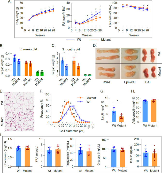
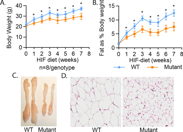
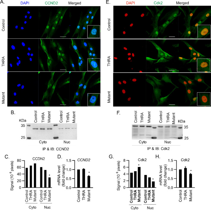
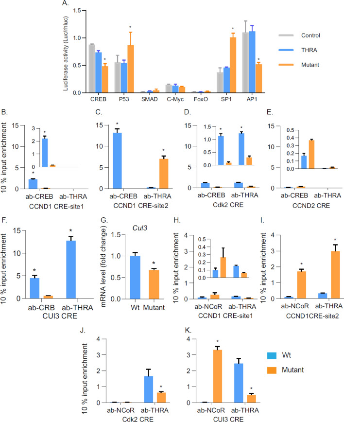
Thyroid hormone (TH) and thyroid hormone receptor (THR) regulate stem cell proliferation and differentiation during development, as well as during tissue renewal and repair in the adult. THR undergoes posttranslational modification by small ubiquitin-like modifier (SUMO). We generated the THRA (K283Q/K288R)-/- mouse model for in vivo studies and used human primary preadipocytes expressing the THRA sumoylation mutant (K283R/K288R) and isolated preadipocytes from mutant mice for in vitro studies. THRA mutant mice had reduced white adipose stores and reduced adipocyte cell diameter on a chow diet, compared to wild-type, and these differences were further enhanced after a high fat diet. Reduced preadipocyte proliferation in mutant mice, compared to wt, was shown after in vivo labeling of preadipocytes with EdU and in preadipocytes isolated from mice fat stores and studied in vitro. Mice with the desumoylated THRA had disruptions in cell cycle G1/S transition and this was associated with a reduction in the availability of cyclin D2 and cyclin-dependent kinase 2. The genes coding for cyclin D1, cyclin D2, cyclin-dependent kinase 2 and Culin3 are stimulated by cAMP Response Element Binding Protein (CREB) and contain CREB Response Elements (CREs) in their regulatory regions. We demonstrate, by Chromatin Immunoprecipitation (ChIP) assay, that in mice with the THRA K283Q/K288R mutant there was reduced CREB binding to the CRE. Mice with a THRA sumoylation mutant had reduced fat stores on chow and high fat diets and reduced adipocyte diameter.


Conflict of interest statement

The authors declare no competing interests.

Figures

Figure 1
Figure 1
Genotype and thyroid function tests in THRA K283Q/K288R mutant mice. (A) A reporter assay was performed to test the T3 induction of the mutant THRA K283Q/K288R on T3-mediated luciferase reporter expression. JEG3 cells were transfected with THRA expression vectors and a luciferase reporter containing consensus 3 × Thyroid Hormone Response Elements. The THRA PV mutant receptor, based on mutations in Resistance to Thyroid Hormone, was used as a negative control since it does not bind to T3. The combination of THRA with THRA K283Q/K288R was used to determine dominant-negative effects of the THRA K283Q/K288R mutant receptor. (B) The presence of the mutation in homozygous THRA K283Q/K288R mutant mice was confirmed using direct DNA sequencing. (C-D) Western blot detection of THRA and sumoylated THRA, based on protein molecular weight. Subcutaneous fat was dissected from mice (n = 3/genotype). Protein lysate (35 μg/lane) was loaded on a 10% SDS gel. Antibody used was ab-THRA (C). (D) Quantification of THRA and sumoylated THRA in Western blot. The antibody detected bands and total protein (from Ponceau stained membrane, see Supplementary Fig. S2 online) were quantified using Li-Cor Image Studio lite. The data was normalized for Ponceau stained protein and presented as % total protein. Each bar in the figure represents the data from three mice. The significance was determined using Student t-test. (E) Fasting serum thyroxine (T4), triiodothyronine (T3) and Thyroid Stimulating Hormone (TSH) concentrations in Wt and THRA K283Q/K288R mutant mice, individual levels shown for Wild type (Wt) (circles) and mutant (squares), with a horizontal line showing the mean (n = 10). Statistical analysis was performed by Student t-test.
Figure 2
Figure 2
THRA K283Q/K288R mutant mice display body composition, fat stores, and metabolic profile. Age-matched wild type (Wt) and THRA K283Q/K288R mutant mice (mutant) were fed with chow diet. (A) Body composition was determined by Echo-MRI (n = 9/group) and average value is shown for body weight (BW), fat and lean body mass. (B) Fat pads (iWAT-inguinal White Adipose Tissue-blue bars, Epi- epididymal fat-orange bars, iBAT- inter-scapular Brown Adipose Tissue-green bars) were dissected and weighed from 6 weeks old mice (n = 6) and from (C) 3 month old mice (n = 6). (D) Images of dissected fat pads from 3 month old Wt and mutant mice (n = 6/group), representative samples are shown. (E) Histological analysis of inguinal WAT, representative slides from wild type and mutant mice are shown (scale bar 100 μm). (F) Fat cell diameter (μM) of inguinal fat was analyzed. A total of 360 cells from each genotype was quantified (see “Materials and methods” section for details). The data is shown as diameter frequency of 360 cells. (G,H) Non-fasting serum leptin and adiponectin levels (n = 7/group, 3 month old mice). (I) Fasting serum cholesterol, triglycerides, free fatty acids (FFA), insulin and glucose concentrations in 3 month old mice. The statistical analysis was performed using paired Student t-test.
Figure 3
Figure 3
THRA K283Q/K288R mice are resistant to High fat diet-induced body fat increase and adipose tissue expansion. Mice (n = 8/genotype) were given High Fat (HIF) diet (40% fat in calories) for 7 weeks starting at age 7 weeks old. The body composition was measured by Eco-MRI. (A) Body weight and (B) body fat as % body weight. (C) Mice were euthanized and fat pads were dissected, rinsed and photographed and representative inguinal fat depot as shown. (D) Representative histological image of inguinal fat from WT and mutant mice. The statistical analysis was perform at each time point using paired Student t-test.
Figure 4
Figure 4
Preadipocyte proliferation in mutant mice determined in vivo and in vitro. (A,B) EdU labeling of proliferating preadipocytes in 3 week old mice. Mice were given EdU, 5 μg/100 g body weight (i.p.) and inguinal fat pads were isolated and fixed in 4% paraformaldehyde 12 h after injection. Frozen sections (12 μM thickness) of inguinal fat depots were stained with β-catenin (red) for cell membrane and DAPI (blue) for nuclei. EdU (Green) incorporation of DNA in dividing cells was detected by Click-IT chemistry. (A) EdU incorporation was imaged using confocal microscope and representative sections of regions with stromal vascular regions are shown. Lower panel shows enlarged view of the area as indicated. The scale bar is 200 μM. (B) EDU incorporation was quantified in three different sections using a × 20 magnification field. (C) Isolated preadipocytes proliferation. Preadipocytes were derived from the stromal vascular fraction of fat from 6 week old wild type (Wt) and mutant mice (n = 6/group). Preadipocytes from each mouse were plated on separate plates and synchronized as described in the “Materials and methods” section. After synchronization, cells were supplemented with 10% serum and labeled with EDU for 4 h, followed by fixation and imaging. Representative images from 3 separate experiments are shown. Scale bar is 50 μM. (D) Quantification of EDU positive cells (green color) and nuclei imaged with DAPI (blue). (E) Quantification of the number of SVF-derived preadipocytes. For each experiment (n = 6 mice). The data shown is the average numbers of preadipocytes in each experiment. (F) q-PCR analysis of Cyclin D1 (CCND1), Cyclin D2 (CCND2) and cyclin-dependent kinase 2 (Cdk2) mRNA levels, obtained from expression PCR array and normalized for four housekeeping genes. Please see “Materials and methods” for quantification shown in (B) and (C). *, p < 0.05 compared to wild type mice.
Figure 5
Figure 5
Mutation of the THRA sumoylation site, K283Q/K288R, associated with reduced nuclear localization of cyclin D1-Cdk4 complex, cyclin D2 and Cdk2 in G1 phase. (A) Human preadipocytes were transfected with empty vector (control) or plasmids expressing THRA or THRA K283Q/K288R. Cells were synchronized to G0/G1 phase by serum starvation for 48 h. Cells were then allowed to re-enter the cell cycle by supplementing the medium with 10% calf serum. After 9 h of serum supplementation, the cells were analyzed for nuclear content of cyclin D1-Cdk4 complex, cyclin D2 and Cdk2. Immunofluorescent (IF) staining of cyclin D1 and Cdk4 nuclear localization in cells. Scale bar, 50 μm. Circled inset shows higher magnification. (B) The nuclear and cytoplasmic fractions were analyzed for Cdk4 protein expression by IP with anti-Cdk4 Ab, followed by immunoblot (IB) with anti-Cdk4 and quantified Cdk4 IB signals using Li-Cor Image Studio Lite. (C) Co-IP detection of cyclin D1 in the cytoplasm and nuclear fractions. Anti-p27kip antibody was used in IP and anti-cyclin D1 antibody in IB. Quantification of WB was done using Image Studio Lite. q-PCR quantification of Cdk4 and cyclin D1 (CCND1) mRNA expression, presented as the mean value and SD, fold above control. *p < 0.05 compare to control by One-way Anova. IP-immunoprecipitation, IB-immunoblot. The blots (B,C) were cut prior to hybridization and the origin blots are provided (see Supplementary Fig. S7 online).
Figure 6
Figure 6
Nuclear localization of Cyclin D2 and Cdk2 in human primary preadipocytes expressing THRA K283Q/K288R. Cell treatment and conditions are the same as described in the Fig. 4 legend. (A,E) IF detection of nuclear cyclin D2 (CCND2) and Cdk2 protein with DAPI stain of nucleus. Circled inset shows higher magnification. (B,F) detection of nuclear CCND2 and Cdk2 protein using IP and followed by IB with anti-cyclin D2 antibody and anti-Cdk2 antibody, and quantified by Li-CoR Imaging Studio lite (C,G). (D,H) q-PCR analysis of CCND2 and Cdk2 mRNA and fold-change from control is shown. Primers were pre-designed (Qiagen). *p < 0.05 as statistically significant by One-way Anova for qPCR data, and by Two-way Anova for IB quantification Scale bar, 50 μm. The blots (panels B and F) were cut prior to hybridization and the origin blots are provided (see Supplementary Fig. S8 online).
Figure 7
Figure 7
Analysis of cell cycle progression in human preadipocytes. (A) Cell cycle progression was analyzed by Propidium iodide Flow Cytometer. Cell synchronization is the same as describe in the Fig. 4 legend. After serum starvation, cells were supplemented with 10% serum for a period of 0, 8, 16, 24 h. At each time point, cells were collected and analyzed by flow cytometry. Percentage of cells in G1 phase (B) and S phase (C), obtained from flow cytometry for each time point is shown. *P < 0.05 as statistically significant compared with control and THRA at the same time point by One-way Anova analysis.
Figure 8
Figure 8
Expression of THRA K283Q/K288R associated with altered CREB binding to the cAMP Response Element (CRE) of cell cycle regulatory genes. (A) Human preadipocytes were transfected by electroporation with luciferase reporter containing multiple copies of a response element for each transcription factor tested. Each transfection had 6 replicates and the data shown is the average with SD. Statistical analysis was formed using One-way Anova. (B–F) ChIP analysis of CREB-THRA interaction on CREs. Preadipocytes were isolated from wild type mice and mutant mice. After synchronization, cells were supplied with serum for 8 h prior to harvest for ChIP assays of CRE on CCND1, CCND2 and Cdk4 promoter. For Cul3 gene promoter, cells were supplemented with serum for 12 h. (G) q-PCR analysis of Cul3 gene expression in cells supplied with 12 h of serum after synchronization. (HK) ChiP assay of THRA requirement of Nuclear Co-Repressor (NCoR) effects on CREs. The antibodies used were anti-THRA, anti-CREB, and anti-NCoR. PCR was performed for 35 cycles. Normal rabbit IgG was used as the negative control in ChIP assays. All ChIP data shown are after deducting the negative control and expressing as % input enrichment. Input was 10% of total pre-cleared lysate. Statistical analysis was performed using Student t-test. CREB, cAMP response element binding protein; P53 tumor suppressor protein, c-Myc cellular homolog of myc oncogene, FoxO1 forkhead box protein 1, Sp1 specificity protein 1, AP1 activator protein 1.

Similar articles

Cited by

References

    1. Brent GA. Mechanisms of thyroid hormone action. J. Clin. Invest. 2012;122:3035–3043. doi: 10.1172/JCI60047. - DOI - PMC - PubMed
    1. Pascual A, Aranda A. Thyroid hormone receptors, cell growth and differentiation. Biochim. Biophys. Acta. 2013;1830:3908–3916. doi: 10.1016/j.bbagen.2012.03.012. - DOI - PubMed
    1. Kress E, Samarut J, Plateroti M. Thyroid hormones and the control of cell proliferation or cell differentiation: Paradox or duality? Mol. Cell Endocrinol. 2009;313:36–49. doi: 10.1016/j.mce.2009.08.028. - DOI - PubMed
    1. White U, Ravussin E. Dynamics of adipose tissue turnover in human metabolic health and disease. Diabetologia. 2019;62:17–23. doi: 10.1007/s00125-018-4732-x. - DOI - PMC - PubMed
    1. Tchoukalova Y, Koutsari C, Jensen M. Committed subcutaneous preadipocytes are reduced in human obesity. Diabetologia. 2007;50:151–157. doi: 10.1007/s00125-006-0496-9. - DOI - PubMed

Publication types

MeSH terms

Substances