Review
. 2021 Nov 30:15:755875.
doi: 10.3389/fncel.2021.755875. eCollection 2021.

The Causes and Long-Term Consequences of Viral Encephalitis

Affiliations
Review

The Causes and Long-Term Consequences of Viral Encephalitis

Karen Bohmwald et al. Front Cell Neurosci. .

Abstract

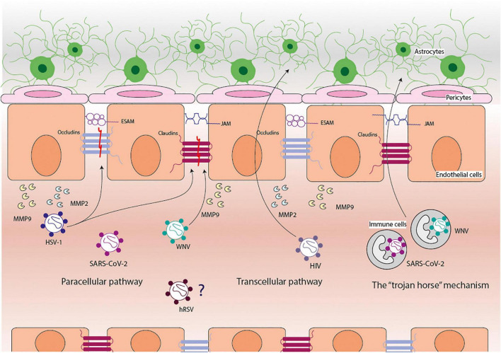
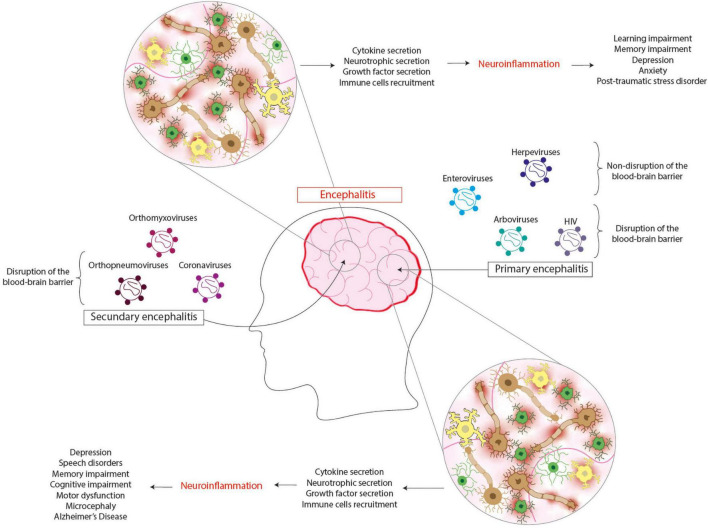
Reports regarding brain inflammation, known as encephalitis, have shown an increasing frequency during the past years. Encephalitis is a relevant concern to public health due to its high morbidity and mortality. Infectious or autoimmune diseases are the most common cause of encephalitis. The clinical symptoms of this pathology can vary depending on the brain zone affected, with mild ones such as fever, headache, confusion, and stiff neck, or severe ones, such as seizures, weakness, hallucinations, and coma, among others. Encephalitis can affect individuals of all ages, but it is frequently observed in pediatric and elderly populations, and the most common causes are viral infections. Several viral agents have been described to induce encephalitis, such as arboviruses, rhabdoviruses, enteroviruses, herpesviruses, retroviruses, orthomyxoviruses, orthopneumovirus, and coronaviruses, among others. Once a neurotropic virus reaches the brain parenchyma, the resident cells such as neurons, astrocytes, and microglia, can be infected, promoting the secretion of pro-inflammatory molecules and the subsequent immune cell infiltration that leads to brain damage. After resolving the viral infection, the local immune response can remain active, contributing to long-term neuropsychiatric disorders, neurocognitive impairment, and degenerative diseases. In this article, we will discuss how viruses can reach the brain, the impact of viral encephalitis on brain function, and we will focus especially on the neurocognitive sequelae reported even after viral clearance.

Keywords: central nervous system; cognitive impairment; encephalitis; inflammation; viral infection.


Conflict of interest statement

The authors declare that the research was conducted in the absence of any commercial or financial relationships that could be construed as a potential conflict of interest.

Figures

FIGURE 1
FIGURE 1
Pathways used by viruses to cross the blood-brain barrier. The blood-brain barrier (BBB) is composed of endothelial cells, pericytes, and astrocytes cells. The viruses use three principal pathways to cross the BBB: paracellular, transcellular, and the “trojan horse” mechanism. West Nile virus (WNV) can reach the central nervous system via the paracellular pathway and the “trojan horse” mechanism, disrupting claudins and promoting the secretion of matrix metalloproteinases (MMPs) 9. Herpes simplex virus (HSV-1) can reach the central nervous system via the paracellular pathway, disrupting claudins and occludins and promoting the secretion of MMP2 and MMP9. Human immunodeficiency virus (HIV) can reach the central nervous system via the paracellular and transcellular pathways and promote the secretion of MMP2 and MMP9. SARS-CoV-2 can reach the central nervous system via the paracellular pathway and the “trojan horse” mechanism; however, it has not been elucidated how and what it promotes. Human respiratory syncytial virus (hRSV) can reach the central nervous system through the disruption of the BBB, but how and what it causes this has not been elucidated yet.
FIGURE 2
FIGURE 2
Viral encephalitis leads to several neurologic sequelae. Primary encephalitis is caused by viruses that infect the brain as their principal target, which can be found in arboviruses, enteroviruses, herpesviruses, and HIV. The infection of cells from the central nervous system induces the cytokine, neurotrophic, and growth factor secretion and the recruitment of immune cells. It is thought that this response can contribute to neuroinflammation, leading to neurologic sequelae such as depression, speech disorders, memory, and cognitive impairment, motor dysfunction, microcephaly, and even Alzheimer’s disease. Secondary encephalitis is caused by viruses that do not infect the brain as their principal target, which can be found in orthomyxovirus, orthopneumoviruses, and coronaviruses. The infection of cells from the central nervous system induces the cytokine, neurotrophic, and growth factor secretion and the recruitment of the immune cells. This response contributes to neuroinflammation, leading to neurologic sequelae such as learning and memory impairment, depression, anxiety, and post-traumatic stress disorder.

References

    1. Abbott N. J., Patabendige A. A. K., Dolman D. E. M., Yusof S. R., Begley D. J. (2010). Structure and function of the blood-brain barrier. Neurobiol. Dis. 37 13–25. 10.1016/j.nbd.2009.07.030 - DOI - PubMed
    1. Acuña-Hinrichsen F., Covarrubias-Pinto A., Ishizuka Y., Stolzenbach M. F., Martin C., Salazar P., et al. (2021). Herpes Simplex Virus Type 1 Neuronal Infection Triggers the Disassembly of Key Structural Components of Dendritic Spines. Front. Cell. Neurosci. 15:36. 10.3389/fncel.2021.580717 - DOI - PMC - PubMed
    1. Adang L. A., Lynch D. R., Panzer J. A. (2014). Pediatric anti-NMDA receptor encephalitis is seasonal. Ann. Clin. Transl. Neurol. 1 921–925. 10.1002/ACN3.100 - DOI - PMC - PubMed
    1. Aksamit A. J. (2021). Treatment of Viral Encephalitis. Neurol. Clin. 39 197–207. 10.1016/j.ncl.2020.09.011 - DOI - PubMed
    1. Ali A., Lopardo G., Scarpellini B., Stein R. T., Ribeiro D. (2020). Systematic review on respiratory syncytial virus epidemiology in adults and the elderly in Latin America. Int. J. Infect. Dis. 90 170–180. 10.1016/J.IJID.2019.10.025 - DOI - PMC - PubMed