

A case report of a severe form of cogan syndrome
- PMID: 34917345
- PMCID: PMC8665300
- DOI: 10.1016/j.amsu.2021.103036
A case report of a severe form of cogan syndrome
Abstract
Introduction: the Cogan syndrome is a very rare, systemic disease that affects young adults. Very few cases are described in the literature. We report the case of a patient with a severe form of Cogan syndrome.
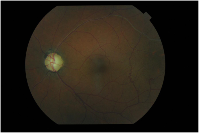
Case presentation: This is a young patient who presented with a painful left red eye and bilateral visual impairment evolving for 5 years with ENT signs such as right hypoacusis and vertigo. Clinical examination in this patient found bilateral hypertensive panuveitis, vertigo of peripheral origin and hypoacusis on the right. The patient is currently on corticosteroid therapy with stabilization of the lesions.
Discussion: This pathology is characterized by ocular and audio-vestibular involvement with sometimes other visceral manifestations. The etiopathogeny is not well known, the evolution is marked by the functional prognosis (visual and auditory) and the vital prognosis (aortic insufficiency). The treatment is essentially based on corticotherapy and the treatment of complications.
Conclusion: It is a very rare condition that should be considered when there are suggestive signs, because the evolution is severe without appropriate care. This underlines the importance of early management and the need for optimal follow-up to avoid the occurrence of complications that are disabling or even fatal.
Keywords: Goniosynechia; Optic atrophy; The cogan syndrome.
© 2021 The Authors.
Conflict of interest statement
The authors declare that there are no conflicts of interest.
Figures


References
-
- Vinceneux P., Pouchot J., Kahn M.F., Peltier A.P., Meyer O., Piette J.C. Flammarion; Paris: 1991. Le syndrome de Cogan.
-
- Agha R.A., Franchi T., Sohrabi C., Mathew G., pour le groupe S.C.A.R.E. Ligne directrice SCARE 2020 : mise à jour des lignes directrices du rapport sur les cas chirurgicaux de consensus (SCARE) Int. J. Surg. 2020;84:226–230. - PubMed
-
- Cundiff J., Kansal S., Kumar A., Goldstein D.A., Tessler H.H. Cogan's syndrome: a cause of progressive hearing deafness. Am. J. Otolaryngol. 2006;27:68–70. - PubMed
-
- Vollertsen R.S., McDonald T.J., Younge B.R., Banks P.M., Stanson A.W., Ilstrup D.M. Cogan's syndrome: 18 cases and a review of the literature. Mayo Clin. Proc. 1986;61:344–361. - PubMed
Publication types
LinkOut - more resources
Full Text Sources
Research Materials
Miscellaneous
