. 2021 Nov 30:8:736226.
doi: 10.3389/fcvm.2021.736226. eCollection 2021.

Plasma Exosomes at the Late Phase of Remote Ischemic Pre-conditioning Attenuate Myocardial Ischemia-Reperfusion Injury Through Transferring miR-126a-3p

Affiliations

Plasma Exosomes at the Late Phase of Remote Ischemic Pre-conditioning Attenuate Myocardial Ischemia-Reperfusion Injury Through Transferring miR-126a-3p

Danni Li et al. Front Cardiovasc Med. .

Abstract

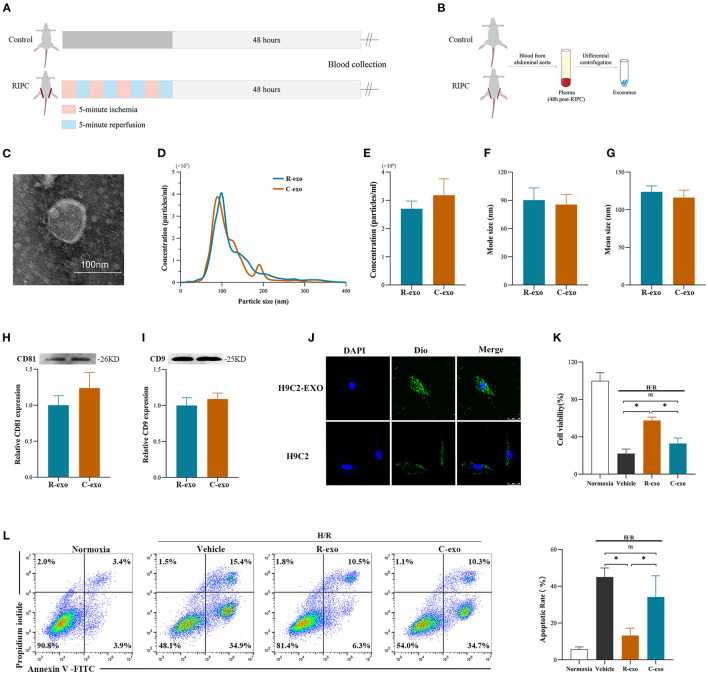
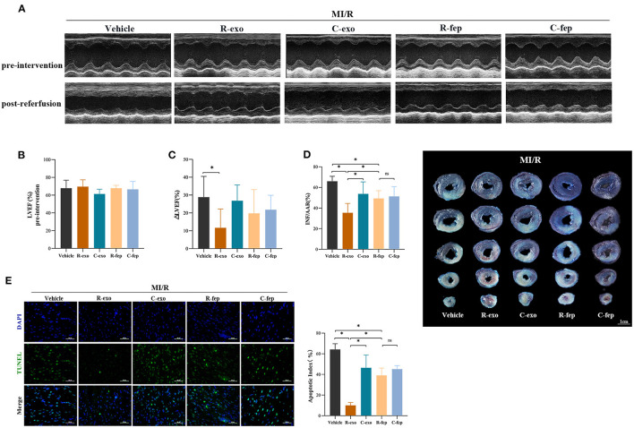
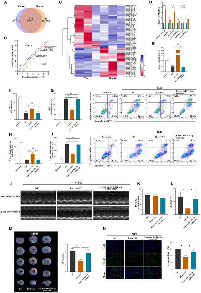
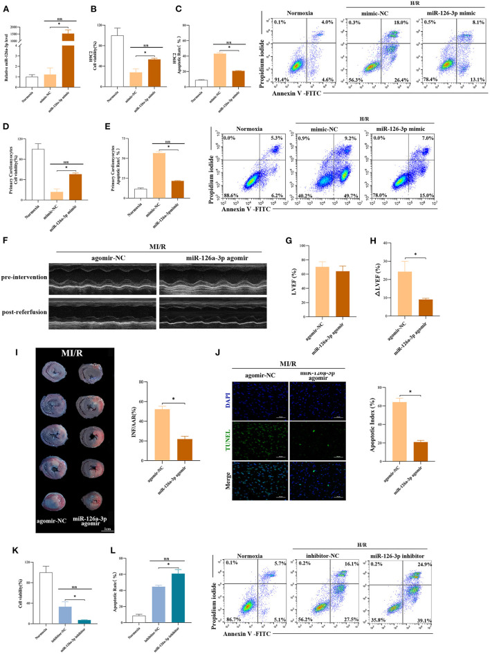
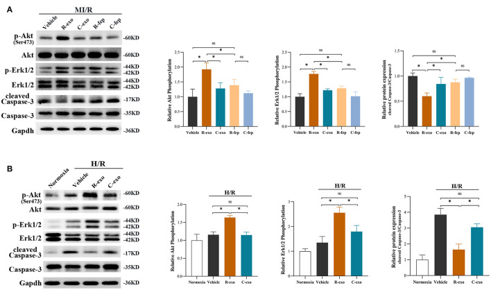
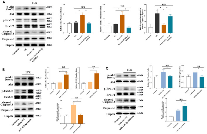
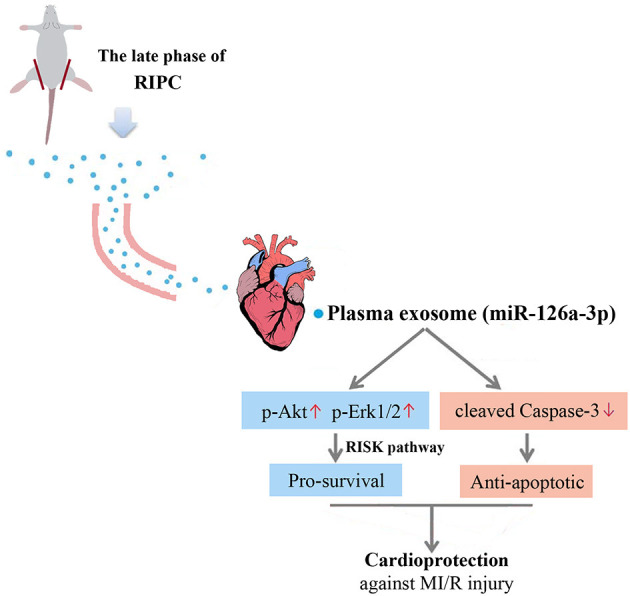
Background: Remote ischemic pre-conditioning (RIPC) alleviated the myocardial ischemia-reperfusion injury, yet the underlying mechanisms remain to be fully elucidated, especially at the late phase. Searching a key component as a transfer carrier may provide a novel insight into RIPC-mediated cardioprotection in the condition of myocardial ischemia-reperfusion. Objective: To investigate the cardioprotective effect of plasma exosomes at the late phase of RIPC and its potential signaling pathways involved. Methods and Results: Exosomes were isolated from the plasma of rats 48 h after the RIPC or control protocol. Although the total plasma exosomes level had no significant change at the late phase of RIPC (RIPC-exosome) compared with the control exosomes (Control-exosome), the RIPC-exosome afforded remarkable protection against myocardial ischemia-reperfusion (MI/R) injury in rats and hypoxia-reoxygenation (H/R) injury in cells. The miRNA array revealed significant enrichment of miR-126a-3p in RIPC-exosome. Importantly, both miR-126a-3p inhibitor and antagonist significantly blunted the cardioprotection of RIPC-exosome in H/R cells and MI/R rats, respectively, while miR-126a-3p mimic and agomir showed significant cardioprotection against H/R injury in cells and MI/R injury in rats. Mechanistically, RIPC-exosome, especially exosomal miR-126a-3p, activated the reperfusion injury salvage kinase (RISK) pathway by enhancing the phosphorylation of Akt and Erk1/2, and simultaneously inhibited Caspase-3 mediated apoptotic signaling. Conclusions: Our findings reveal a novel myocardial protective mechanism that plasma exosomes at the late phase of RIPC attenuate myocardial ischemia-reperfusion injury via exosomal miR-126a-3p.

Keywords: cardiomyocyte apoptosis; cardioprotection; exosomes; miR-126a-3p; microRNA; myocardial ischemia-reperfusion; remote ischemic pre-conditioning.


Conflict of interest statement

The authors declare that the research was conducted in the absence of any commercial or financial relationships that could be construed as a potential conflict of interest.

Figures

Figure 1
Figure 1
Identification and functional verification of plasma exosomes at the late phase of RIPC in vitro. (A) Establishment of the late-phase RIPC model in rats. (B) The process to obtain the plasma exosomes from rats with or without the late-phase RIPC protocol. (C) Representative electron micrograph of isolated plasma exosomes. Scale bar: 100 nm. (D) Representative results of NTA (nanoparticle tracking analysis) demonstrating size distribution and concentration in Control-exosomes (C-exo) and RIPC-exosomes (R-exo). (E) The average concentration in Control-exosomes and RIPC-exosomes (n = 4). (F) Mode size in Control-exosomes and RIPC-exosomes (n = 4). (G) Mean size in Control-exosomes and RIPC-exosomes (n = 4). (H) Representative images of Western blot (top) and quantified data (bottom) in exosomal marker proteins CD81 in Control-exosomes and RIPC-exosomes (n = 4). (I) Representative images of Western blot (top) and quantified data (bottom) in exosomal marker proteins CD9 in Control-exosomes and RIPC-exosomes (n = 4). (J) Representative images of H9C2 cells that were incubated with the presence (H9C2-EXO) or absence (H9C2) of Dio-labeled exosomes (green). Cardiomyocyte nuclei were stained with DAPI (blue). Scale bar: 25 μm. (K) The data of cell viability showed that RIPC-exosomes improved cell viability with 24 h co-incubation and following H/R injury. (L) The data and representative images of cell apoptosis by flow cytometric analysis showed that RIPC-exosomes decreased cell apoptosis with 24 h co-incubation and following H/R injury (n = 3). RIPC, remote ischemic pre-conditioning; R-exo, exosomes isolated from the plasma of the late-phase RIPC rats; C-exo, exosomes isolated from the plasma of the control rats; H/R, Hypoxia/Reoxygenation (24/6 h). *p < 0.05 and nsp > 0.05.
Figure 2
Figure 2
Plasma exosomes at the late phase of RIPC exerted cardioprotection against MI/R injury in vivo. (A) Representative echocardiographic images of rats before the intervention (pre-intervention) and after the reperfusion (post-reperfusion) in each group of rats. (B) Left ventricular ejection fraction (LVEF) of rats in pre-intervention (n = 8). (C) ΔLVEF (the difference between the LVEF in pre-intervention and that in post-reperfusion) in each group. RIPC-exosome injected into rats for 24 h before MI/R improved cardiac function as evidence by significantly narrowed the ΔLVEF. (D) Quantified data and presentative photographs of heart sections. RIPC-exosome decreased infarct size in rats subjected to MI/R (n = 8). INF/AAR, infarct size as a percentage of myocardial area at risk. (E) Representative images and quantified data of TUNEL assay in each group showed that RIPC-exosome decreased myocardial apoptosis in rats subjected to MI/R. Myocardial apoptosis in rats determined by a percent of TUNEL-positive nuclei (green)/total nuclei (blue) (n = 4). Scale bar: 50 μm. MI/R, myocardial ischemia/reperfusion (30/180 min). R-exo, exosomes isolated from the plasma of the late-phase RIPC rats. C-exo, exosomes isolated from the plasma of the control rats. R-fep, the supernatant collected after the first ultracentrifugation from the plasma of the late-phase RIPC rats. C-fep, the supernatant collected after the first ultracentrifugation from the plasma of the control rats. *p < 0.05, nsp > 0.05.
Figure 3
Figure 3
miR-126a-3p was essential in the cardioprotection provided by plasma exosomes at the late phase of RIPC. (A) Venn diagram showing the numbers of overlapping and unique miRNAs detected in Control-exosome (C-exo) and RIPC-exosome (R-exo) (n =3). (B) The expression level of miRNAs in Control-exosome and RIPC-exosome. Yellow dots represented up-regulated miRNAs in RIPC-exosome and blue dots represented up-regulated miRNAs in Control-exosome (fold change >2 and p < 0.05, n =3). (C) Heat map representing the miRNA profiling assays between Control-exosome and RIPC-exosome (fold change >2 and p < 0.05, n =3). (D) qRT-PCR analysis of the six differentially expressed miRNAs in Control-exosome and RIPC-exosome. Data are normalized to cel-miR-39 (n =3). (E) The qRT-PCR analysis of miR-126a-3p level in H9C2 cells treated with inhibitor negative control (NC), R-exo + NC, or R-exo + miR-126a-3p inhibitor followed by H/R. Data were presented as a fold change of the normoxia. U6 was used as an internal control (n = 3). (F) The data of cell viability showed that inhibition of miR-126a-3p attenuated the protective effects of RIPC-exosome in H9C2 cells subjected to H/R (n = 3). (G) The data and representative images of apoptosis of H9C2 cells subjected to H/R by flow cytometric analysis (n = 3). The protective effects of RIPC-exosome were eliminated by inhibition of miR-126a-3p. (H) The data of cell viability showed that the protective effects of RIPC-exosomal miR-126a-3p were reduced by a miR-126a-3p inhibitor in primary cardiomyocytes following H/R injury (n =3). (I) The data and representative images of apoptosis of primary cardiomyocytes subjected to H/R by flow cytometric analysis (n = 3). The anti-apoptosis effects of RIPC-exosomal miR-126a-3p were eliminated by inhibition of miR-126a-3p (n =3). (J) Representative echocardiographic images of rats in pre-intervention and post-reperfusion in each group of rats (n = 5). (K) Left ventricular ejection fraction (LVEF) of rats in pre-intervention. (L) ΔLVEF in each group showed that miR-126a-3p antagomir blunted the protective effects of RIPC-exosome in cardiac function. (M) Representative photographs of heart sections and quantified data demonstrated that miR-126a-3p antagomir observably attenuated the protective effects of RIPC-exosome in myocardial infarct size (n = 5). (N) Representative images and quantified data of TUNEL assay in each group demonstrated that miR-126a-3p antagomir observably blunted the protective effects of RIPC-exosome in myocardial apoptosis (n = 4). Scale bar: 50 μm. R-exo, exosomes isolated from the plasma of the late-phase RIPC rats. C-exo, exosomes isolated from the plasma of the control rats. H/R, Hypoxia/Reoxygenation (24/6 h). MI/R, myocardial ischemia/reperfusion (30/180 min). *p < 0.05.
Figure 4
Figure 4
miR-126a-3p was an important molecule in cardioprotection. (A) The qRT-PCR analysis of miR-126a-3p level in H9C2 cells transfected with miR-126a-3p mimic or negative control (mimic-NC) for 24 h followed by H/R. Data were presented as a fold change of the normoxia. U6 was used as an internal control (n = 3). (B) The increasing level of miR-126a-3p improved cell viability in H9C2 cells subjected to H/R (n = 3). (C) Flow cytometric data and representative images of apoptosis in H9C2 cells subjected to H/R (n = 3). The increasing level of miR-126a-3p decreased cell apoptosis in H9C2 cells subjected to H/R. (D) Cell viability in primary cardiomyocytes transfected with a miR-126a-3p mimic or mimic-NC followed by H/R injury (n =3). (E) Flow cytometric data and representative images of the anti-apoptosis effect of miR-126a-3p in primary cardiomyocytes treated with a miR-126a-3p mimic or mimic-NC followed by H/R injury (n =3). (F) Representative echocardiographic images of rats in pre-intervention and post-reperfusion in each group of rats (n = 5). (G) Left ventricular ejection fraction (LVEF) of rats in pre-intervention. (H) ΔLVEF in each group showed that miR-126a-3p agomir improved cardiac function. (I) Representative photographs of heart sections and quantified data demonstrated that miR-126a-3p agomir reduced myocardial infarct size (n = 5). (J) Representative images and quantified data of TUNEL assay in each group demonstrated that miR-126a-3p agomir reduced myocardial apoptosis (n = 4). Scale bar: 50 μm. (K) Cell viability in H9C2 cells treated with a miR-126a-3p inhibitor or inhibitor-NC followed by H/R (n = 3). (L) Flow cytometric data and representative images of apoptosis in H9C2 cells treated with a miR-126a-3p inhibitor or inhibitor-NC followed by H/R (n = 3). H/R, Hypoxia/Reoxygenation (24/6 h). MI/R, myocardial ischemia/reperfusion (30/180 min). *p < 0.05.
Figure 5
Figure 5
The signaling pathways involved in plasma exosomes at the late phase of RIPC mediated effects. (A) Representative images of Western blot and quantified data in rats received R-exo, C-exo, R-fep, C-fep, or vehicle for 24 h followed by MI/R. (n = 3). (B) Representative images of Western blot and quantified data in H9C2 cells preincubated with R-exo, C-exo, or vehicle for 24 h followed by H/R (n = 3). GAPDH was used as a loading control. Quantified data showed phosphorylation level of Akt, expressed as the ratio of p-Akt to Akt; phosphorylation level of Erk1/2, expressed as the ratio of p-Erk1/2 to Erk1/2; the activation of Caspase-3 expressed as the ratio of cleaved Caspase-3 to Caspase-3. MI/R, myocardial ischemia/reperfusion (30/180 min). H/R, Hypoxia/Reoxygenation (24/6 h). R-exo, exosomes isolated from the plasma of the late-phase RIPC rats; C-exo, exosomes isolated from the plasma of the control rats; R-fep, the supernatant collected after the first ultracentrifugation from the plasma of the late-phase RIPC rats; C-fep, the supernatant collected after the first ultracentrifugation from the plasma of the control rats. *p < 0.05, nsp > 0.05.
Figure 6
Figure 6
The signaling pathways involved in plasma exosomes at the late phase of RIPC via miR-126-3p mediated effects. (A) Representative images of Western blot and quantified data in H9C2 cells treated with inhibitor-NC, R-exo + NC, or R-exo + miR-126a-3p inhibitor followed by H/R (n = 3). (B) Representative images of Western blot and quantified data in H9C2 cells transfected with miR-126a-3p mimics or mimic-NC for 24 h followed by H/R (n = 3). (C) Representative images of Western blot and quantified data in H9C2 cells transfected with miR-126a-3p inhibitors or negative control (inhibitor-NC) for 24 h followed by H/R (n = 3). GAPDH was used as a loading control. Quantified data showed phosphorylation level of Akt, expressed as the ratio of p-Akt to Akt; phosphorylation level of Erk1/2, expressed as the ratio of p-Erk1/2 to Erk1/2; the activation of Caspase-3 expressed as the ratio of cleaved Caspase-3 to Caspase-3. H/R, Hypoxia/Reoxygenation (24/6 h). R-exo, exosomes isolated from the plasma of the late-phase RIPC rats. *p < 0.05.
Figure 7
Figure 7
Proposed mechanisms by which plasma exosomes at the late phase of RIPC protect the heart against MI/R injury through transferring miR-126a-3p. Plasma exosomes could be used as a carrier to transfer the myocardial protective effect of RIPC at the late phase against MI/R injury, activate the RISK pathway by increasing the phosphorylation level of Akt, and Erk1/2, and inhibit the activation of apoptotic protein Caspase-3 by reducing the expression level of cleaved Caspase-3.

References

    1. Birnbaum Y, Hale SL, Kloner RA. Ischemic preconditioning at a distance: reduction of myocardial infarct size by partial reduction of blood supply combined with rapid stimulation of the gastrocnemius muscle in the rabbit. Circulation. (1997) 96:1641–6. 10.1161/01.CIR.96.5.1641 - DOI - PubMed
    1. Murry CE, Jennings RB, Reimer KA. Preconditioning with ischemia: a delay of lethal cell injury in ischemic myocardium. Circulation. (1986) 74:1124–36. 10.1161/01.CIR.74.5.1124 - DOI - PubMed
    1. Mauser M, Hoffmeister HM, Nienaber C, Schaper W. Influence of ribose, adenosine, and “AICAR” on the rate of myocardial adenosine triphosphate synthesis during reperfusion after coronary artery occlusion in the dog. Circ Res. (1985) 56:220–30. 10.1161/01.RES.56.2.220 - DOI - PubMed
    1. Loukogeorgakis SP, Panagiotidou AT, Broadhead MW, Donald A, Deanfield JE, MacAllister RJ. Remote ischemic preconditioning provides early and late protection against endothelial ischemia-reperfusion injury in humans: role of the autonomic nervous system. J Am Coll Cardiol. (2005) 46:450–6. 10.1016/j.jacc.2005.04.044 - DOI - PubMed
    1. Gho BC, Schoemaker RG, van den Doel MA, Duncker DJ, Verdouw PD. Myocardial protection by brief ischemia in noncardiac tissue. Circulation. (1996) 94:2193–200. 10.1161/01.CIR.94.9.2193 - DOI - PubMed