

Case Reports
doi: 10.1016/j.jdcr.2021.11.001.
eCollection 2022 Jan.
Cutaneous Mycobacterium chelonae infection presenting clinically as a mycetoma
Affiliations
- PMID: 34917726
- PMCID: PMC8669244
- DOI: 10.1016/j.jdcr.2021.11.001
Item in Clipboard
Case Reports
Cutaneous Mycobacterium chelonae infection presenting clinically as a mycetoma
JAAD Case Rep.
.
No abstract available
Keywords: AFB, acid-fast bacilli; Mycobacterium chelonae; NTM, nontuberculous mycobacteria; infectious disease; mycetoma; mycobacteria; nontuberculous mycobacteria; rapidly growing.
Conflict of interest statement
None disclosed.
Figures
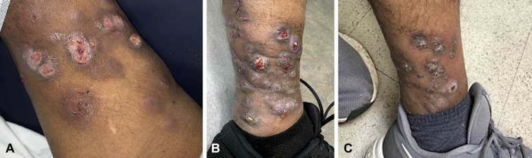
Cutaneouos Mycobacterium chelonae infection. A, Initial clinical presentation showing violaceous, friable, ulcerated nodules scattered on the left medial lower extremity. B, Clinical image taken at 2-month follow-up showing worsening of lesions. There are multiple, grouped, erythematous nodules on the left medial lower extremity with ulceration, purulent crusting, and sinus tract formation. C, Improvement of patient's lesions seen after 3 months of combined antibiotic therapy with tobramycin, linezolid, and azithromycin.
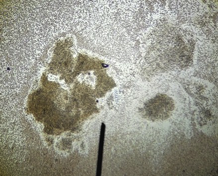
Dark brown granule seen on wet preparation of purulence aspirated from one of the patient's draining sinus tracts.
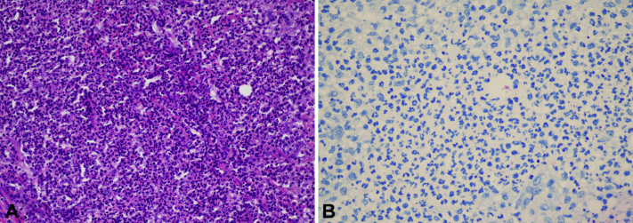
Cutaneous Mycobacterium chelonae infection. A, Neutrophilic dermal inflammation intermixed with scattered lymphocytes and histiocytes (hematoxylin-eosin stain). B, Acid-fast bacilli apparent on acid-fast bacilli staining.
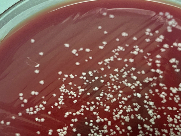
Mycobacterial colonies growing on the blood agar plate used for acid-fast bacilli tissue culture. Mycobacterium chelonae was identified via mass spectrometry.
References
Publication types
LinkOut - more resources
Full Text Sources