. 2021 Oct 6;4(1):100386.
doi: 10.1016/j.jhepr.2021.100386. eCollection 2022 Jan.

ROCK2 inhibition attenuates profibrogenic immune cell function to reverse thioacetamide-induced liver fibrosis

Affiliations

ROCK2 inhibition attenuates profibrogenic immune cell function to reverse thioacetamide-induced liver fibrosis

Christina Nalkurthi et al. JHEP Rep. .

Abstract

Background & aims: Fibrosis, the primary cause of morbidity in chronic liver disease, is induced by pro-inflammatory cytokines, immune cell infiltrates, and tissue resident cells that drive excessive myofibroblast activation, collagen production, and tissue scarring. Rho-associated kinase 2 (ROCK2) regulates key pro-fibrotic pathways involved in both inflammatory reactions and altered extracellular matrix remodelling, implicating this pathway as a potential therapeutic target.

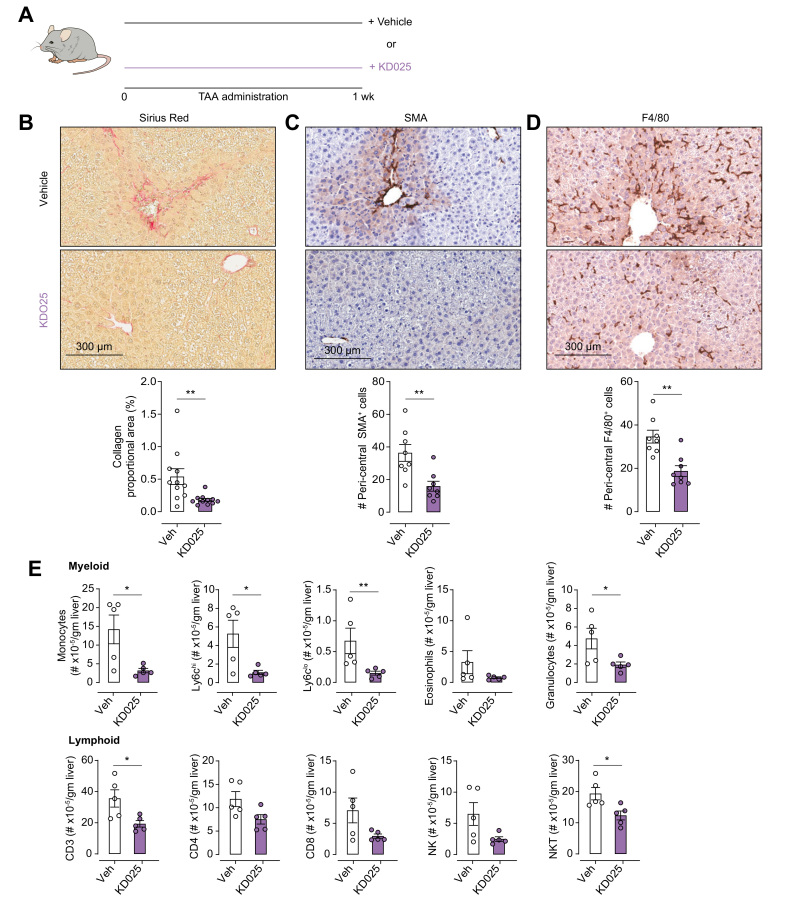
Methods: We used the thioacetamide-induced liver fibrosis model to examine the efficacy of administration of the selective ROCK2 inhibitor KD025 to prevent or treat liver fibrosis and its impact on immune composition and function.

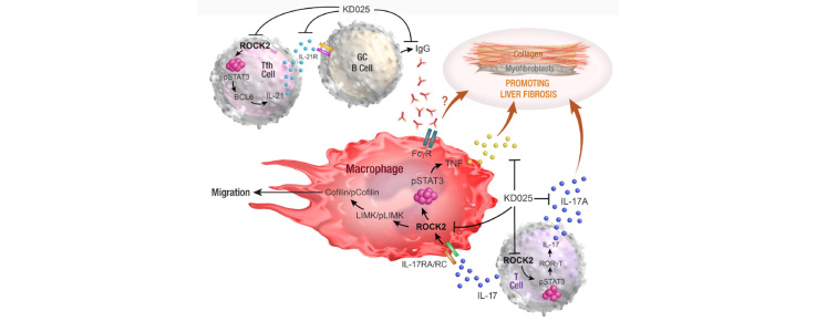
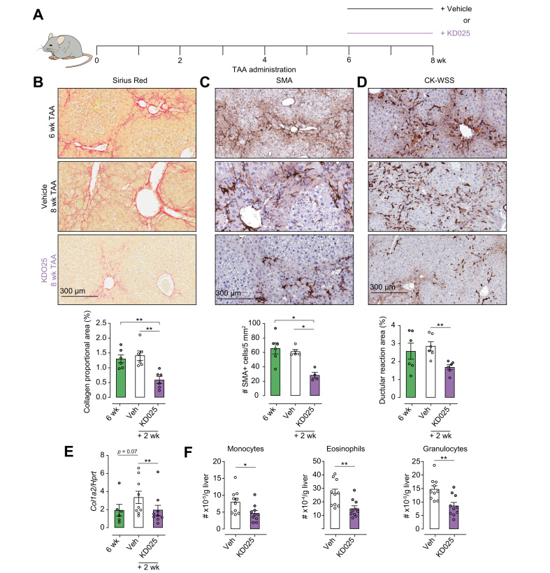
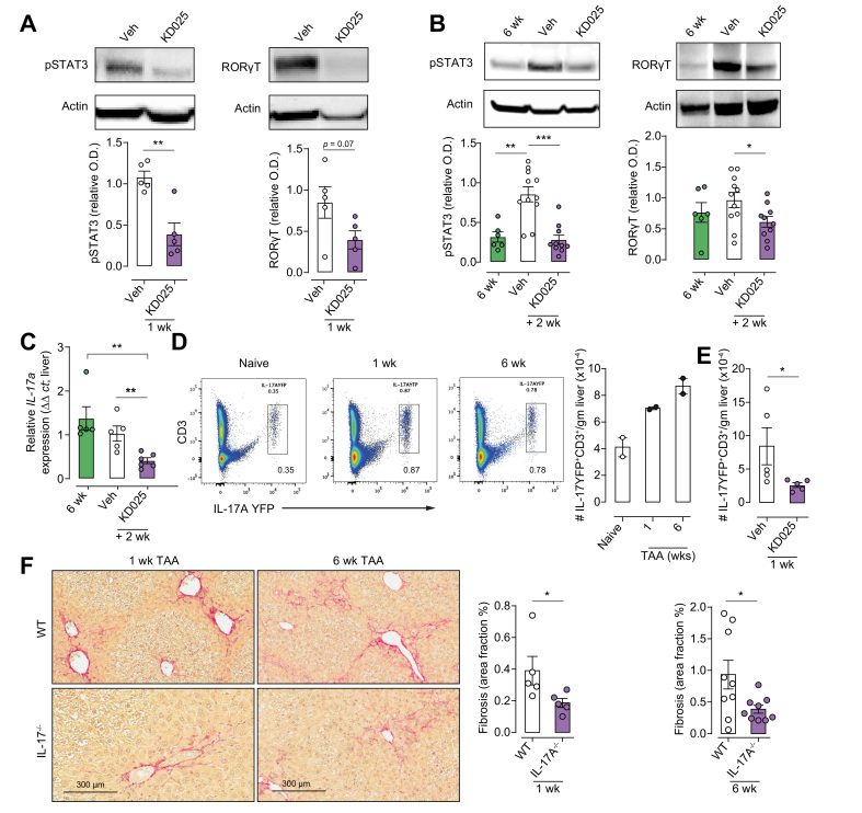
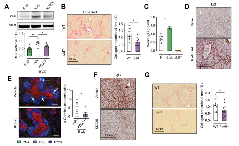
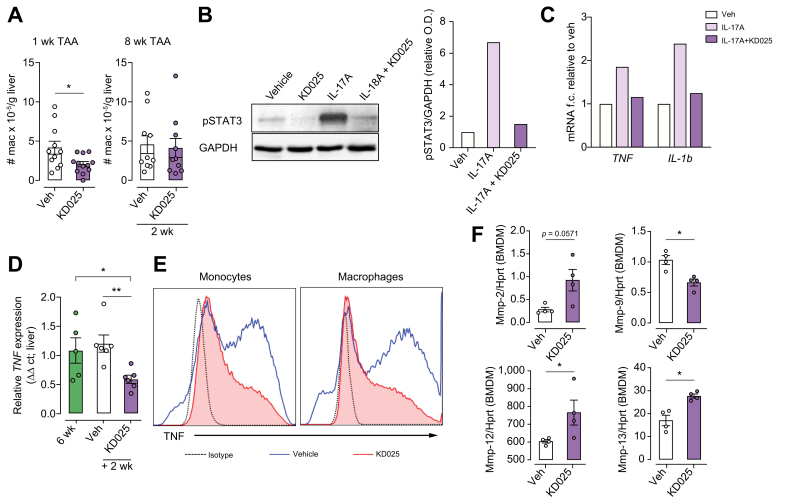
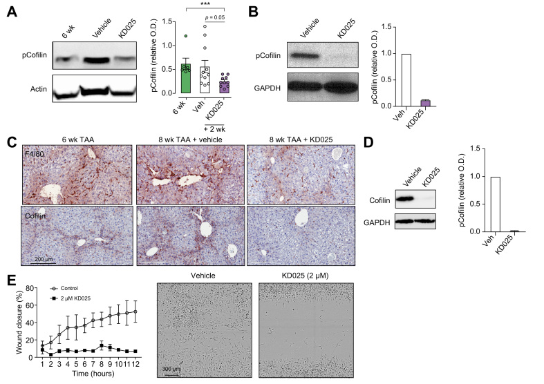
Results: Prophylactic and therapeutic administration of KD025 effectively attenuated thioacetamide-induced liver fibrosis and promoted fibrotic regression. KD025 treatment inhibited liver macrophage tumour necrosis factor production and disrupted the macrophage niche within fibrotic septae. ROCK2 targeting in vitro directly regulated macrophage function through disruption of signal transducer and activator of transcription 3 (STAT3)/cofilin signalling pathways leading to the inhibition of pro-inflammatory cytokine production and macrophage migration. In vivo, KDO25 administration significantly reduced STAT3 phosphorylation and cofilin levels in the liver. Additionally, livers exhibited robust downregulation of immune cell infiltrates and diminished levels of retinoic acid receptor-related orphan receptor gamma (RORγt) and B-cell lymphoma 6 (Bcl6) transcription factors that correlated with a significant reduction in liver IL-17, splenic germinal centre numbers and serum IgG.

Conclusions: As IL-17 and IgG-Fc binding promote pathogenic macrophage differentiation, together our data demonstrate that ROCK2 inhibition prevents and reverses liver fibrosis through direct and indirect effects on macrophage function and highlight the therapeutic potential of ROCK2 inhibition in liver fibrosis.

Lay summary: By using a clinic-ready small-molecule inhibitor, we demonstrate that selective ROCK2 inhibition prevents and reverses hepatic fibrosis through its pleiotropic effects on pro-inflammatory immune cell function. We show that ROCK2 mediates increased IL-17 production, antibody production, and macrophage dysregulation, which together drive fibrogenesis in a model of chemical-induced liver fibrosis. Therefore, in this study, we not only highlight the therapeutic potential of ROCK2 targeting in chronic liver disease but also provide previously undocumented insights into our understanding of cellular and molecular pathways driving the liver fibrosis pathology.

Keywords: ALT, alanine aminotransferase; AST, aspartate aminotransferase; B cells; BMDM, bone marrow-derived macrophages; Bcl6, B-cell lymphoma 6; CLD, chronic liver disease; Col1a2, collagen type α1; DR, ductular reaction; ECM, extracellular matrix; GC, germinal centre; HCC, hepatocellular carcinoma; HSC, hepatic stellate cell; IHC, immunohistochemical; IL-17; Inflammation; LPS, lipopolysaccharide; Liver fibrosis; MMP, matrix metalloproteinase; Macrophages; NASH, non-alcoholic steatohepatitis; RAR, retinoic acid receptor; ROCK, Rho-associated coiled-coil forming protein kinases; ROCK2; ROCK2, Rho-associated kinase 2; RORγt, RAR-related orphan receptor gamma; SR, Sirius red; STAT3, signal transducer and activator of transcription 3; TAA, thioacetamide; TGF-β, transforming growth factor-beta; TNF, tumour necrosis factor; Tfh, T follicular helper; Th17, T helper 17; Therapy; cGVHD, chronic graft-vs-host disease; pCofilin, phosphorylated cofilin; pMac, peritoneal macrophages; pSTAT3, phosphorylated signal transducer and activator of transcription; qRT-PCR, quantitative real-time PCR; α-SMA, alpha smooth muscle actin.


Conflict of interest statement

The authors declare no competing financial interests. BRB receives remuneration as an advisor to Magenta Therapeutics and BlueRock Therapeutics; received research funding from BlueRock Therapeutics, Rheos Medicines, Childrens' Cancer Research Fund, and KidsFirst Fund; and is a co-founder of Tmunity. MN, WC, and AZZ are full-time employees of Kadmon Pharmaceuticals, LLC. Please refer to the accompanying ICMJE disclosure forms for further details.

Figures

None
Graphical abstract
Fig. 1
Fig. 1
KDO25 inhibits TAA-induced liver fibrosis. (A) C57BL/6 mice were treated for 1 week with TAA and vehicle or TAA and KD025 co-administration. Histochemical staining and Aperio quantification of liver sections were undertaken for (B) SR, (C) SMA, and (D) F4/80 (n = 8–11 animals/group; combined from 2 independent experiments). Representative images are shown. (E) Absolute number of liver myeloid and lymphoid cells in mice treated as in (A) was determined by flow cytometry (n = 5 animals/group). Data are presented as mean ± SEM. ∗p <0.05, ∗∗p <0.01 Mann–Whitney U test. SMA, smooth muscle actin; SR, Sirius red; TAA, thioacetamide.
Fig. 2
Fig. 2
KDO25 promotes regression of TAA-induced liver fibrosis. (A) C57BL/6 mice were treated with TAA for 6 weeks followed by 2 weeks of TAA and vehicle or TAA and KD025 co-administration. Histochemical staining and Aperio quantification of (B) SR, (C) SMA, and (D) pan-keratin (CK-WSS) in liver sections were undertaken (n = 4–6 animals/group). Representative images are shown. (E) Expression of col-1a mRNA in whole liver was determined by qRT-PCR and normalised to HPRT mRNA levels (n = 6–9 animals/group; combined from 2 independent experiments). (F) Absolute number of monocytes, eosinophils, and granulocytes was determined by flow cytometry (n = 10 animals/group; combined from 2 independent experiments). Representative images, and data are presented as mean ± SEM. ∗p <0.05, ∗∗p <0.01 Mann–Whitney U test. qRT-PCR, quantitative real-time PCR; SMA, smooth muscle actin; SR, Sirius red; TAA, thioacetamide; WSS, wide spectrum screening.
Fig. 3
Fig. 3
KDO25 inhibits pSTAT3, RORγt, and IL-17A expression in liver. (A) Liver lysates of C57BL/6 mice, treated for 1 week with TAA and vehicle or with TAA and KD025, were analysed by immunoblot using antibodies specific for phosphorylated STAT3Y705 (pSTAT3), RORγt, and actin. Densitometric analysis of pSTAT3 and RORγt normalised to actin is shown (n = 5 animals/group). (B) Mice treated with TAA for 6 weeks or treated with TAA for 6 weeks followed by 2 weeks of coadministration with 0.4% methylcellulose (veh) or 100 mg/kg KDO25, were analysed as in (A) (n = 6–11 animals/group). (C) IL-17a mRNA expression in mice treated as in (B) was determined by qRT-PCR and normalised to HPRT mRNA levels (n = 5–6 animals/group). (D) Livers of IL-17AYFP fate reporter mice left untreated (naïve) or treated with TAA for 1 or 6 weeks were analysed for IL-17A-YFP and CD3 expression by flow cytometry. Representative dot plots and the number of IL-17-YFP+/CD3+ cells are shown (n = 2 animals/group). (E) IL-17A-YFP fate reporter mice treated for 1 week with TAA and 0.4% methylcellulose (veh) or with TAA and 100 mg/kg KDO25 were analysed as in (D) (n = 5 animals/group). (F) WT and IL-17A−/− mice treated with TAA for 1 or 6 weeks, and liver sections were analysed by histochemical staining and Aperio quantification of Sirius red. Representative liver sections and quantitation of collagen are shown (n = 5–9 animals/group; combined from 2 independent experiments). Data are presented as mean ± SEM. ∗p <0.05, ∗∗p <0.01, ∗∗∗p <0.001 Mann–Whitney U test. O.D., optical density; pSTAT3, signal transducer and activator of transcription 3 phosphorylation; qRT-PCR, quantitative real-time PCR; RORγt, retinoic acid receptor-related orphan receptor gamma; SMA, smooth muscle actin; SR, Sirius red; TAA, thioacetamide; WT, wild type.
Fig. 4
Fig. 4
KDO25 inhibits Bcl6 expression, germinal centre formation, and Ig deposition in B cells. (A) Mice treated with TAA for 6 weeks or treated with TAA for 6 weeks followed by 2 weeks of co-administration with vehicle or KD025 were analysed by immunoblot for Bcl6 and actin expression. Densitometric analysis of Bcl6 levels normalised to actin is shown (n = 6–11 animals/group). (B). C57BL/6 WT and μMT−/− mice were treated with TAA for 6 weeks, and liver sections were analysed by histochemical staining and Aperio quantification of SR. Representative liver sections and quantitation of collagen are shown (n = 7–10 animals/group; combined from 2 separate experiments). (C) C57BL/6 WT and μMT mice were treated with TAA for 6 weeks. These and untreated WT mice (N) were analysed by ELISA for serum Ig levels (n = 4–6 animals/group). (D) C57BL/6 mice were left untreated (naïve) or were treated with TAA for 6 weeks, and liver sections were analysed by histochemical staining for Igs. (E) Mice were treated with TAA for 6 weeks or treated with TAA for 6 weeks followed by 2 weeks of co-administration with vehicle or KD025. Spleens were analysed for germinal centre formation by immunofluorescent staining with anti-CD3 (red), anti-B220 (blue), and carbohydrate PNA (green) followed by confocal microscopy (n = 6 animals/group). (F) Histochemical staining for immunoglobulin deposition (IgG) in the liver of mice treated for 6 weeks with TAA co-administered with vehicle or KD025. (G) WT and FcγR−/− mice were treated with TAA for 6 weeks followed by histochemical staining and Aperio quantification of SR. Representative liver sections are shown with SR quantification presented as mean ± SEM (n = 9–12 animals/group; combined from 2 independent experiments). ∗p <0.05, ∗∗p <0.01 Mann–Whitney U test. Bcl6, B-cell lymphoma 6; O.D., optical density; PNA, peanut agglutin; SR, Sirius red; WT, wild type.
Fig. 5
Fig. 5
KDO25 alters macrophage function. (A) C57BL/6 mice were treated for 1 week with TAA and vehicle or with TAA and KD025 (left) or were treated with TAA for 6 weeks followed by 2 weeks of TAA co-administration with vehicle or KD025 (right). The number of liver macrophages were quantified (n = 10−12 animals/group; combined from 2 independent experiments). (B) BMDM were treated with DMSO (vehicle), KD025 (10 μM), IL-17A (10 μg/ml), or KD025 + IL-17A for 20 min. Lysates were analysed by immunoblot using antibodies specific for pSTAT3 and GAPDH, with densitometric analyses shown. (C) BMDM were treated with DMSO (vehicle), IL-17A (10 μg/ml), or KD025 (10 μM) + IL-17A for 6 h. qRT-PCR analysis of TNF and IL-1β mRNA normalised to HPRT mRNA is shown. Data shown in (B) and (C) are representative of 2 independent experiments (n = 1/condition). (D) Mice were treated with TAA for 6 weeks or treated with TAA for 6 weeks followed by 2 weeks of co-administration with vehicle or KD025. qRT-PCR analysis of TNF mRNA expression in whole liver (n = 5–6 animals/group) is shown. (E) Isolated hepatic leucocytes from mice treated as in (D) were treated in vitro with LPS (100 ng/ml) and analysed by flow cytometry for monocyte (left) and macrophage (right) TNF production. (F) BMDM were treated with KD025 (10 μM) or DMSO control for 6 h, and the expression of mRNAs encoding MMPs, normalised to HPRT mRNA, was determined by qRT-PCR (n = 4 animals/group). Data are presented as mean ± SEM. ∗p <0.05, ∗∗p <0.01 Mann–Whitney U test. BMDM, bone marrow-derived macrophages; MMP, matrix metalloproteinase; O.D., optical density; pSTAT3, signal transducer and activator of transcription 3 phosphorylation; qRT-PCR, quantitative real-time PCR; TAA, thioacetamide.
Fig. 6
Fig. 6
KDO25 alters macrophage migration. (A) C57BL/6 mice were treated with TAA for 6 weeks or treated with TAA for 6 weeks followed by 2 weeks of co-administration with vehicle or KD025. Livers lysates were analysed for the level of pCofilin and actin. A representative immunoblot and densitometric analysis of pCofilin normalized to actin is shown (n = 6–11 animals/group). Data are presented as mean ± SEM.  ∗∗∗p <0.001 Mann–Whitney U test. (B) BMDM were treated with 10 μM KD025 or with DMSO (veh) for 20 min. Levels of pCofilin and GAPDH were determined by immunoblot. A representative blot and densitometric analysis are shown (n = 2 animals/group). (C) Mice were treated as in (A), and liver sections were analysed by histochemical staining for total cofilin. (D) BMDM were treated with KD025 (2 μM) of DMSO (veh) for 6 h, and total cofilin and GAPDH levels were assessed by immunoblot. A representative blot and densitometric analysis of cofilin normalized to GAPDH is shown (n = 2 animals/group). (E) Migration of BMDM in the presence of DMSO (veh) or 2 μM KD025 was assessed over 12 h by scratch wound assay and Incucyte microscopic analysis (n = 3 animals/group). Representative images at 8 h are shown. BMDM, bone marrow-derived macrophages; pCofilin, phosphorylated cofilin; O.D., optical density; TAA, thioacetamide.

References

    1. Ishizaki T., Maekawa M., Fujisawa K., Okawa K., Iwamatsu A., Fujita A., et al. The small GTP-binding protein Rho binds to and activates a 160 kDa Ser/Thr protein kinase homologous to myotonic dystrophy kinase. EMBO J. 1996;15:1885–1893. - PMC - PubMed
    1. Hoon J.L., Tan M.H., Koh C.-G. The regulation of cellular responses to mechanical cues by Rho GTPases. Cells. 2016;5:17. - PMC - PubMed
    1. Riches D.W., Backos D.S., Redente E.F. ROCK and Rho: promising therapeutic targets to ameliorate pulmonary fibrosis. Am J Pathol. 2015;185:909–912. - PMC - PubMed
    1. Riento K., Ridley A.J. Rocks: multifunctional kinases in cell behaviour. Nat Rev Mol Cell Biol. 2003;4:446–456. - PubMed
    1. Zhou Y., Huang X., Hecker L., Kurundkar D., Kurundkar A., Liu H., et al. Inhibition of mechanosensitive signaling in myofibroblasts ameliorates experimental pulmonary fibrosis. J Clin Invest. 2013;123:1096–1108. - PMC - PubMed