. 2022 Mar 26;14(1):mjab079.
doi: 10.1093/jmcb/mjab079.

LRRK2 regulates actin assembly for spindle migration and mitochondrial function in mouse oocyte meiosis

Affiliations

LRRK2 regulates actin assembly for spindle migration and mitochondrial function in mouse oocyte meiosis

Zhen-Nan Pan et al. J Mol Cell Biol. .

Erratum in

  • Erratum.
    [No authors listed] [No authors listed] J Mol Cell Biol. 2022 May 25;14(2):mjac030. doi: 10.1093/jmcb/mjac030. J Mol Cell Biol. 2022. PMID: 35639540 Free PMC article. No abstract available.

Abstract

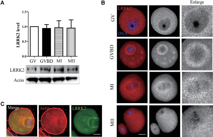
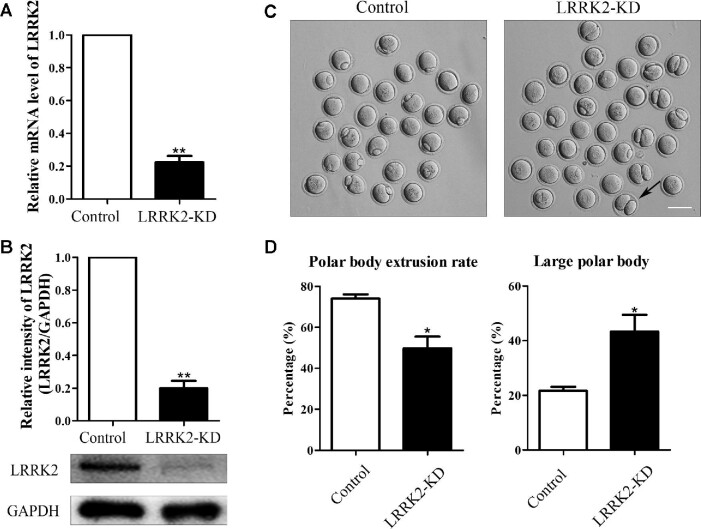
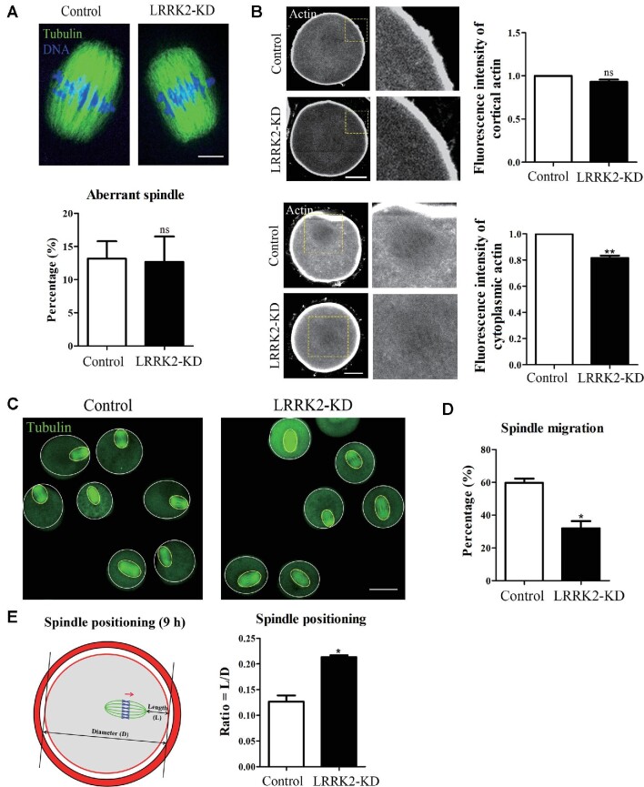
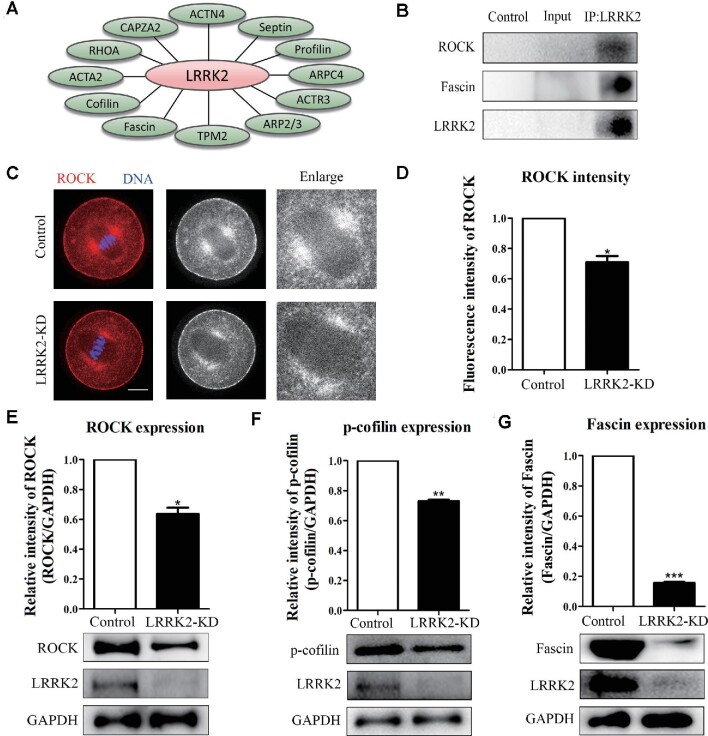
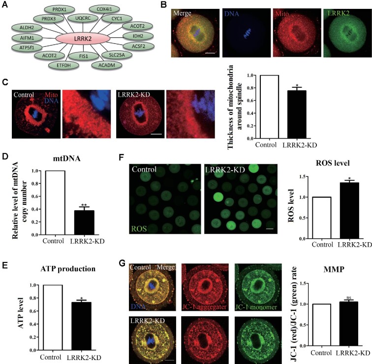
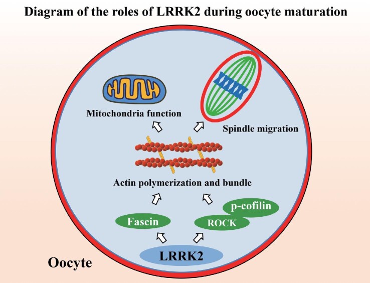
Leucine-rich-repeat kinase 2 (LRRK2) belongs to the Roco GTPase family and is a large multidomain protein harboring both GTPase and kinase activities. LRRK2 plays indispensable roles in many processes, such as autophagy and vesicle trafficking in mitosis. In this study, we showed the critical roles of LRRK2 in mammalian oocyte meiosis. LRRK2 is mainly accumulated at the meiotic spindle periphery during oocyte maturation. Depleting LRRK2 led to the polar body extrusion defects and also induced large polar bodies in mouse oocytes. Mass spectrometry analysis and co-immunoprecipitation results showed that LRRK2 was associated with several actin-regulating factors, such as Fascin and Rho-kinase (ROCK), and depletion of LRRK2 affected the expression of ROCK, phosphorylated cofilin, and Fascin. Further analysis showed that LRRK2 depletion did not affect spindle organization but caused the failure of spindle migration, which was largely due to the decrease of cytoplasmic actin filaments. Moreover, LRRK2 showed a similar localization pattern to mitochondria, and LRRK2 was associated with several mitochondria-related proteins. Indeed, mitochondrial distribution and function were both disrupted in LRRK2-depleted oocytes. In summary, our results indicated the critical roles of LRRK2 in actin assembly for spindle migration and mitochondrial function in mouse oocyte meiosis.

Keywords: LRRK2; actin; meiosis; oocyte; spindle.


Figures

Figure 1
Figure 1
Expression and localization of LRRK2 in mouse oocyte meiosis. (A) The expression level of LRRK2 at different stages (GV, GVBD, MI, and MII) was examined by western blotting in mouse oocytes. (B) The localization of LRRK2 was certificated by LRRK2 antibody in mouse oocytes. LRRK2 dispersed in the cytoplasm at the GV stage and was mainly distributed around the spindle after GVBD. Red, LRRK2; blue, DNA; scale bar, 20 μm. (C) LRRK2 was colocalized with actin around the spindle in the MI-stage oocytes. Red, actin; green, LRRK2; blue, DNA; scale bar, 20 μm.
Figure 2
Figure 2
LRRK2 depletion affects mouse oocyte maturation. (A) The LRRK2 mRNA expression level was tested by RT-PCR in the LRRK2 siRNA-injected and control oocytes. (B) The LRRK2 protein expression level was examined by western blotting in the LRRK2 siRNA-injected and control oocytes. GAPDH served as the control. Band intensities were analyzed by Image J software. (C) LRRK2 depletion caused polar body extrusion defects and large polar bodies in mouse oocytes. The black arrow indicates an oocyte extruding a large polar body after 12 h culture. Scale bar, 100 μm. (D) The rate of polar body extrusion was lower, whereas the rate of the large polar body was higher in the LRRK2 siRNA-injected group compared with that in the control group. *P < 0.05 and **P < 0.01.
Figure 3
Figure 3
LRRK2 depletion affects cytoplasmic actin assembly for spindle migration in mouse oocytes. (A) Immunofluorescence staining showed that the spindle morphology of the LRRK2 siRNA-injected oocytes was normal and there was no difference in the percentage of aberrant spindle compared with the control oocytes. Green, tubulin; blue, DNA; scale bar, 10 μm. (B) LRRK2 depletion did not affect the fluorescence intensity of cortical actin but decreased that of cytoplasmic actin in the LRRK2 siRNA-injected oocytes. Gray, actin; scale bar, 20 μm. (C) Representative images of the spindle positioning in the control and LRRK2 siRNA-injected oocytes after 9 h culture. Green, tubulin; scale bar, 50 μm. (D) The meiotic spindles migrated under the cortex after 9 h culture in the control oocytes, while rates of spindle to the cortex were significantly decreased in the LRRK2 siRNA-injected oocytes. (E) Quantitative analysis of the extent of spindle migration showed that the L/D ratio was higher in the LRRK2 siRNA-injected oocytes than in the control oocytes. Red, actin; green, spindle; arrow, the direction of spindle migration. *P < 0.05, **P < 0.01, and ns, no significant difference.
Figure 4
Figure 4
LRRK2 depletion affects the expression of ROCK‒cofilin and Fascin in mouse oocytes. (A) Screening actin-related proteins associated with LRRK2 by MS analysis. (B) Co-IP results showed that LRRK2 was correlated with ROCK and Fascin. (C) Immunofluorescence staining displayed the localization of ROCK in the control and LRRK2 siRNA-injected oocytes. Red, ROCK; blue, DNA; scale bar, 20 μm. (D) Statistical analysis showed that the fluorescence intensity of ROCK was significantly decreased at the spindle periphery after LRRK2 depletion. (E) ROCK protein expression was significantly decreased in the LRRK2 siRNA-injected oocytes. (F) The phosphorylation level of cofilin was significantly decreased in the LRRK2 siRNA-injected oocytes. (G) Fascin protein expression was significantly decreased in the LRRK2 siRNA-injected oocytes. *P < 0.05, **P < 0.01, and ***P < 0.001.
Figure 5
Figure 5
LRRK2 depletion affects the distribution and function of mitochondria in mouse oocytes. (A) Screening mitochondria-related proteins associated with LRRK2 by MS analysis. (B) LRRK2 showed a similar localization pattern to mitochondria in mouse oocytes. Red, mitochondria; green, LRRK2; blue, DNA; scale bar, 20 μm. (C) The accumulation of mitochondria around the spindle in the LRRK2 siRNA-injected oocytes was decreased compared with that in the control oocytes. Red, mitochondria; blue, DNA; scale bar, 20 μm. (D) The relative content of mtDNA copy number in the LRRK2 siRNA-injected oocytes was significantly decreased compared with that in the control oocytes. (E) The relative content of ATP in the LRRK2 siRNA-injected oocytes was significantly decreased compared with that in the control oocytes. (F) The relative fluorescence intensity of ROS in the LRRK2 siRNA-injected oocytes was significantly increased compared with that in the control oocytes. Scale bar, 50 μm. (G) JC-1 staining showed no significant difference in MMP between the LRRK2 siRNA-injected and control oocytes. Red, JC-1 aggregates; green, JC-1 monomer; blue, DNA; scale bar, 20 μm. *P < 0.05, **P < 0.01, and ns, no significant difference.
Figure 6
Figure 6
Diagram for the LRRK2 functions during mouse oocyte maturation. The cellular actin cytoskeleton is formed by the assembly of globular actin (G-actin) into double helical filaments (F-actin). LRRK2 modulates ROCK kinase and cofilin for actin assembly and Fascin for actin bundles and further affects mitochondrial function and spindle migration in mouse oocytes.

References

    1. Alegre-Abarrategui J., Christian H., Lufino M.M.et al. (2009). LRRK2 regulates autophagic activity and localizes to specific membrane microdomains in a novel human genomic reporter cellular model. Hum. Mol. Genet. 18, 4022–4034. - PMC - PubMed
    1. Almonacid M., Terret M.E., Verlhac M.H. (2014). Actin-based spindle positioning: new insights from female gametes. J. Cell Sci. 127, 477–483. - PubMed
    1. Bavister B.D., Squirrell J.M. (2000). Mitochondrial distribution and function in oocytes and early embryos. Hum. Reprod. 15 (Suppl 2), 189–198. - PubMed
    1. Biskup S., Moore D.J., Celsi F.et al. (2006). Localization of LRRK2 to membranous and vesicular structures in mammalian brain. Ann. Neurol. 60, 557–569. - PubMed
    1. Breitsprecher D., Koestler S.A., Chizhov I.et al. (2011). Cofilin cooperates with Fascin to disassemble filopodial actin filaments. J. Cell Sci. 124, 3305–3318. - PMC - PubMed

Publication types

Substances