

Parathyroid Hormone-Related Changes of Bone Structure
- PMID: 34918524
- PMCID: PMC8884379
- DOI: 10.33549/physiolres.934779
Parathyroid Hormone-Related Changes of Bone Structure
Abstract
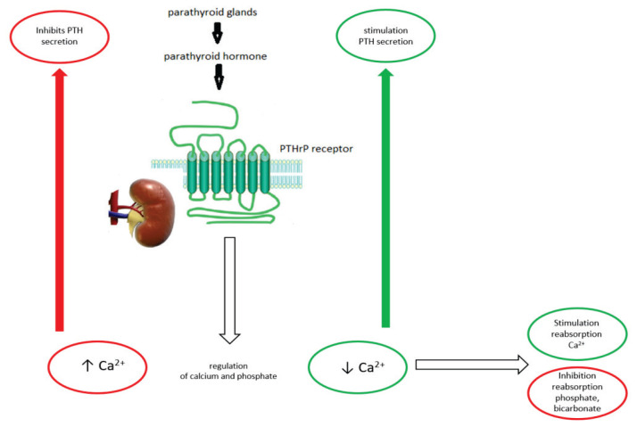
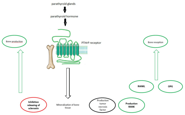
Parathyroid hormone (PTH) increases the release of serum calcium through osteoclasts, which leads to bone resorption. Primary, PTH stimulates osteoblasts leading to increase RANKL (receptor activator for nuclear factor kappa-B ligand) expression and thus differentiation of osteoclasts. In kidneys, PTH increases calcium and decrease phosphate reabsorption. In kidneys, PTH stimulates 1alpha-hydroxylase to synthesize active vitamin D. Primary hyperparathyroidism (PHPT) is characterized by skeletal or renal complications. Nowadays, the classical form of PHPT is less seen and asymptomatic or subclinical (oligo symptomatic) forms are more frequent. Previously, it was thought that cortical bone is preferably affected by PHPT and that predispose bones to fracture at sites with a higher amount of cortical bone. However, an increased risk of vertebral fractures has been found by most of the studies showing that also trabecular bone is affected. Bone Mass measurement (BMD) at all skeletal sites is advised, but another specific tool for fracture assessment is needed. Trabecular bone score (TBS), an indirect measure of trabecular bone, maybe a useful method to estimate fracture risk. TBS is associated with vertebral fractures in PHPT regardless of BMD, age, BMI and gender. Furthermore, there is an association between TBS and high resolution peripheral quantitative computed tomography (HR-pQCT) parameters in the trabecular and cortical compartment. However, studies considering the effect of PHPT treatment on TBS are more conflicting. Secondary hyperparathyroidism caused by vitamin D deficiency was associated with impaired bone microarchitecture in all age categories, as measured by TBS and Hr-pQCT with further improvement after treatment with vitamin D.
Conflict of interest statement
There is no conflict of interest.
Figures
References
-
- BILEZIKIAN JP, BRANDI ML, EASTELL R, SILVERBERG SJ, UDELSMAN R, MARCOCCI C, POTTS JT., Jr Guidelines for the management of asymptomatic primary hyperparathyroidism: summary statement from the Fourth International Workshop. J Clin Endocrin Metab. 2014;99:3561–3569. doi: 10.1210/jc.2014-1413. - DOI - PMC - PubMed