. 2022 Jan 15;135(2):jcs258818.
doi: 10.1242/jcs.258818. Epub 2022 Jan 26.

The histone H4 lysine 20 demethylase DPY-21 regulates the dynamics of condensin DC binding

Affiliations

The histone H4 lysine 20 demethylase DPY-21 regulates the dynamics of condensin DC binding

Laura Breimann et al. J Cell Sci. .

Abstract

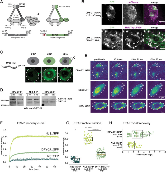
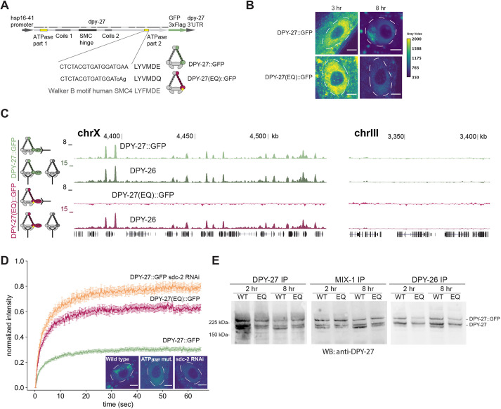
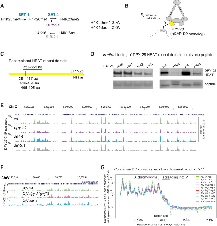
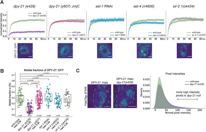
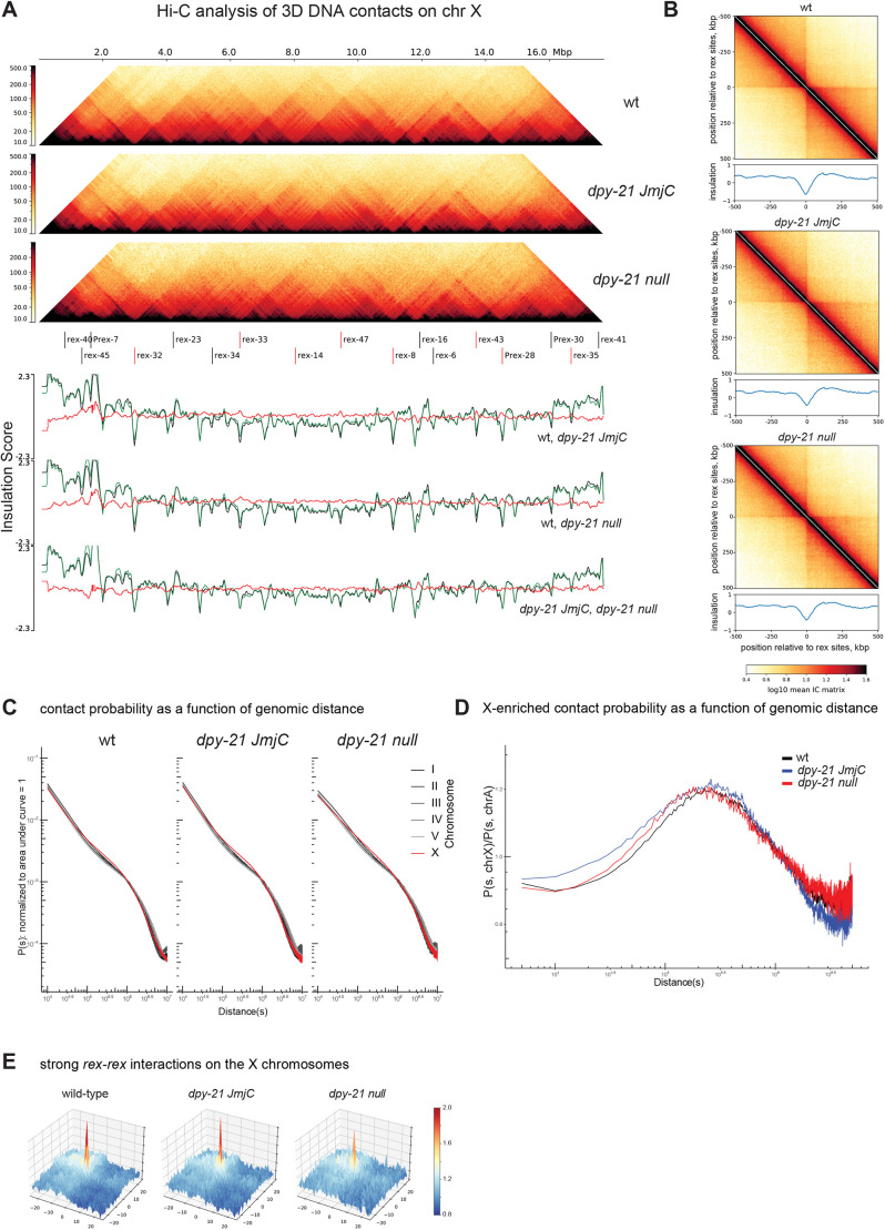
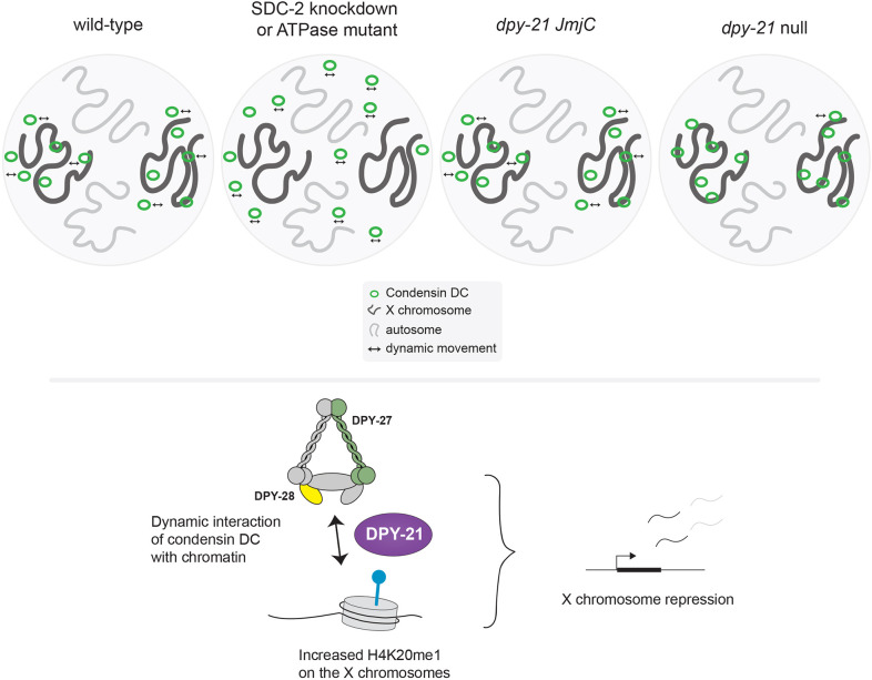
Condensin is a multi-subunit structural maintenance of chromosomes (SMC) complex that binds to and compacts chromosomes. Here, we addressed the regulation of condensin binding dynamics using Caenorhabditis elegans condensin DC, which represses X chromosomes in hermaphrodites for dosage compensation. We established fluorescence recovery after photobleaching (FRAP) using the SMC4 homolog DPY-27 and showed that a well-characterized ATPase mutation abolishes DPY-27 binding to X chromosomes. Next, we performed FRAP in the background of several chromatin modifier mutants that cause varying degrees of X chromosome derepression. The greatest effect was in a null mutant of the H4K20me2 demethylase DPY-21, where the mobile fraction of condensin DC reduced from ∼30% to 10%. In contrast, a catalytic mutant of dpy-21 did not regulate condensin DC mobility. Hi-C sequencing data from the dpy-21 null mutant showed little change compared to wild-type data, uncoupling Hi-C-measured long-range DNA contacts from transcriptional repression of the X chromosomes. Taken together, our results indicate that DPY-21 has a non-catalytic role in regulating the dynamics of condensin DC binding, which is important for transcription repression.

Keywords: C. elegans; Condensin; FRAP; Hi-C; Histone modifications; Transcription.


Conflict of interest statement

Competing interests The authors declare no competing or financial interests.

Figures

Fig. 1.
Fig. 1.
FRAP analysis of condensin DC binding. (A) Left panel illustrates condensin DC along with the rest of the DCC subunits. The right panel indicates the expression of GFP-tagged DPY-27 under the control of a heat shock-inducible promoter at the chromosome II MosSCI site. (B) DPY-27::GFP subnuclear localization to the X chromosomes in intestinal cells 8 h after heat-induced expression (top row) was validated by colocalization with the endogenously tagged DPY-27::Halo stained with JF635 HaloTag ligand (bottom row). Nuclei are outlined by white dashed lines. Images are representative of three biological replicates with a minimum of 20 images. Scale bars: 5 µm. (C) Illustration of the heat-shock protocol (top). Young adult worms were heat shocked for 1 h at 35°C, and fluorescence was followed in the large intestinal cells. DPY-27::GFP subnuclear localization indicative of X chromosome binding is apparent after 8 h of recovery. Representative example images from a total of three biological replicates with a minimum of 30 images are shown for each time point, with the nuclear area marked using a white dashed line. Scale bars: 5 µm. (D) DPY-27::GFP interaction with condensin DC subunits was validated by co-immunoprecipitation with MIX-1 and DPY-26. Young adult worms were used for immunoprecipitation (IP) either 2 h or 8 h after heat shock at 35°C for 1 h and analyzed by western blotting (WB) using an anti-DPY-27 antibody. The intensity of the GFP-tagged DPY-27 and endogenous protein bands in the DPY-27 IP lane indicates the relative abundance of each protein. The intensity of the GFP-tagged DPY-27 and the endogenous protein bands in the other lanes indicates the relative interaction of endogenous and DPY-27::GFP with the immunoprecipitated subunit. Blots are representative of three experiments. (E) FRAP sequence for intestine nuclei of adult C. elegans worms expressing either DPY-27::GFP, NLS::GFP or H2B::GFP. The first column of images depicts the first image of the pre-bleach series (a total of 20 images). The second column shows the first image after the single point bleach, with the bleached area indicated by the dotted circle. The third and fourth columns depict two time points after the bleach point: t100 (21 s) and t320 (70 s), respectively. Nuclei are outlined by white dashed lines. (F) Mean FRAP recovery curves from worms expressing wild-type DPY-27::GFP, H2B::GFP or NLS::GFP. Data are mean±s.e.m. Numbers of bleached single intestine nuclei (from at least three biological replicates) for each experiment are n=81 for DPY-27::GFP, n=48 for NLS::GFP and n=61 for H2B::GFP. (G) Mobile fractions for the different GFP-tagged proteins or free GFP. The mobile fraction is the lowest for H2B::GFP and the highest for NLS::GFP. The mobile fraction for DPY-27::GFP is ∼28%. P-values are from an two-tailed independent two-sample t-test. (H) FRAP half time recovery (T-half) values for the bleach curves shown in Fig. 1F. The half time recovery for NLS::GFP shows a shorter diffusion time than DPY-27::GFP. H2B::GFP is not shown due to the very low recovery of the fluorescence signal during the experimental time frame. Boxplots in G and H show the median (line), interquartile range (box) and whiskers at the 5th and 95th percentile of the dataset. The median values (med) and number of nuclei analyzed are shown for each group.
Fig. 2.
Fig. 2.
The effect of a conserved SMC ATPase mutation on DPY-27 binding, function, protein stability and complex formation. (A) Sequence encoding heat shock-inducible GFP-tagged DPY-27(EQ). The DNA sequence coding for the conserved Walker B motif and the E-to-Q mutation are shown below. (B) Localization of the wild-type and EQ ATPase mutant DPY-27::GFP proteins in intestine cells. Adults were heat shocked at 35°C for 1 h and recovered for either 3 h or 8 h. Unlike DPY-27::GFP, the ATPase EQ mutant did not show subnuclear localization. Nuclei are outlined by white dashed lines. Images are representative of three replicates (quantified in Fig. S3E). Scale bars: 5 µm. (C) ChIP-seq analysis of wild-type and ATPase mutant DPY-27::GFP using an anti-GFP antibody in embryos. ChIP against DPY-26 was used as a positive control in the same extracts. Unlike the wild-type protein, the ATPase mutant failed to bind to the X chromosome (chrX), and both did not localize to the autosomes. A representative region from chromosome III is shown in the right panel. ChIP profiles show normalized read coverage (y-axis) for representative regions on chromosome X and III in a UCSC genome browser snapshot. Data are representative of three replicate experiments. (D) Mean FRAP recovery curves from DPY-27::GFP, DPY-27(EQ)::GFP and DPY-27::GFP upon SDC-2 RNAi. FRAP was performed ∼8 h after the heat shock. Data are mean±s.e.m. Numbers of bleached single intestine nuclei (from at least three biological replicates) for each experiment are n=81 for DPY-27::GFP, n=37 for DPY-27(EQ)::GFP and n=32 for DPY-27::GFP sdc-2 RNAi. The images depict examples of intestine nuclei used for FRAP analysis. Unlike DPY-27::GFP, the ATPase EQ mutant did not show subnuclear localization, similar to when condensin DC recruiter SDC-2 was knocked down. Nuclei are outlined by white dashed lines. Scale bars: 5 µm. (E) Co-immunoprecipitation analysis of condensin DC subunits. Protein extracts were prepared from larvae that were heat shocked for 1 h at 35°C and recovered at 20°C for 2 h or 8 h. Immunoprecipitation (IP) of condensin DC subunits DPY-27, DPY-26 and MIX-1 was performed, and immunoprecipitated DPY-27::GFP and endogenous protein were analyzed by western blotting (WB) with an anti-DPY-27 antibody. The intensity of the DPY-27::GFP and endogenous protein bands in the DPY-27 IP lane indicates the relative abundance of each protein. The intensity of DPY-27::GFP and endogenous protein bands in other lanes indicates their relative interaction with each subunit. Blots are representative of two experiments.
Fig. 3.
Fig. 3.
Condensin DC may interact with histone tails, but set-4, sir-2.1 and catalytic activity of DPY-21 do not regulate condensin DC binding as measured by ChIP-seq. (A) Enzymes that regulate H4K20 methylation and H4K16 acetylation. In hermaphrodites, H4K20me1 is increased and H4K16ac is reduced on the dosage compensated X chromosomes (X) compared to autosomes (A). The dpy-21 null mutant is the (e418) allele with a premature stop codon that eliminates the protein (Yonker and Meyer, 2003), the dpy-21(JmjC) mutant is the (y607) allele, a point mutation that nearly abolishes H4K20me2 demethylase activity without eliminating the protein itself (Brejc et al., 2017). The set-4 null mutant is (n4600), a knockout allele that eliminates H4K20me2 and H4K20me3 (Delaney et al., 2017). The sir-2.1 null mutant is (ok434), a knockout allele that increases H4K16ac (Wells et al., 2012). (B) Cartoon depicting possible interaction of HEAT repeat-containing domain of DPY-28 (homologous to human hCAP-D2) with histone tail modifications. (C) Three HEAT repeats annotated by Pfam are shown as tick marks. The amino acids (aa) 351–661 were purified and used in peptide binding assays. (D) In-solution peptide binding assay was performed using GST-tagged DPY-28 HEAT domain and biotinylated histone N-terminal tail peptides with the indicated modifications (H3ac and H4ac indicate tetra-acetylated histone H3 and H4 peptides, respectively). The recombinant protein was incubated with peptides bound to magnetic streptavidin beads, and bound fractions were analyzed using western blot. The streptavidin signal below indicates the amount of peptide in each fraction. Methyl modified histone peptide blots representative of two replicates; acetyl and unmodified histone peptides representative of two replicates. (E) UCSC genome browser (https://genome.ucsc.edu/) shot of a representative region of the X chromosome (ChrX) showing similar DPY-27 ChIP-seq patterns in the sir-2.1 null mutant. Data from wild-type N2, the dpy-21 null mutant and the set-4 null mutant are from Kramer et al. (2015) and are plotted for comparison. Chromosome locations are marked in kb. (F) Genome browser view of DPY-27 ChIP-seq enrichment across the fusion site on the autosomal region of the X;V chromosome in X;V wild-type, dpy-21(JmjC) and set-4 null backgrounds. (G) A moving average of the DPY-27 ChIP enrichment score is plotted with a window size of 500 kb and step size of 50 kb in X;V fusion strains with wild-type, dpy-21(JmjC) and set-4 null backgrounds. DPY-27 ChIP-seq data was normalized to reduce variability between replicates by z-score standardization of ChIP/input ratios to the background from autosomes I–IV, followed by equalization of total ChIP-seq signal to 1 in X;V.
Fig. 4.
Fig. 4.
The dpy-21 null mutant but not the dpy-21(JmjC) catalytic mutant reduces the proportion of mobile condensin DC. (A) Mean FRAP recovery curves of DPY-27::GFP in either wild-type (green) or different mutant conditions. Data are mean±s.e.m. Numbers of bleached single intestine nuclei (from at least three biological replicates) for each experiment are n=81 for wild type, n=72 for the dpy-21 null mutant [dpy-21 (e428)], n=102 for the dpy-21(JmjC) mutant [dpy-21 (y607)], n=28 for set-1 RNAi, n=45 for the set-4 null mutant [set-4 (n4600)] and n=41 for the sir-2.1 null mutant [sir-2.1 (ok434)]. Corresponding images of intestine nuclei for each mutant condition are depicted under each FRAP curve. Nuclei are outlined by dashed lines. Scale bars: 5 µm. (B) Mobile fractions calculated from individual replicate FRAP recovery curves as shown in A. P-values are from a two-tailed independent two-sample t-test. Boxplots show the median (line), interquartile range (box). Whiskers are at the 5th and 95th percentile of the dataset. The number of images of nuclei analyzed is noted under each boxplot, along with the median values (med). (C) Analysis of endogenous DPY-27::Halo fluorescence intensity on the X chromosome in wild-type and dpy-21 null worms. The HaloTag signal of DPY-27 was segmented in 3D and quantified in adult intestine cells in two biological replicates (Fig. S4C). The left panel depicts two example nuclei (marked by dashed lines). Scale bars: 5 µm. For the wild-type worms, 27 images were analyzed, for the dpy-21(e428) mutant images of 35 nuclei were analyzed. The right panel shows the binned mean pixel fluorescence intensity for the two conditions in a smoothed density plot. The distributions of pixel intensities are significantly different in the two conditions, with a P-value of 1.46×10−114 (Mann–Whitney U-test).
Fig. 5.
Fig. 5.
Hi-C analysis of 3D DNA contacts in dpy-21(JmjC) and dpy-21 null mutant embryos. (A) Hi-C heatmap (top) and insulation scores (bottom) of the X chromosome (chr X) showing wild type (wt), the dpy-21(JmjC) mutant, and the dpy-21 null mutant. ‘The fall’ colormap, adapted from cooltools, is used to depict the strength of relative contact probability between pairs of genomic bins. The 17 strong rex sites indicated are as annotated by Albritton et al. (2017), eight of which were annotated as DCC-dependent boundary rex sites (indicated by red lines) by Anderson et al. (2019). The insulation scores and their subtractions for three possible pairwise comparisons are shown in the lower panels, The insulation scores for the three pair-wise comparison are as follows: top: wild-type (black), dpy-21(JmjC) (green); middle wild-type (black), dpy-21 null (green); bottom: dpy-21(JmjC) (black), dpy-21 null (green); the red lines indicate per bin subtraction of green minus black. (B) Pile-up analysis showing the average Hi-C map and the insulation scores ±500 kb surrounding the annotated 17 strong rex sites for the indicated genotypes. IC or ‘iteratively corrected' matrix’ is a type of matrix balancing used to correct for different bins having sequencing/representation bias. (C) Distance decay curve showing the relationship between 5-kb binned genomic separation, s, and average contact probability, P(s) computed per chromosome for the indicated genotypes. (D) X chromosome-enriched chromosomal contacts for the indicated genotypes are visualized using an X chromosome minus autosome (X−A)-normalized distance decay curve. For every genomic separation s, the unity-normalized contact probability of the X chromosome, P(s, chrX), is divided by that of autosomes, P(s, chrA). Distances in C and D are shown in bp. (E) Meta-dot plot showing the average strength of interactions between pairs of rex sites on a 10 kb distance-normalized (observed divided by expected) matrix. A total of ±25 bins (±250kb) regions surrounding each rex site are shown. For the 17 strong rex sites, a total of 33 rexrex pairs located within 3 Mb of each other were used. More blue coloring indicates interaction strengths weaker than expected and more red coloring indicates strength greater than expected.
Fig. 6.
Fig. 6.
Summary of results and DPY-21 function in condensin DC-mediated X chromosome repression. In a wild-type hermaphrodite cell, condensin DC binds dynamically and specifically to the X chromosomes. This binding is disrupted by knockdown of the condensing DC recruiter SDC-2 or a single amino acid mutation in the ATPase domain of DPY-27. Condensin DC may interact with histone tails through HEAT repeats within DPY-28. The H4K20me2 demethylase DPY-21 has a dual function in X chromosome repression. The catalytic activity reduces H4K20me2 and H4K20me3 and increases H4K20me1 on the X chromosome. This leads to reduced H4K16ac and contributes to repression. The non-catalytic activity of DPY-21 increases the mobility of condensin DC molecules, which is important for transcription repression. In the dpy-21 null condition, both catalytic and non-catalytic activities are eliminated, resulting in stronger X chromosome derepression.

References

    1. Albritton, S. E. and Ercan, S. (2018). Caenorhabditis elegans dosage compensation: insights into condensin- mediated gene regulation. Trends Genet. 34, 41-53. 10.1016/j.tig.2017.09.010 - DOI - PMC - PubMed
    1. Albritton, S. E., Kranz, A.-L., Winterkorn, L. H., Street, L. A. and Ercan, S. (2017). Cooperation between a hierarchical set of recruitment sites targets the X chromosome for dosage compensation. eLife 6, e23629-e23645. 10.7554/eLife.23645 - DOI - PMC - PubMed
    1. Amemiya, H. M., Kundaje, A. and Boyle, A. P. (2019). The ENCODE blacklist: identification of problematic regions of the genome. Sci. Rep. 9, 9354. 10.1038/s41598-019-45839-z - DOI - PMC - PubMed
    1. Anders, S., Pyl, P. T. and Huber, W. (2015). HTSeq—a Python framework to work with high-throughput sequencing data. Bioinformatics 31, 166-169. 10.1093/bioinformatics/btu638 - DOI - PMC - PubMed
    1. Anderson, E. C., Frankino, P. A., Higuchi-Sanabria, R., Yang, Q., Bian, Q., Podshivalova, K., Shin, A., Kenyon, C., Dillin, A. and Meyer, B. J. (2019). X Chromosome domain architecture regulates caenorhabditis elegans lifespan but not dosage compensation. Dev. Cell 51, 192-207.e6. 10.1016/j.devcel.2019.08.004 - DOI - PMC - PubMed

Publication types

MeSH terms