. 2021 Dec 17;6(1):428.
doi: 10.1038/s41392-021-00849-0.

SARS-CoV-2-triggered mast cell rapid degranulation induces alveolar epithelial inflammation and lung injury

Affiliations

SARS-CoV-2-triggered mast cell rapid degranulation induces alveolar epithelial inflammation and lung injury

Meng-Li Wu et al. Signal Transduct Target Ther. .

Abstract

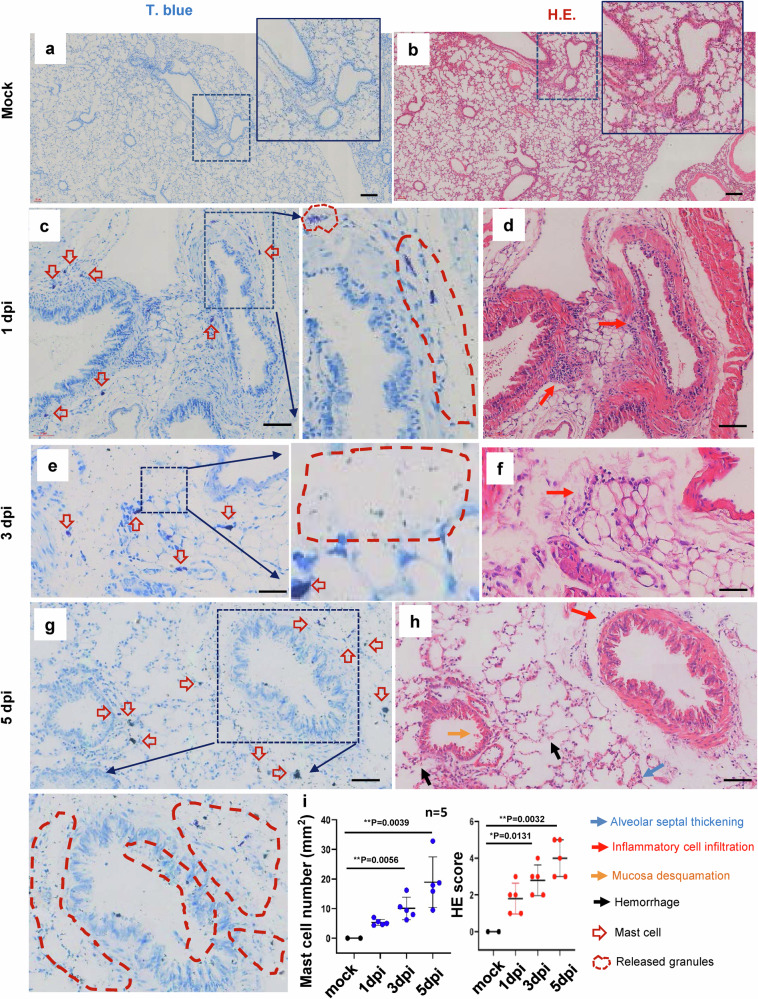
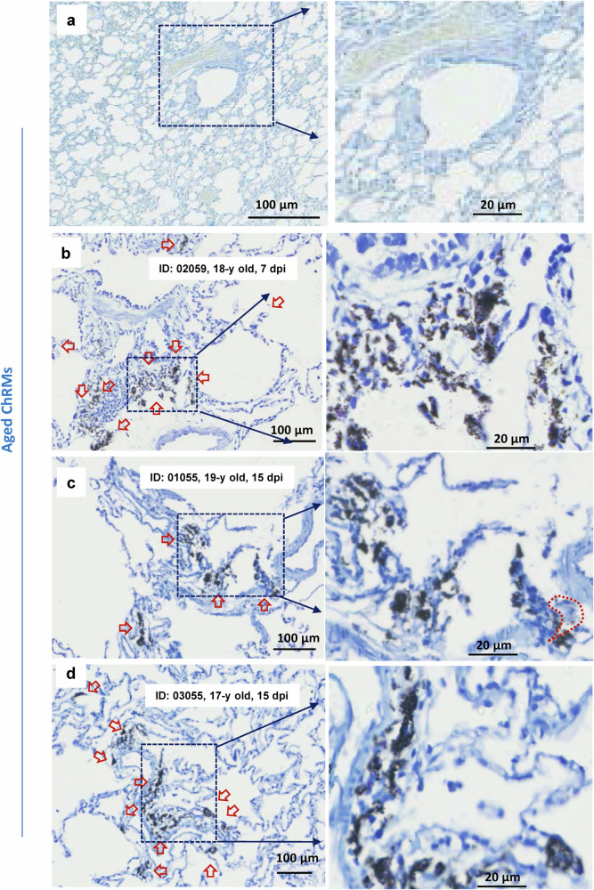
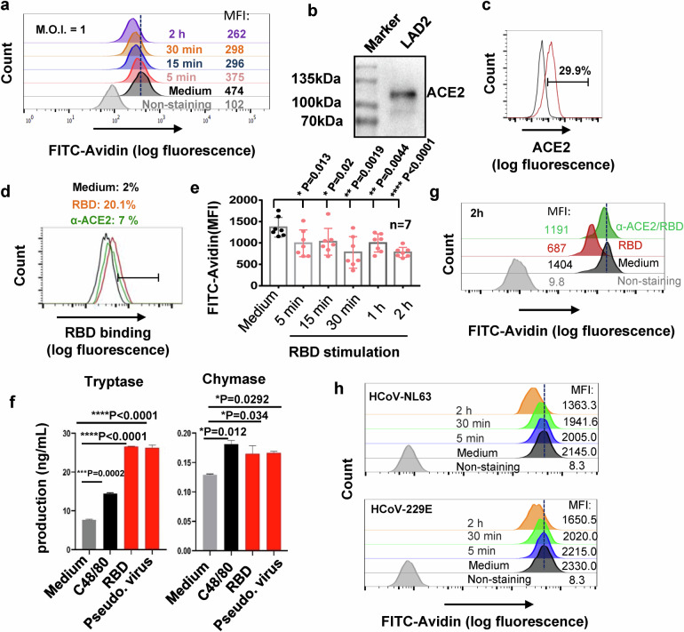
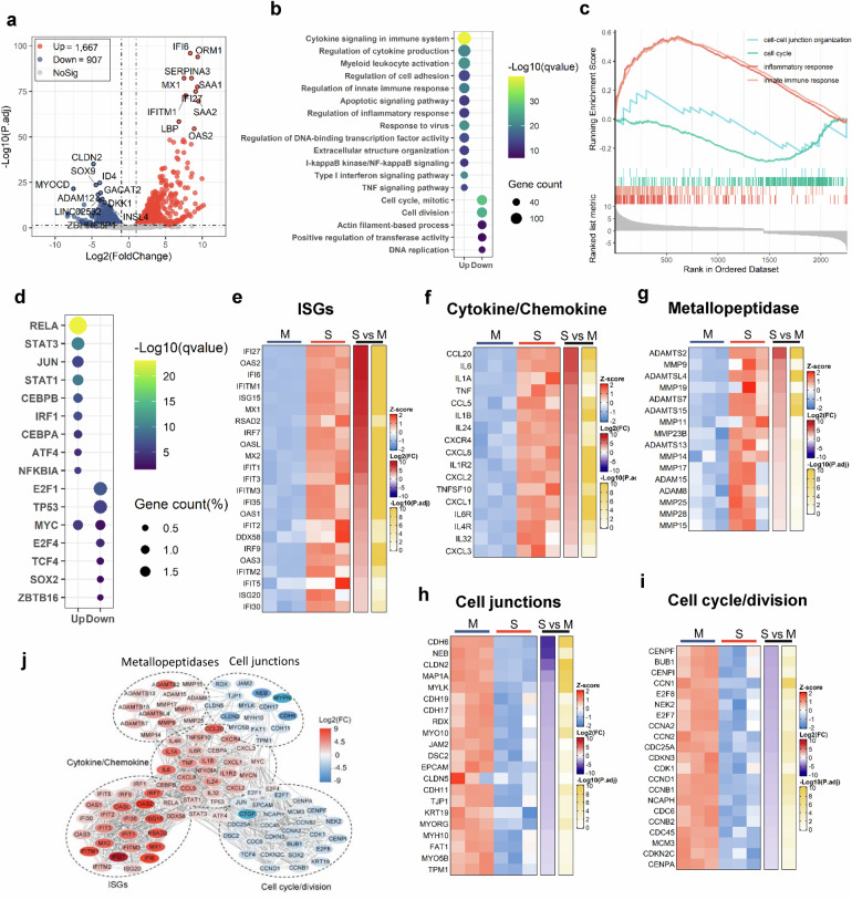
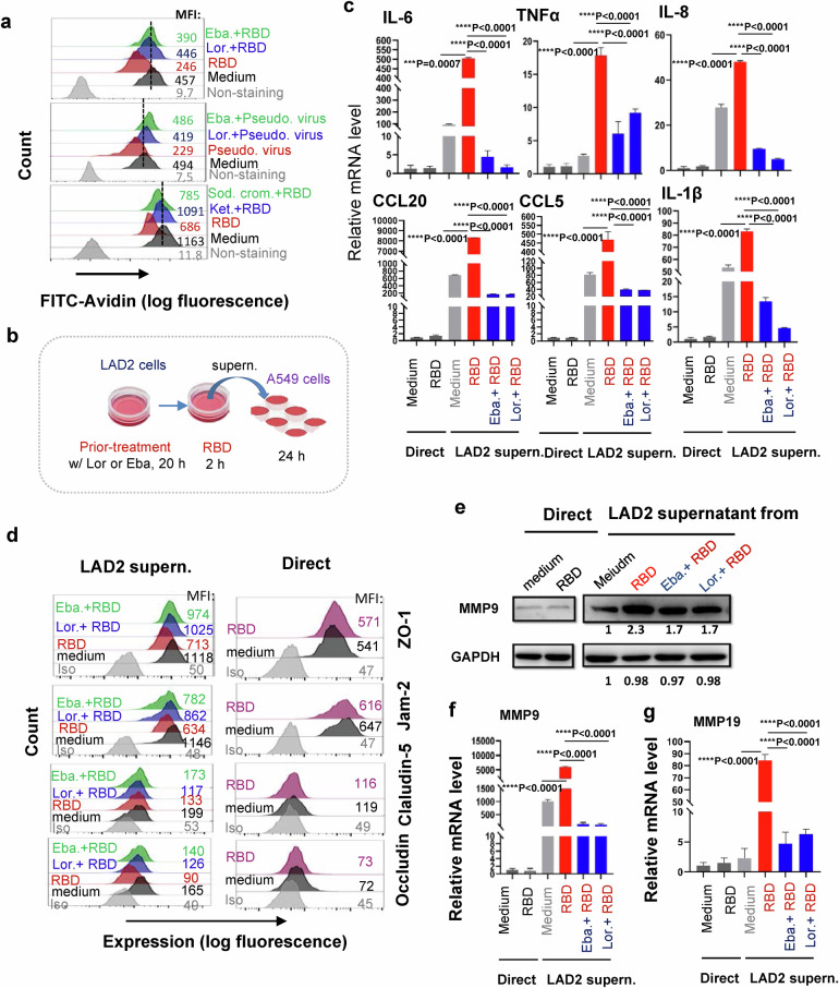
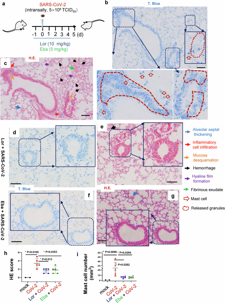
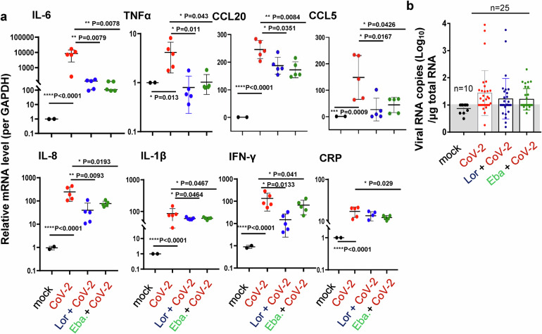
SARS-CoV-2 infection-induced hyper-inflammation links to the acute lung injury and COVID-19 severity. Identifying the primary mediators that initiate the uncontrolled hypercytokinemia is essential for treatments. Mast cells (MCs) are strategically located at the mucosa and beneficially or detrimentally regulate immune inflammations. In this study, we showed that SARS-CoV-2-triggered MC degranulation initiated alveolar epithelial inflammation and lung injury. SARS-CoV-2 challenge induced MC degranulation in ACE-2 humanized mice and rhesus macaques, and a rapid MC degranulation could be recapitulated with Spike-RBD binding to ACE2 in cells; MC degranulation altered various signaling pathways in alveolar epithelial cells, particularly, the induction of pro-inflammatory factors and consequential disruption of tight junctions. Importantly, the administration of clinical MC stabilizers for blocking degranulation dampened SARS-CoV-2-induced production of pro-inflammatory factors and prevented lung injury. These findings uncover a novel mechanism for SARS-CoV-2 initiating lung inflammation, and suggest an off-label use of MC stabilizer as immunomodulators for COVID-19 treatments.


Conflict of interest statement

The authors declare no competing interests.

Figures

Fig. 1
Fig. 1. SARS-CoV-2 induces mast cell degranulation and lung injury in hACE2-humanized mice.
Five C57BL/6N-Ace2em2(hACE2-WPRE,pgk-puro)/CCLA mice were intranasally infected with SARS-CoV-2 (strain 107) at a dose of 2 × 106 TCID50, two mice were used as the mock-infections. The mice were euthanized at the 1 dpi, 3 dpi and 5 dpi, and the lung tissues were harvested for histological analysis. Toluidine blue staining was used to observe MCs and their degranulation (a, c, e and g), and the lung injury was observed by H.E. staining (b, d, f and h), scale bar: 100 μm. (i) The pathological score was assessed according to the degree of lung tissue lesions and MC count in lung sections was calculated. *p < 0.05 and **p < 0.01 are considered significant differences
Fig. 2
Fig. 2. SARS-CoV-2 induces mast cell degranulation in young chRMs.
Young chRMs (3- to 6-year old) were anaesthetized by Zoletil 50 and intratracheally inoculated with SARS-CoV-2 (1 × 107 TCID50) in a 2 mL volume by bronchoscope. a Mock infection. The animals were euthanized at 7 dpi (b, c, d) or 15 dpi (e, f) and the lung lobes were collected for histology analysis. Toluidine blue staining was used to observe MCs and their degranulation. Red arrow indicates the MCs
Fig. 3
Fig. 3. SARS-CoV-2 induces mast cell degranulation in aged chRMs.
Aged chRMs (17- to 19-year old) were anaesthetized by Zoletil 50 and intratracheally inoculated with SARS-CoV-2 (1 × 107 TCID50) in a 2 mL volume by bronchoscope. a Mock infection. The animals were euthanized at 7 dpi (b) or 15 dpi (c, d) and the lung lobes were collected for histology analysis. Toluidine blue staining was used to observe MCs and their degranulation. Red arrow indicates the MCs
Fig. 4
Fig. 4. The binding of Spike-RBD to ACE2 triggers a rapid MC degranulation.
a SARS-CoV-2 (M.O.I. = 1)-induced LAD2 degranulation, detected by flow cytometry with immunostaining the intracellular avidin. b, c ACE2 expression in LAD2 cells, detected with Western blotting and flow cytometry with immunostaining with specific antibodies. d Prior-blocking with anti-ACE2 antibody reduces RBD binding. LAD2 cells were incubated with anti-ACE2 antibody (5 μg/ml) at 37 °C for 1 h, then Spike-RBD (5 μg/ml) were added for binding at 4 °C for 1 h, and the binding of Spike-RBD to LAD2 cells was detected with flow cytometry. e, f, g Spike-RBD induces LAD2 degranulation. LAD2 cells were incubated with Spike-RBD (5 μg/ml) at 37 °C for the indicated time, then cells were fixed with 4% paraformaldehyde and permeabilized and immunostained with anti-avidin-FITC at 4 °C for 1 h, and analyzed with flow cytometry (e, g), results from 7 independent repeats were summarized and presented (e). The degranulated components Tryptase and Chymase were detected by ELISA (f). LAD2 cells were prior-treated with anti-ACE2 antibody (5 μg/ml) at 37 °C for 1 h before the Spike-RBD stimulation (g). h HCoV-NL63 and HCoV-229E induced LAD2 degranulation. LAD2 cells were incubated with HCoV-NL63 or HCoV-229E (M.O.I = 1) for the indicated time, and cell degranulation was detected as above. One representative data from 3 (a, b, f, h) or 4 (c, d, g) independent repeats are shown. Data are presented as mean ± SD. *p < 0.05, **p < 0.01, ***p < 0.001 and *p < 0.0001 are considered significant differences. MFI mean fluorescence intensity
Fig. 5
Fig. 5. Transcriptome analysis of A549 cells treated with LAD2/RBD supernatant.
a Volcano plot of DEGs comparing LAD2/RBD supernatant versus medium. The symbols of top 10 up-regulated or down-regulated genes are shown. b GO functional enrichment analysis of DEGs. The color bar indicates the minus logarithm of q values, and bubble size indicates the absolute gene counts enriched in a GO term. c GSEA showing the distribution of gene sets that related to inflammatory response, innate immune response, cell–cell junction organization or cell cycle and the enrichment scores based on DEGs. d Transcription-factor enrichment analysis of DEGs. The color bar indicates the minus logarithm of q values, and bubble size indicates the gene enrichment ratio regulated by a transcription factor. ei Heatmaps showing relative expression level (left panel), fold change (middle panel), and adjusted p values (right panel) for sets of ISGs (e), cytokine- and chemokine-related genes (f), Metallopeptidase (g), cell junction-related genes (h), cell cycle- and division-related genes (i). M medium; S, LAD2/RBD supernatant. j A Protein–Protein interaction network analysis of the core DEGs. The color bar represents the fold change of protein-coded genes at transcriptome level
Fig. 6
Fig. 6. Inhibition of MC degranulation abolishes alveolar epithelial inflammation.
a Lor., Eba., Ket., or Sod. Crom. inhibits Spike-RBD or pseudotyped lentivirus-induced LAD2 cell degranulation. Cells were prior-treated with Lor. (5 μg/mL), Eba. (3 μg/mL), Ket. (40 μg/mL), or Sod. Crom. (10 μg/mL) for 20 h, then were incubated with Spike-RBD (5 μg/ml) or Spike-pseudotyped lentivirus (5 ng p24Gag) at 37 °C for the 2 h, and cell degranulation was detected with Flow cytometry. b The illustration for treatments. LAD2 cells were prior-treated with or without Lor. (5 μg/mL) or Eba. (3 μg/mL) for 20 h, then cells were treated with Spike-RBD (5 μg/ml) for 2 h, and the culture supernatants were harvested to treat A549 cells for additional 24 h, or A549 cells were directly treated with or without Spike-RBD for 24 h, c the mRNA levels of IL-6, TNF-α, IL-8, CCL20, CCL5 and IL-1β were quantified with real time q(RT-) PCR, and normalized to gapdh mRNA, d the expressions of ZO-1, Jam-2, Claudin-5 and Occludin were detected by immunstaining with specific antibodies and analyzed with flow cytometry, and (e, f), the expressions of MMP9 and MMP19 in A549 cells were measured with Western blotting or real time q(RT-) PCR, and the gray intensity of gel was calculated by Image J and normalized (e). One representative data from 3 (c, d, e, g) or 5 (a, f) independent repeats are shown. Data are presented as mean ± SD. ***p < 0.0001 and ****p < 0.0001 are considered significant difference (c, f). MFI mean fluorescence intensity (a, d)
Fig. 7
Fig. 7. MC stabilizers prevent lung injury in mice.
a The illustration of mice treatment. C57BL/6N-Ace2em2(hACE2-WPRE,pgk-puro)/CCLA mice were prior- administered with or without Eba. (5 mg/kg) or Lor. (10 mg/kg) via i.p. 1 day before intranasal infection with SARS-CoV-2 (strain 107) at a dose of 2 × 106 TCID50, and the Eba. and Lor. treatments were continued each day over the couse of infection. 5 mice for each treatment groups, and 2 mice without infection and drug treatment were used as the mock controls. Mice were euthanized and lung lobes were harvested for pathological analysis: Toluidine blue staining was used to observe MC degranulation, and the lung injury was observed via H.E. staining. Scale bar: 100 μm. h H.E. scores and (i) MC counts in lung section (5 dpi). *p < 0.05 and **p < 0.01 are considered significant differences
Fig. 8
Fig. 8. MC stabilizers reduce SARS-CoV-2-induced inflammation.
C57BL/6N-Ace2em2(hACE2-WPRE,pgk-puro)/CCLA mice were prior- administered with or without Eba. (5 mg/kg) or Lor. (10 mg/kg) via i.p. 1 day before intranasal infection with SARS-CoV-2 (strain 107) at a dose of 2 × 106 TCID50, and the Eba. and Lor. treatments were continued each day over the couse of infection. 5 mice for each treatment groups, and 2 mice without infection and drug treatment were used as the mock controls. Mice were euthanized and lung lobes were harvested for analysis. a The mRNA levels of IL-6, TNF-α, CCL20, CCL5, IL-8, IL-1β, IFN-γ and CRP were quantified with q(RT-) PCR, and normalized to gapdh mRNA. b Viral replication was monitored by quantifying the expression of nucleocapsid gene. Data are presented as mean ± SD. *p < 0.05, **p < 0.01, ***p < 0.001 and ****p < 0.0001 are considered significant differences

Similar articles

Cited by

References

    1. O’Driscoll M, et al. Age-specific mortality and immunity patterns of SARS-CoV-2. Nature. 2021;590:140–145. - PubMed
    1. Rubin D, Chan-Tack K, Farley J, Sherwat A. FDA approval of remdesivir - a step in the right direction. N. Engl. J. Med. 2020;383:2598–2600. - PubMed
    1. Mahase E. Covid-19: UK becomes first country to authorise antiviral molnupiravir. BMJ. 2021;375:n2697. - PubMed
    1. Mahase E. Covid-19: Molnupiravir reduces risk of hospital admission or death by 50% in patients at risk, MSD reports. BMJ. 2021;375:n2422. - PubMed
    1. Sneed TJ, Bible GH. An administrative procedure for improving staff performance in an institutional setting for retarded persons. Ment. Retard. 1979;17:92–94. - PubMed

Publication types

MeSH terms

Substances