. 2021 Dec 17;11(1):24194.
doi: 10.1038/s41598-021-03679-w.

Dual role of neutrophils in modulating liver injury and fibrosis during development and resolution of diet-induced murine steatohepatitis

Affiliations

Dual role of neutrophils in modulating liver injury and fibrosis during development and resolution of diet-induced murine steatohepatitis

Andrea D Kim et al. Sci Rep. .

Abstract

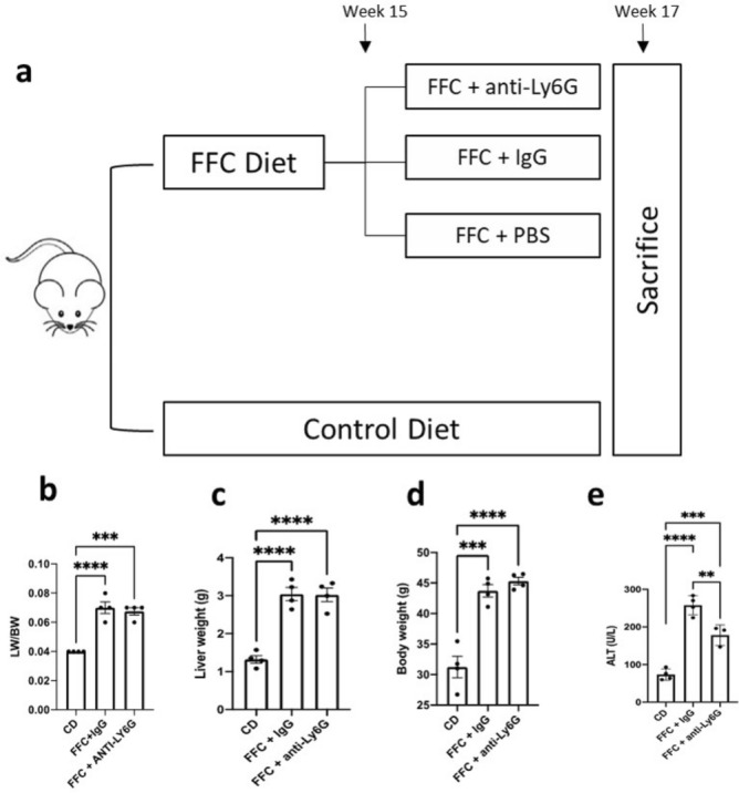
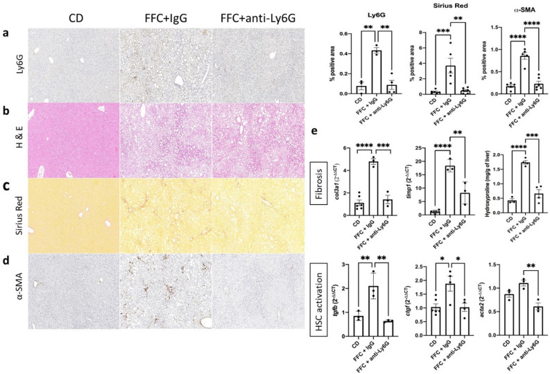
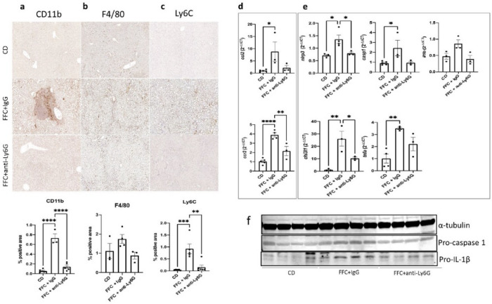
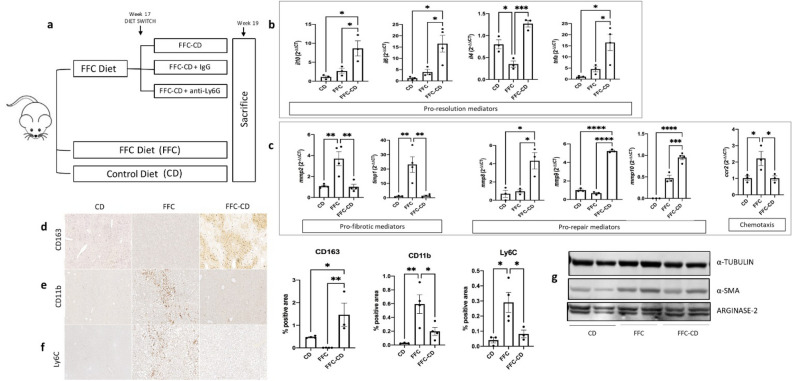
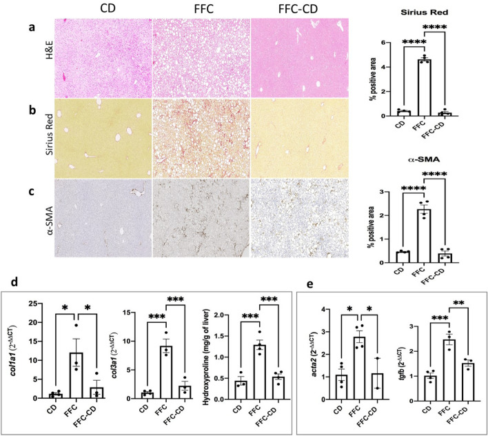
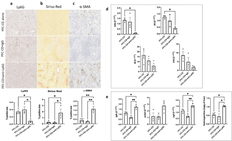
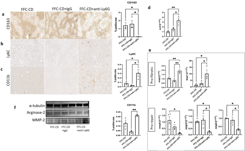
Inflammatory changes in the liver represent a key feature of non-alcoholic steatohepatitis (NASH), the progressive form of non-alcoholic fatty liver disease (NAFLD). Innate immune activation including hepatic neutrophilic infiltration acts as an important inflammatory trigger as well as a potential mediator of inflammation resolution. In this study, we dissected the effects of neutrophil depletion via anti-lymphocyte antigen 6 complex locus G6D (Ly6G) antibodies administration during ongoing high fat-fructose-cholesterol (FFC) diet-induced murine NASH and during inflammation resolution by switching into a low-fat control diet. During NASH progression, protective effects were shown as HSC activation, cell infiltration and activation of pro-inflammatory macrophages were ameliorated. Furthermore, these changes were contrasted with the effects observed when neutrophil depletion was performed during the resolution phase. Impaired resolving mechanisms, such as a failure to balance the pro and anti-inflammatory cytokines ratio, deficient macrophage phenotypic switch into a pro-restorative profile, and defective repair and remodeling processes were observed when neutrophils were depleted in this scenario. This study described phase-dependent contrasting roles of neutrophils as triggers and pro-resolutive mediators of liver injury and fibrosis associated with diet-induced NASH in mice. These findings have important translational implications at the time of designing NASH therapeutic strategies.


Conflict of interest statement

The authors declare no competing interests.

Figures

Figure 1
Figure 1
Neutrophil depletion treatment after 15 weeks of high fat-fructose-cholesterol diet (FFC). (a) Mice were fed with FFC diet for 15 weeks and were administered with IgG (FFC + IgG, n = 6) or anti-Ly6G (FFC + anti-Ly6G, n = 6) intraperitoneal injections for 14 days until sacrifice. Control diet was used as a NASH negative control (CD, n = 6). Liver/body weight ratio (b), liver weight (c), body weight (d) and serum alanine transaminase (e) were compared between the mentioned groups with 1-way analysis of variance (ANOVA) followed by the Turkey post hoc test. P < .05 was considered a significant difference. Ly6G lymphocyte antigen 6 complex locus G6D, LW/BW liver/body weight ratio, ALT alanine aminotransferase.
Figure 2
Figure 2
Hepatic steatosis, fibrosis and hepatic stellate cell (HSC) activation are ameliorated in neutrophil depleted mice during NASH development. Neutrophil marker Ly6G (a), steatosis (b) and fibrosis (c) levels were assessed via IHC, H&E and Picrosirius Red stainings IHC respectively on paraffin-embedded tissue sections. Activated α-SMA+ HSCs (d) were detected via IHC. Quantification and ANOVA comparison were made between the control diet and IgG or anti-Ly6G regimen receiving groups during ongoing NASH development. (e) mRNA expression of hepatic fibrosis and HSC activation related genes and hydroxyproline levels were analyzed between the mentioned groups and compared with ANOVA IHC. CD control diet, α-SMA/acta2 alpha smooth muscle actin, IHC immunohistochemistry, col3a1 collagen type 3 alpha 1 chain, timp1 tissue inhibitor of metalloproteinases 1, tgfb transforming growth factor beta, ctgf connective tissue growth factor.
Figure 3
Figure 3
Cellular infiltration and pro-inflammatory macrophage activity are abrogated in neutrophil depleted mice during NASH development. IHC for detection of monocyte derived macrophages and other CD11b+ infiltrating myeloid cells (a), hepatic resident F4/80+ Kupffer cells (b) and pro-inflammatory Ly6Chi monocytes/macrophages was performed and quantified for comparison among the control diet receiving group and the mice receiving either IgG or anti-Ly6G antibodies during ongoing FFC diet . mRNA expression was analyzed via RT-qPCR and fold-change was compared between the mentioned groups regarding the main monocyte chemotactic pathway CCL2/CCR2 (d) and pro-inflammatory macrophage activity markers such as components of the NLRP3 inflammasome pathway, chitinase-3 like 1 protein (CHI3L1) and TNF-α (e). Data was compared by using ANOVA analysis followed by the Turkey post hoc test. Western Blots targeting components of the NLRP3 downstream cascade were performed to compare the mentioned groups in a protein expression level (f). α-tubulin (band imported from Supplementary Fig. 1b) was used as housekeeping protein in both assessments. Full-length blottings of pro-caspase 1 and pro-IL-1β are shown in Supplementary Fig. 1b and 2, respectively. Ccl2 C–C motif chemokine ligand 2, ccr2 C–C motif chemokine receptor 2, NLRP3 NLR family pyrin domain containing 3, CHI3L1 chitinase 3-like 1, TNF-α/tnfa tumor necrosis factor alpha.
Figure 4
Figure 4
NASH inflammation resolution induced by a diet reversal from high fat diet to control diet. (a) After 17 weeks of high fat-fructose-cholesterol diet (FFC), mice underwent a 2-week diet reversal into control diet (CD), allowing spontaneous resolution of inflammation. During the diet reversal period, mice received treatment with anti-Ly6G (FFC-CD + anti-Ly6G, n = 3), IgG (FFC-CD + IgG, n = 3) or no treatment (FFC-CD, n = 4). Groups receiving 19 weeks of FFC (n = 4) or CD (n = 4) were used as positive and negative NASH controls respectively. mRNA expression was measured via RT-qPCR and fold change was analyzed between control diet, FFC and diet reversal receiving groups through ANOVA comparison: expression of mediators with pro-resolutive roles (b), fibrotic mediators (mmp2, timp1) vs. pro-repairing matrix metalloproteinases (mmp8, mmp9 and mmp10) and chemotactic marker ccr2 were compared (c) among the mentioned groups. Detection of pro-resolution profile CD163+ macrophages (d), monocyte-derived macrophages and other infiltrating CD11b+myeloid cells (e) and pro-inflammatory Ly6Chi monocytes/macrophages (f) was performed via IHC and quantification. (g) Western blot was used to assess differences in protein expression of hepatic stellate cell activation marker α-SMA (cropped from Supplementary Fig. 3a) and pro-resolutive M2 macrophage marker arginase-2 (cropped from Supplementary Fig. 3b) between the mentioned groups. α-tubulin expression (band imported from Supplementary Fig. 3a) was used as housekeeping control. Mmp matrix metalloproteinases.
Figure 5
Figure 5
Hepatic steatosis, fibrosis and hepatic stellate cell (HSC) activation are ameliorated in NASH resolution induced by diet reversal. Steatosis (a), fibrosis (b) and activated HSC activation (c) were detected via hematoxylin, Sirius Red and α-SMA IHC stainings respectively in paraffin embedded tissue sections. Hydroxyproline levels and mRNA expression of fibrosis-related genes (d) and HSC activation markers (e) were analyzed through RT-qPCR among the control diet, FFC and diet reversal receiving groups. Data was analyzed with ANOVA comparison followed by the Turkey post hoc test.
Figure 6
Figure 6
Fibrosis and hepatic stellate cell (HSC) activation are exacerbated in neutrophil depleted mice during inflammation resolution. Neutrophil depletion was achieved via administration of anti-Ly6G antibodies during the 2-week diet reversal-induced NASH inflammation resolution. Ly6G+ neutrophils (a), fibrosis (b) and α-SMA+ activated HSCs (c) were detected through paraffin-embedded tissue sections stainings and analyzed by ANOVA comparison between mice undergoing diet reversal alone (FFC-CD), and diet reversal in association with either IgG (FFC-DC + IgG) or anti-Ly6G antibodies (FFC-CD + anti-Ly6G) administration. mRNA expression of genes associated with inflammation resolution and HSC activation was assessed comparing fold-changes between the mentioned groups. Hydroxyproline levels were also compared among the mentioned groups by ANOVA followed by the Turkey post hoc test (d,e).
Figure 7
Figure 7
Phenotypic switch into pro-resolution macrophage population and release of pro-repairing mediators are impaired in neutrophil depleted mice during inflammation resolution. Detection of CD163+ pro-restorative macrophages (a), pro-inflammatory Ly6Chi monocytes/macrophages (b) and infiltrating CD11b+ myeloid cells (c) were detected via IHC and quantified for ANOVA comparison between the groups receiving diet reversal alone (FFC-CD), and diet reversal in association with either IgG (FFC-CD + IgG) or anti-Ly6G (FFC-CD + anti-Ly6G). mRNA expression was compared and expressed as fold changes. Monocyte chemoattractant receptor ccr2 (d) and changes in the expression of pro- (mmp2, timp1) versus anti-fibrotic (mmp8, mmp9, mmp10) mediators were compared among the mentioned groups (e). (f) Western blots for protein expression assessment was performed targeting pro-restorative macrophage marker arginase 2 (cropped from Supplementary Fig. 4) and pro-fibrotic MMP-2 (cropped from Supplementary Fig. 5b). α-tubulin expression (band imported from Supplementary Fig. 4) was illustrated as housekeeping control.

Similar articles

Cited by

References

    1. Younossi Z, et al. Global burden of NAFLD and NASH: Trends, predictions, risk factors and prevention. Nat. Rev. Gastroenterol. Hepatol. 2018;15:11–20. doi: 10.1038/nrgastro.2017.109. - DOI - PubMed
    1. Satapathy SK, Sanyal AJ. Epidemiology and natural history of nonalcoholic fatty liver disease. Semin. Liver Dis. 2015;35:221–235. doi: 10.1055/s-0035-1562943. - DOI - PubMed
    1. Wu L, et al. The role of neutrophils in innate immunity-driven nonalcoholic steatohepatitis: Lessons learned and future promise. Hepatol. Int. 2020;14:652–666. doi: 10.1007/s12072-020-10081-7. - DOI - PubMed
    1. van der Windt DJ, et al. Neutrophil extracellular traps promote inflammation and development of hepatocellular carcinoma in nonalcoholic steatohepatitis. Hepatology. 2018;68:1347–1360. doi: 10.1002/hep.29914. - DOI - PMC - PubMed
    1. Jones HR, Robb CT, Perretti M, Rossi AG. The role of neutrophils in inflammation resolution. Semin. Immunol. 2016;28:137–145. doi: 10.1016/j.smim.2016.03.007. - DOI - PubMed

Publication types