. 2022 Mar;43(4):1196-1213.
doi: 10.1002/hbm.25697. Epub 2021 Dec 17.

Prevalence of white matter pathways coming into a single white matter voxel orientation: The bottleneck issue in tractography

Affiliations

Prevalence of white matter pathways coming into a single white matter voxel orientation: The bottleneck issue in tractography

Kurt G Schilling et al. Hum Brain Mapp. 2022 Mar.

Abstract

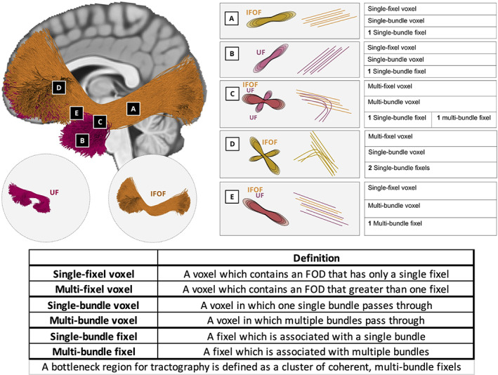
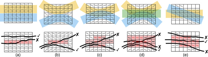
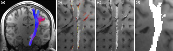
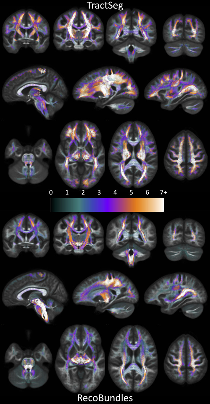
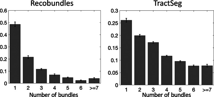
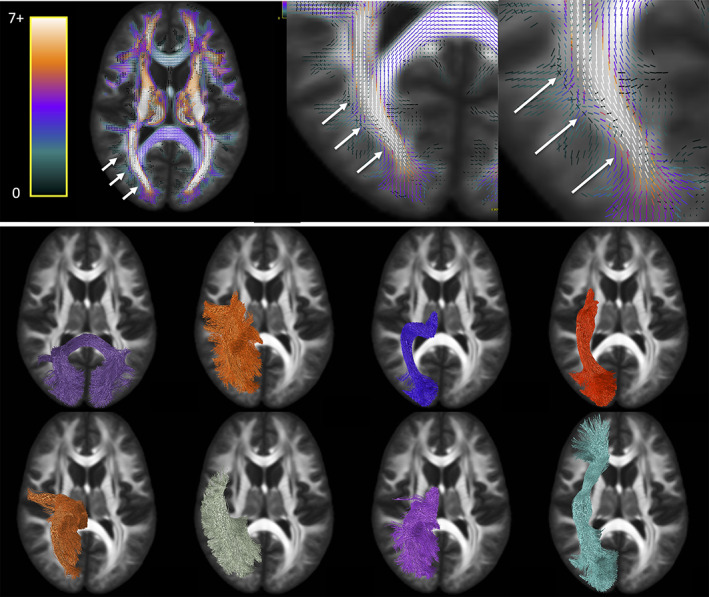
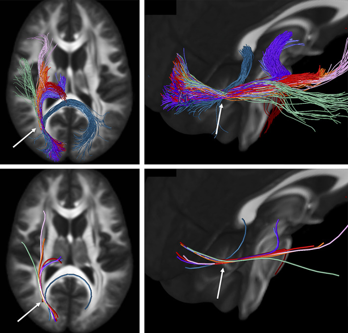
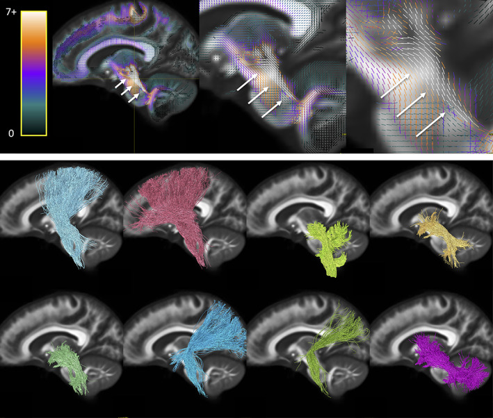
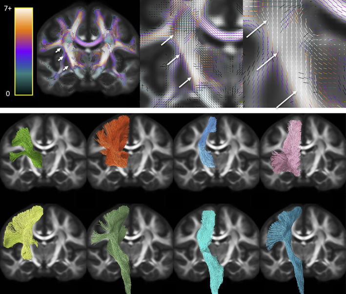
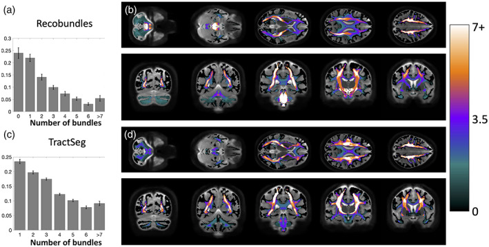
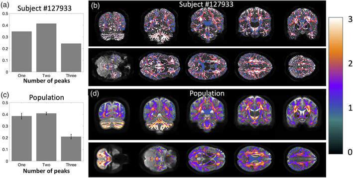
Characterizing and understanding the limitations of diffusion MRI fiber tractography is a prerequisite for methodological advances and innovations which will allow these techniques to accurately map the connections of the human brain. The so-called "crossing fiber problem" has received tremendous attention and has continuously triggered the community to develop novel approaches for disentangling distinctly oriented fiber populations. Perhaps an even greater challenge occurs when multiple white matter bundles converge within a single voxel, or throughout a single brain region, and share the same parallel orientation, before diverging and continuing towards their final cortical or sub-cortical terminations. These so-called "bottleneck" regions contribute to the ill-posed nature of the tractography process, and lead to both false positive and false negative estimated connections. Yet, as opposed to the extent of crossing fibers, a thorough characterization of bottleneck regions has not been performed. The aim of this study is to quantify the prevalence of bottleneck regions. To do this, we use diffusion tractography to segment known white matter bundles of the brain, and assign each bundle to voxels they pass through and to specific orientations within those voxels (i.e. fixels). We demonstrate that bottlenecks occur in greater than 50-70% of fixels in the white matter of the human brain. We find that all projection, association, and commissural fibers contribute to, and are affected by, this phenomenon, and show that even regions traditionally considered "single fiber voxels" often contain multiple fiber populations. Together, this study shows that a majority of white matter presents bottlenecks for tractography which may lead to incorrect or erroneous estimates of brain connectivity or quantitative tractography (i.e., tractometry), and underscores the need for a paradigm shift in the process of tractography and bundle segmentation for studying the fiber pathways of the human brain.

Keywords: bottleneck; crossing fibers; fiber pathways; tractography; tractometry; white matter.


Figures

FIGURE 1
FIGURE 1
Nomenclature. Two bundles, the UF and IFOF, are used to highlight classifications of voxels (a–e), and fixels within the voxels. Voxels in a and b are examples of single‐fixel voxel and single‐bundle voxels and also single‐bundle fixel. Because the UF and IFOF diverge in Voxel c, this is an example of a multi‐fixel voxel and multi‐bundle voxel, with one fixel classified as a single‐bundle fixel and the other a multi‐bundle fixel. Voxel d highlights the fanning of the IFOF, which results in a multi‐fixel voxel and single‐bundle voxel, and both fixels are single‐bundle fixels. Finally, both the IFOF and UF pass through voxel E following the same orientation, thus Voxel e is a single‐orientation voxel but multi‐bundle voxel, and also a multi‐bundle fixel. This fixel, and thus also this voxel, represents a bottleneck for tractography
FIGURE 2
FIGURE 2
Bottlenecks and when they become a problem. Anytime two connections of the brain start and end at different locations, but at some point, are within the same voxel (spatial partial‐volume), and share an orientation not resolvable by diffusion (angular partial‐volume), diffusion tractography faces a bottleneck. Cartoon examples illustrate increasing levels of pathway complexity: parallel pathways (a), kissing or touching pathways (b), two overlapping pathways (c), three overlapping pathways (d), or bundles crossing at sharp angles not resolvable by diffusion techniques (e). Multi‐bundle voxels are shaded red, and multi‐bundle fixels are also colored red. Bottleneck regions are apparent as spatial clusters of multi‐bundle fixels. In these regions, bundles run nearly parallel, and as tractography streamlines enter and exit these regions they may generate either valid (✓) or invalid (X) connections
FIGURE 3
FIGURE 3
Assigning bundles to voxels and fixels. Each segmented white matter bundle (a) was assigned to each fixel by counting the number of streamlines aligned with each fixel to create fixel‐density map (b) which was thresholded to generate the binary fixel‐based profile of each bundle (c). This allows us to query the number of known bundles per fixel. Next, this map was projected to the voxel level, and binary voxel‐based profiles of each bundle (d) were generated, which allows us to query the number of known bundles per voxel. White matter bundles were derived from TractSeg (N = 72 bundles) and Recobundles (N = 66 bundles)
FIGURE 4
FIGURE 4
There is a high prevalence of bundles assigned to each fixel in the brain. Fixels, in template‐space, are shown as vectors, colored by the number of associated bundles, and averaged across the population (note continuous color‐map due to population‐averaging). TractSeg results are shown on top, Recobundles on bottom. Fixels with more than one bundle traversing through them represent bottleneck regions for tractography
FIGURE 5
FIGURE 5
Number of bundles assigned to fixels in the brain, averaged across the population. Most fixels had greater than one bundle traversing through their designated orientation. Note that fixels which were assigned to 0 bundles are not shown. Y‐axis is shown as a fraction of fixels. Error bars represent variation across the studied population
FIGURE 6
FIGURE 6
Bottleneck region in the anterior–posterior oriented white matter of the occipital lobe (arrows) contains a large number of white matter bundles with unique starting and ending connections. Colormap ranges from 0 to 7+ bundles. Pathways (derived from TractSeg) from left to right, top to bottom: CC7, ST_par, OR, ST_OCC, POPT, MdLF, T_PAR, IFO (for full names see acronyms at end of document)
FIGURE 7
FIGURE 7
Illustration of the bottleneck in the anterior–posterior oriented white matter of the occipital lobe. Fiber bundles from Figure 5 (same color scheme) were filtered to select only streamlines (top) traversing a small 2 × 2 × 2 voxel region of interest (arrow). A single representative streamline from each sub‐bundle is also shown (bottom). This example emphasizes that streamlines belonging to many fiber bundles may traverse through the same small region, in the same orientation
FIGURE 8
FIGURE 8
Bottleneck region in the superior–inferior oriented white matter of the brain‐stem (arrow) contains a large number of white matter bundles with unique starting and ending connections. Hot‐cold colormap ranges from 0 to 7 bundles. Pathways (derived from Recobundles) from top to bottom, left to right: CST, FPT, LL, MLL, CTT, OPT, TPT, and SCP (for full names see acronyms at end of document)
FIGURE 9
FIGURE 9
Bottleneck region in the superior–inferior oriented white matter of the internal capsule (arrow) contains a large number of white matter bundles with unique starting and ending connections. Hot‐cold colormap ranges from 0 to 7 bundles. Pathways (derived from TractSeg) from left to right, top to bottom: T_PREM, T_PAR, STR, ST_PREF, ST_POST, POPT, FPT, and CST (for full names see acronyms at end of document)
FIGURE 10
FIGURE 10
Many voxels in the white matter contain multiple known bundles. Prevalence of voxels with 1–7+ bundles averaged across the population is quantified for Recobundles (a) and visualized in template space (b), and also quantified for TractSeg bundles (c) and visualized overlaid in template space (d). Note that many voxels contain 0 bundles (i.e., are not associated with known bundles in our atlas) and are thus not quantified. Visualization in template space is mapped to a continuous color‐scale due to population‐averaging
FIGURE 11
FIGURE 11
There is a high prevalence of multi‐orientation voxels throughout the brain. Prevalence of voxels with one fixel, two fixels, and three fixels is quantified for a single subject (a) and visualized overlaid on an anatomical image (b) and also averaged across the population (c) and visualized overlaid across the population template (d). Note that the number of fixels is discrete on a single subject but continuous when averaged across all subjects

References

    1. Aganj, I. , Lenglet, C. , Sapiro, G. , Yacoub, E. , Ugurbil, K. , & Harel, N. (2010). Reconstruction of the orientation distribution function in single‐ and multiple‐shell q‐ball imaging within constant solid angle. Magnetic Resonance in Medicine, 64(2), 554–566. 10.1002/mrm.22365 - DOI - PMC - PubMed
    1. Alexander, D. C. , & Seunarine, K. K. (2010). Mathematics of crossing fibers. In Jones D. K. (Ed.), Diffusion MRI: theory, methods, and application (pp. 451–464). Oxford; New York: Oxford University Press.
    1. Ambrosen, K. S. , Eskildsen, S. F. , Hinne, M. , Krug, K. , Lundell, H. , Schmidt, M. N. , … Dyrby, T. B. (2020). Validation of structural brain connectivity networks: The impact of scanning parameters. NeuroImage, 204, 116207. 10.1016/j.neuroimage.2019.116207 - DOI - PubMed
    1. Aranda, R. , Rivera, M. , & Ramirez‐Manzanares, A. (2014). A flocking based method for brain tractography. Medical Image Analysis, 18(3), 515–530. 10.1016/j.media.2014.01.009 - DOI - PubMed
    1. Barakovic, M. , Tax, C. M. W. , Rudrapatna, U. , Chamberland, M. , Rafael‐Patino, J. , Granziera, C. , … Jones, D. K. (2021). Resolving bundle‐specific intra‐axonal T2 values within a voxel using diffusion‐relaxation tract‐based estimation. NeuroImage, 227, 117617. 10.1016/j.neuroimage.2020.117617 - DOI - PMC - PubMed

Publication types

MeSH terms