

More passive internal tibial rotation with posterior cruciate ligament retention than with excision in a medial pivot TKA implanted with unrestricted caliper verified kinematic alignment
- PMID: 34921630
- PMCID: PMC9958185
- DOI: 10.1007/s00167-021-06840-0
More passive internal tibial rotation with posterior cruciate ligament retention than with excision in a medial pivot TKA implanted with unrestricted caliper verified kinematic alignment
Abstract
Purpose: Excision of the posterior cruciate ligament (PCL) is recommended when implanting a medial pivot (MP) total knee arthroplasty (TKA) to reduce the risk of limiting flexion by over-tensioning the flexion space. The present study determined whether PCL retention (1) limits internal tibial rotation and (2) causes anterior lift-off of the insert in 90° flexion after implantation of an MP design with unrestricted caliper verified kinematic alignment (KA).
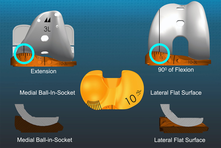
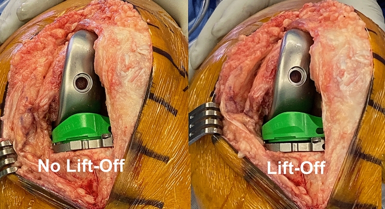
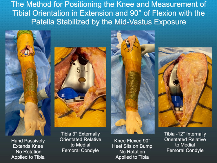
Methods: Four surgeons implanted an MP TKA design with medial ball-in-socket and lateral flat tibial insert in ten fresh-frozen cadaveric knees. Before and after PCL excision, trial inserts with medial goniometric markings measured the angular I-E tibial orientation relative to the trial femoral component's medial condyle in extension and at 90° flexion, and the surgeon recorded the occurrence of anterior lift-off of the insert at 90° flexion.
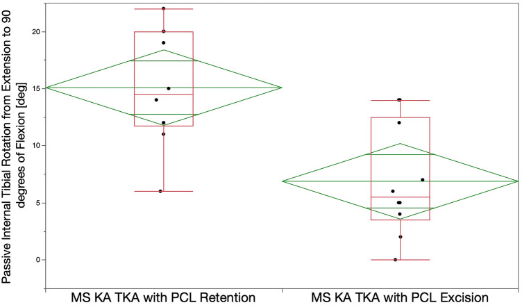
Results: PCL retention resulted in greater internal tibial rotation than PCL excision, with mean values of 15° vs 7° degrees from maximum extension to 90° flexion, respectively (p < 0.0007). At 90° flexion, no TKAs with PCL retention and one TKA with PCL excision had anterior lift-off of the insert (N.S.).
Conclusions: This preliminary study of ten cadaveric knees showed that PCL retention restored more passive internal tibial rotation than PCL excision with a negligible risk of anterior lift-off. However, in vivo analysis from multiple authors with a larger sample size is required to recommend PCL retention with an MP TKA design implanted with unrestricted caliper verified KA.
Keywords: Calipered; Insert thickness; Posterior cruciate ligament; Tibial rotation; Total knee arthroplasty; Total knee replacement.
© 2021. The Author(s).
Conflict of interest statement
A. J. Nedopil is a paid consultant for Medacta USA, Inc., S. M. Howell is a paid consultant for THINK Surgical and Medacta International., M. L. Hull receives research support from Medacta USA, Inc.
Figures
Similar articles
-
Adjusting Insert Thickness and Tibial Slope Do Not Correct Internal Tibial Rotation Loss Caused by PCL Resection: In Vitro Study of a Medial Constraint TKA Implanted with Unrestricted Calipered Kinematic Alignment.J Knee Surg. 2023 Apr;36(5):507-514. doi: 10.1055/s-0041-1739147. Epub 2021 Nov 15. J Knee Surg. 2023. PMID: 34781395
-
Ball-in-socket medial conformity with posterior cruciate ligament retention neither limits internal tibial rotation and knee flexion nor lowers clinical outcome scores after unrestricted kinematically aligned total knee arthroplasty.Int Orthop. 2023 Jul;47(7):1737-1746. doi: 10.1007/s00264-023-05834-6. Epub 2023 May 17. Int Orthop. 2023. PMID: 37195465
-
An insert with less than spherical medial conformity causes a loss of passive internal rotation after calipered kinematically aligned TKA.Arch Orthop Trauma Surg. 2021 Dec;141(12):2287-2294. doi: 10.1007/s00402-021-04054-0. Epub 2021 Jul 15. Arch Orthop Trauma Surg. 2021. PMID: 34264381 Free PMC article.
-
An Insert Goniometer Can Help Select the Optimal Insert Thickness When Performing Kinematically Aligned Total Knee Arthroplasty with a Medial 1:1 Ball-in-Socket and Lateral Flat Surface Insert and Posterior Cruciate Ligament Retention.Bioengineering (Basel). 2024 Sep 12;11(9):910. doi: 10.3390/bioengineering11090910. Bioengineering (Basel). 2024. PMID: 39329652 Free PMC article. Review.
-
Comparison between gaits after a medial pivot and posterior stabilized primary total knee arthroplasty: a systematic review of the literature.Arthroplasty. 2023 Mar 17;5(1):15. doi: 10.1186/s42836-023-00165-8. Arthroplasty. 2023. PMID: 36927464 Free PMC article. Review.
Cited by
-
Measurement of Tibial Orientation Helps Select the Optimal Insert Thickness to Personalize PCL Tension in a Medial Ball-in-Socket TKA.J Pers Med. 2022 Aug 31;12(9):1427. doi: 10.3390/jpm12091427. J Pers Med. 2022. PMID: 36143212 Free PMC article.
-
What is the "safe zone" for transition of coronal alignment from systematic to a more personalised one in total knee arthroplasty? A systematic review.Knee Surg Sports Traumatol Arthrosc. 2022 Feb;30(2):419-427. doi: 10.1007/s00167-021-06811-5. Epub 2022 Jan 1. Knee Surg Sports Traumatol Arthrosc. 2022. PMID: 34973095 Free PMC article.
-
PCL retained is safe in medial pivot TKA-a prospective randomized trial.Knee Surg Sports Traumatol Arthrosc. 2023 Dec;31(12):5856-5863. doi: 10.1007/s00167-023-07634-2. Epub 2023 Nov 14. Knee Surg Sports Traumatol Arthrosc. 2023. PMID: 37962615 Free PMC article. Clinical Trial.
-
A new tibial insert design with ball-in-socket medial conformity and posterior cruciate ligament retention has low tibial baseplate migration after unrestricted kinematically aligned total knee arthroplasty: a cohort study using radiostereometric analysis.Acta Orthop. 2024 Dec 23;95:758-764. doi: 10.2340/17453674.2024.42489. Acta Orthop. 2024. PMID: 39713914 Free PMC article.
-
Medial Congruent and Medial Pivot Inserts in Total Knee Arthroplasty: A Scoping Review.Medicina (Kaunas). 2025 May 3;61(5):844. doi: 10.3390/medicina61050844. Medicina (Kaunas). 2025. PMID: 40428802 Free PMC article.
References
-
- Delman CM, Ridenour D, Howell SM, Hull ML. The posterolateral upslope of a low-conforming insert blocks the medial pivot during a deep knee bend in TKA: a comparative analysis of two implants with different insert conformities. Knee Surg Sports Traumatol Arthrosc. 2021 doi: 10.1007/s00167-021-06668-8. - DOI - PubMed
-
- Emerson RH, Jr, Barrington JW, Olugbode SA, Alnachoukati OK. A comparison of 2 tibial inserts of different constraint for cruciate-retaining primary total knee arthroplasty: an additional tool for balancing the posterior cruciate ligament. J Arthroplasty. 2016;31:425–428. doi: 10.1016/j.arth.2015.09.032. - DOI - PubMed
MeSH terms
LinkOut - more resources
Full Text Sources
Medical
Miscellaneous
