Review
. 2021 Dec 18;12(1):189.
doi: 10.1186/s13244-021-01129-9.

Imaging features of primary sites and metastatic patterns of angiosarcoma

Affiliations
Review

Imaging features of primary sites and metastatic patterns of angiosarcoma

Basrull N Bhaludin et al. Insights Imaging. .

Abstract

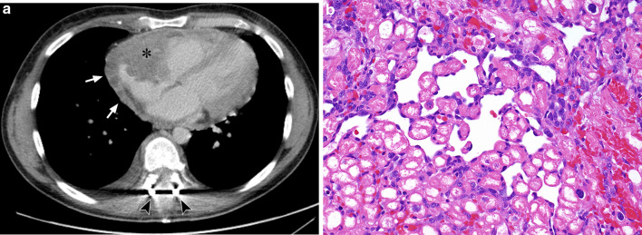
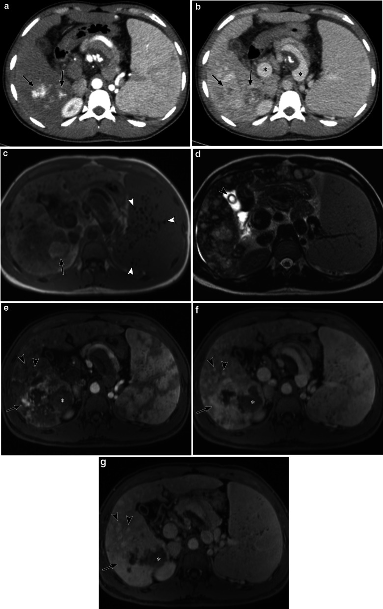
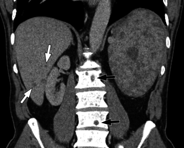
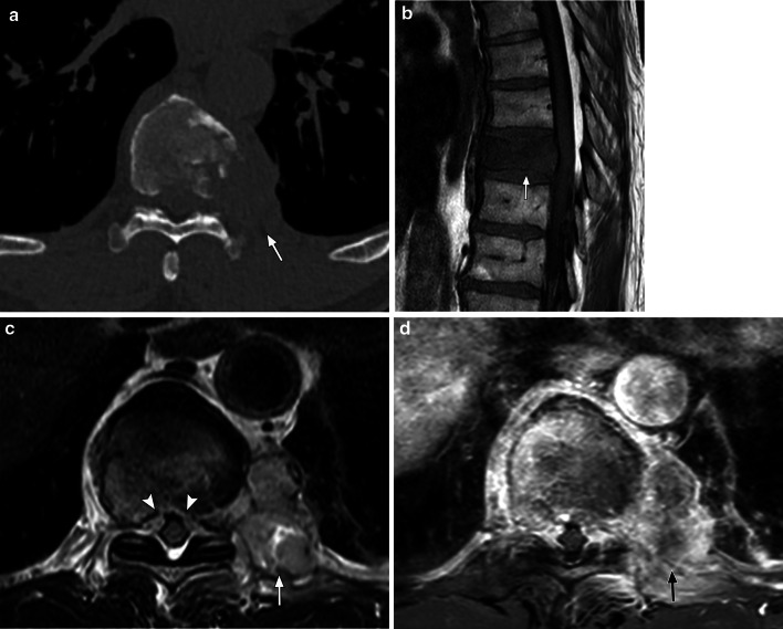
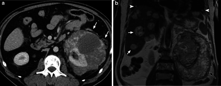
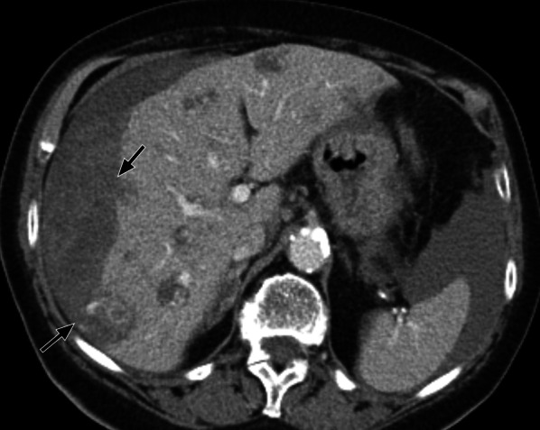
Angiosarcomas are rare, aggressive soft tissue sarcomas originating from endothelial cells of lymphatic or vascular origin and associated with a poor prognosis. The clinical and imaging features of angiosarcomas are heterogeneous with a wide spectrum of findings involving any site of the body, but these most commonly present as cutaneous disease in the head and neck of elderly men. MRI and CT are complementary imaging techniques in assessing the extent of disease, focality and involvement of adjacent anatomical structures at the primary site of disease. CT plays an important role in the evaluation of metastatic disease. Given the wide range of imaging findings, correlation with clinical findings, specific risk factors and patterns of metastatic disease can help narrow the differential diagnosis. The final diagnosis should be confirmed with histopathology and immunohistochemistry in combination with clinical and imaging findings in a multidisciplinary setting with specialist sarcoma expertise. The purpose of this review is to describe the clinical and imaging features of primary sites and metastatic patterns of angiosarcomas utilising CT and MRI.

Keywords: Angiosarcoma; CT; MRI; Metastasis; Radiation.


Conflict of interest statement

RLJ is in receipt of grants/research support from MSD and GSK. RLJ is in receipt of consultation fees from Adaptimmune, Athenex, Bayer, Boehringer Ingelheim, Blueprint, Clinigen, Eisai, Epizyme, Daichii, Deciphera, Immunedesign, Lilly, Merck, Pharmamar, Springworks, Tracon & UpToDate.

Figures

Fig. 1
Fig. 1
Histopathological features of angiosarcoma. a Photomicrograph of histologic specimen shows extensive, poorly formed, anastomosing vascular channels containing prominent blood. The channels are lined by moderately atypical ovoid cells with vesicular nuclei and prominent nucleoli (H and E, × 100). b Photomicrograph of histologic specimen shows a predominantly solid architecture without apparent vascular channel formation, but shows diffuse and strong expression of the vascular endothelial marker CD31. CD31 immunohistochemistry is seen to highlight small vascular channels within the mostly solid configuration of the tumour (immunohistochemistry, × 200)
Fig. 2
Fig. 2
77-year-old man with cutaneous scalp angiosarcoma. a Axial contrast-enhanced CT images show a lobulated heterogeneous cutaneous mass overlying the left scalp (arrow) with infiltration of the subcutaneous tissues and contacting the outer table of the skull. b Axial CT (bone windows) image shows no evidence of bony destruction (arrowheads). c Coronal contrast-enhanced T1-weight MR image performed 3 months later shows enlargement of the cutaneous mass. There is heterogeneous peripheral enhancement of the mass lesion with central necrosis (asterisk) and ulceration (arrowhead). d Photomicrograph shows a specimen mostly composed of large, anaplastic epithelioid cells with extensive surrounding haemorrhage. Only focally (left of field), vasoformation is evident, with variably sized, sometimes compressed vessels lined by similarly markedly atypical ovoid cells with prominent nuclei (H and E, × 200)
Fig. 3
Fig. 3
53-year-old woman with angiosarcoma associated with congenital lymphoedema of the left leg (Stewart–Treves syndrome). a Sagittal T2-weighted fat-suppressed image shows diffuse circumferential oedematous changes within the subcutaneous tissues and skin thickening in the left leg. There are multiple well-circumscribed nodules in the anterior proximal leg which are of predominantly low signal intensity compared to the surrounding tissues (arrow). bd Axial images of the left leg show skin thickening and multiple soft tissue nodules (white arrows) within the oedematous subcutaneous tissues with a further intramuscular nodule (black arrow) within the lateral muscular compartment of the leg. b Axial T2-weighted image shows the nodules to be of heterogeneously lower signal compared to the adjacent oedematous subcutaneous tissues and higher signal compared to the skeletal muscles with central low signal striations. c Axial T1-weighted fat-suppressed pre-contrast image shows the nodules to be isointense compared to the skeletal muscles. d Axial T1-weighted fat-suppressed post-contrast image shows avid heterogeneous enhancement of the nodules within the subcutaneous tissues (white arrows) and the lateral muscular compartment (black arrow). e Photomicrograph shows a specimen composed predominantly of epithelioid cells, is seen to prominently infiltrate the dermal collagen and the subcutaneous adipose tissue (top right). Much of the tumour is disposed in solid nests, but small areas of vasoformation are discernible (H and E, × 200)
Fig. 4
Fig. 4
27-year-old woman with primary left breast angiosarcoma. a Contrast-enhanced axial CT image shows a lesion in the medial left breast with peripheral rim enhancement (arrow). b Axial T1-weighted fat-suppressed pre-contrast image shows a large mass (arrowheads) occupying much of the left medial and central left breast which displaces the normal fibroglandular tissues laterally. There is a small area of high T1 signal within the mass suggestive of haemorrhage (arrow). c Axial T1-weighted fat-suppressed early dynamic post-contrast image shows multifocal areas of variable heterogeneous peripheral enhancement (arrows). The extent of disease was underestimated on CT
Fig. 5
Fig. 5
62-year-old female with radiation-associated angiosarcoma of the right breast. She had a previous grade 1 invasive ductal carcinoma of the right breast 10 years earlier which was treated with breast-conserving surgery and radiotherapy. a Axial T2-weighted image shows unilateral cutaneous thickening with areas of high signal intensity (arrows) in the right breast. b Axial T1-weighted fat-suppressed pre-contrast image shows isointense thickening of the right breast compared to the left. c Axial T1-weighted fat-suppressed early post-contrast subtracted image shows multifocal areas of avid enhancement within the thickened cutaneous layer. d Photomicrograph of the skin of the breast shows small, compressed slightly angulated vessels with minimally atypical hyperchromatic ovoid to spindle nuclei, with nuclear debris. These vessels are seen to dissect the surrounding dermal collage (H and E, × 400)
Fig. 6
Fig. 6
35-year-old man with cardiac angiosarcoma. a Axial contrast-enhanced CT shows an irregular mass arising from the right atrial wall protruding into the cardiac chamber (asterisk) with nodular soft tissue extending into the adjacent pericardium (arrows). The patient previously presented with cord compression from a thoracic vertebral metastasis which has been decompressed surgically, as shown by the presence of the spinal fixation rods (arrowheads). b Photomicrograph of histologic specimen shows markedly infiltrative tumour extensively permeating cardiac tissue, with dissection of the cardiac myocytes. The minimally atypical spindle and ovoid malignant endothelial cells are seen to line ill-defined, angulated vascular channels insinuating between individual cardiac muscle cells (H and E, × 400)
Fig. 7
Fig. 7
30-year-old man with primary hepatic angiosarcoma and longstanding portal hypertension from microvascular veno-occlusive disease. a, b Axial contrast-enhanced CT images of the abdomen show a dominant mass in the posterior right hepatic lobe with smaller multifocal nodules. a Arterial phase CT image shows central foci of avid enhancement within the dominant lesion (arrows). b Portal venous phase CT shows progressive heterogeneous enhancement (arrows). There are features of portal hypertension with distension of the portal and hepatic veins (asterisks) and splenomegaly. c Axial T1-weighted in-phase image shows multifocal predominantly low signal intensity lesions compared to the adjacent liver parenchyma. There is a focus of high signal intensity within the large lesion posteriorly suggestive of haemorrhage (arrow). Small low T1 signal nodules within the enlarged spleen represent haemosiderin deposition related to portal hypertension (arrowheads). d Axial T2-weighted image shows multifocal heterogeneous hepatic lesions which are of predominantly high signal intensity. There is an incidental calculus within the gallbladder (arrowhead). eg Axial T1-weighted dynamic contrast-enhanced MR images of the liver. Arterial (e), portal venous (f) and delayed-phase (g) images show peripheral nodular enhancement of the dominant lesion with progressive enhancement (arrow) and a persistent area of non-enhancement (asterisk). Smaller nodules elsewhere in the liver also show progressive enhancement but appear relatively homogeneous compared to the dominant lesion (arrowheads)
Fig. 8
Fig. 8
66-year-old man with primary splenic angiosarcoma. Coronal contrast-enhanced CT shows multiple low attenuation lesions of varying sizes within an enlarged spleen. There are multiple low attenuation nodules within the liver (white arrows) and lytic lesions (black arrows) within the vertebral bodies in keeping with liver and skeletal metastases
Fig. 9
Fig. 9
61-year-old man with primary bone angiosarcoma. a Axial CT (bone windows) image shows a destructive lesion centred at the left T9 vertebral body with extra-osseous soft tissue (arrow). b Sagittal T1-weighted image shows low signal change of the T9 vertebral body (arrow) and bulging of the posterior cortex in keeping with malignant infiltration. c Axial T2-weighted image shows infiltration of the T9 vertebral body with a heterogeneous, predominantly high signal intensity left paravertebral mass (arrow) involving the adjacent rib and pleura. There is extra-osseous soft tissue narrowing the spinal canal (arrowheads). d Axial T1 fat-suppressed post-contrast image shows avid heterogeneous enhancement of the T9 vertebral body and the left paravertebral mass (arrow)
Fig. 10
Fig. 10
74-year-old man with angiosarcoma of the left kidney. a Axial contrast-enhanced CT image shows a large heterogeneously enhancing mass surrounding the left kidney occupying much of the peri-renal space displacing the adjacent mesenteric fat and bowel loops (arrows). There are numerous areas of low attenuation within the mass in keeping with necrosis. b Coronal T2-weighted image shows a large mass within the left peri-renal space (black arrow) consisting of predominantly high signal intensity, with low signal intensity striations within the mass. There are multiple metastatic deposits in the liver (white arrows) and lungs (arrowheads)
Fig. 11
Fig. 11
65-year-old with radiation-associated angiosarcoma of the parotid gland and pulmonary metastases. a Axial and (b) coronal CT (lung windows) images show multiple nodule and cysts in the lungs, some of which are surrounded with ground-glass changes. There is a small left-sided apical pneumothorax (arrows). There is also a chest drain within the right pleural space due to a previous right pneumothorax (arrowhead)
Fig. 12
Fig. 12
79-year-old woman with radiation-associated angiosarcoma of the breast. Axial contrast-enhanced CT shows multiple liver metastases which are of predominantly low attenuation with central and peripheral areas of enhancement. Haemoperitoneum is seen adjacent to the peripheral metastases (arrows)

Similar articles

Cited by

References

    1. Young RJ, Brown NJ, Reed MW, Hughes D, Woll PJ. Angiosarcoma. Lancet Oncol. 2010;11:983–991. doi: 10.1016/S1470-2045(10)70023-1. - DOI - PubMed
    1. Antonescu C. Malignant vascular tumorsan update. Mod Pathol. 2014;27(Suppl 1):S30–S38. doi: 10.1038/modpathol.2013.176. - DOI - PubMed
    1. Wang L, Lao IW, Yu L, Wang J. Clinicopathological features and prognostic factors in angiosarcoma: a retrospective analysis of 200 patients from a single Chinese medical institute. Oncol Lett. 2017;14:5370–5378. doi: 10.3892/ol.2017.6892. - DOI - PMC - PubMed
    1. Buehler D, Rice SR, Moody JS, et al. Angiosarcoma outcomes and prognostic factors: a 25-year single institution experience. Am J Clin Oncol. 2014;37:473–479. doi: 10.1097/COC.0b013e31827e4e7b. - DOI - PMC - PubMed
    1. Abraham JA, Hornicek FJ, Kaufman AM, et al. Treatment and outcome of 82 patients with angiosarcoma. Ann Surg Oncol. 2007;14:1953–1967. doi: 10.1245/s10434-006-9335-y. - DOI - PubMed

LinkOut - more resources