. 2022 May;31(5):764-774.
doi: 10.1111/exd.14515. Epub 2022 Jan 15.

Effects of amniotic fluid on human keratinocyte gene expression: Implications for wound healing

Affiliations

Effects of amniotic fluid on human keratinocyte gene expression: Implications for wound healing

Erika Nyman et al. Exp Dermatol. 2022 May.

Abstract

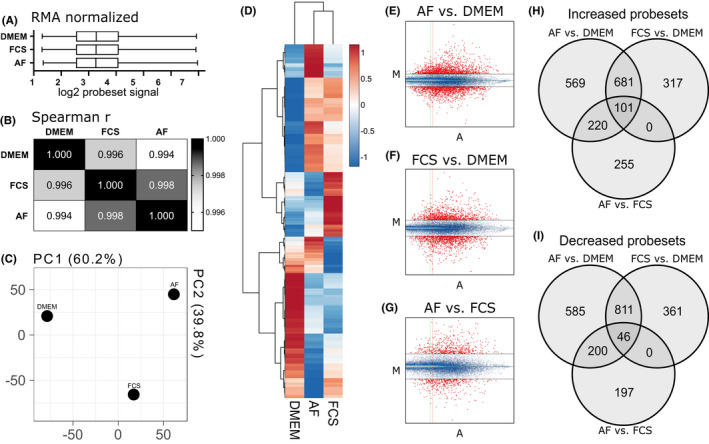
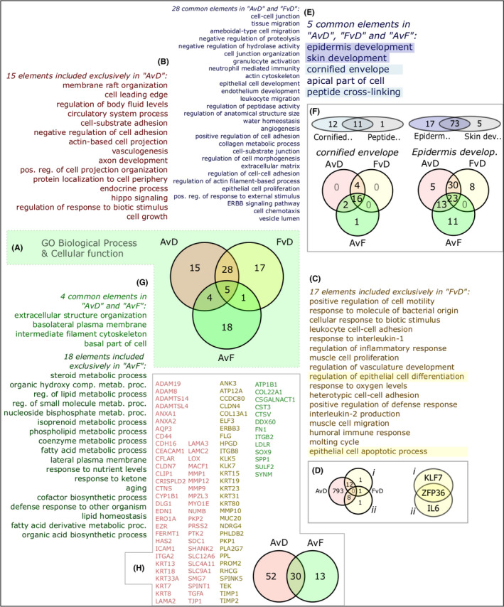
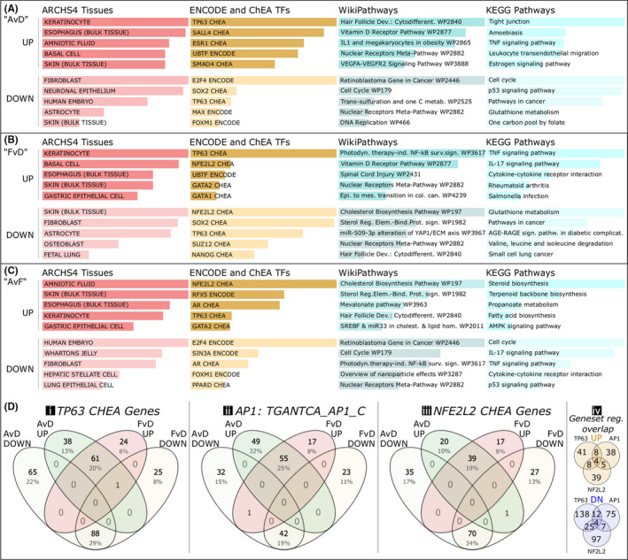
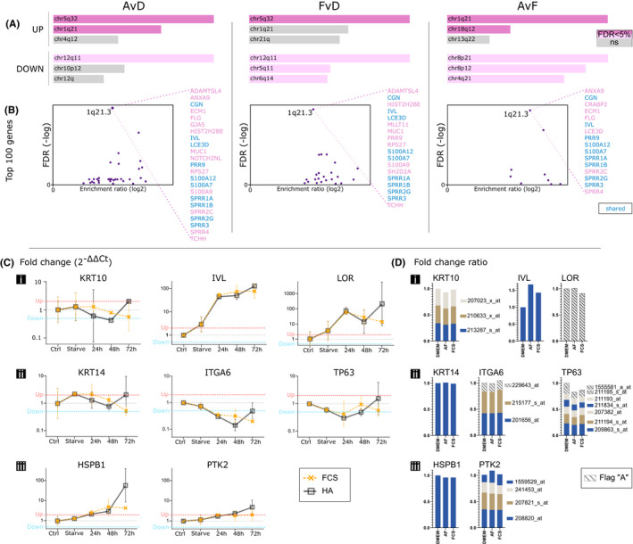
Cutaneous wounds can lead to huge suffering for patients. Early fetal wounds have the capacity to regenerate without scar formation. Amniotic fluid (AF), containing hyaluronic acid (HA), may contribute to this regenerative environment. We aimed to analyse changes in gene expression when human keratinocytes are exposed to AF or HA. Human keratinocytes were cultured to subconfluence, starved for 12 h and then randomised to be maintained in (1) Dulbecco's modified Eagle's medium (DMEM), (2) DMEM with 50% AF, or (3) DMEM with 50% fetal calf serum (FCS). Transcriptional changes were analysed using microarray and enriched with WebGestalt and Enrichr. Additionally, eight diagnostic genes were analysed using semiquantitative real-time PCR to investigate epidermal differentiation and cellular stress after HA exposure as an alternative for AF exposure. The AF and FCS treatments resulted in enrichment of genes relating to varied aspects of epidermal and keratinocyte biology. In particular, p63-, AP1- and NFE2L2- (Nrf2) associated genes were found significantly regulated in both treatments. More genes regulated by FCS treatment were associated with inflammatory signalling, whilst AF treatment was dominantly associated with molecular establishment of epidermis and lipid metabolic activity. HA exposure mostly resulted in gene regulation that was congruent with the AF microarray group, with increased expression of ITGA6 and LOR. We conclude that AF exposure enhances keratinocyte differentiation in vitro, which suggests that AF constituents can be beneficial for wound-healing applications.

Keywords: PCR; fetal wound healing; human skin cells; in vitro; microarray.


Conflict of interest statement

The authors declare that there is no conflict of interest regarding the publication of this paper.

Figures

FIGURE 1
FIGURE 1
Overview of array experimental data. (A) RMA normalised log2 values across samples. (B) Spearman's correlation between groups. (C) Principal component analysis showing PC1 and PC2. (D) Heatmap with hierarchical clustering of top 1000 genes and all groups. (E‐G) MvA plots for each comparison: (E) amniotic fluid (AF) vs. DMEM, (F) FCS vs. DMEM and (G) AF vs. FCS—probesets above or below 2‐fold change are shown in red, and the threshold of A = 3 is shown as a vertical line; (H) Venn diagram showing probesets that are marked as “Increased”, meaning upregulated, in all the three comparisons. (I) Venn diagram showing “Decreased” probesets. A = ½ (log2 (experiment ×reference); AF, amniotic fluid–exposed cultures; DMEM, serum‐starved cultures; FCS, fetal calf serum–exposed cultures; M = log2 (experiment/reference); PC, principal component; RMA, robust multiarray averaging
FIGURE 2
FIGURE 2
Gene Ontology (GO) gene set differential analysis and interpretation. (A) Significant GO terms among upregulated genes shown as sets for all three comparisons. (B) Unique terms for amniotic fluid (AF) treatment vs DMEM group (AvD). (C) Unique terms for fetal calf serum (FCS) treatment vs DMEM (FvD). (D) Genes representing epithelial‐related terms, (i) “regulation of epithelial cell differentiation”; (ii) “epithelial cell apoptotic process”). (E) Enriched GO terms in AF and FCS groups compared with DMEM (28 and 5, respectively). (F) Sets of genes related to terms “cornified envelope” and “epidermis development”. (G) Enriched terms in the AF versus FCS comparison (4 and 18 terms, respectively) of which 18 were not also increased in the AvD comparison. (H) Expansion of the four terms in AvD intersecting AvF into gene sets shows 13 genes (in green) that signify AF over FCS treatment and 52 (in red) that are increased in AF vs. DMEM, with an overlap of 30 (yellowish) satisfying both criteria. AvD, AF vs. DMEM; FvD, FCS vs. DMEM; AvF, AF vs. FCS
FIGURE 3
FIGURE 3
Categories of genes regulated “up” or “down” (>2‐fold change) in the three comparisons, with examples of major keratinocyte transcription factor network associations. (A) Amniotic fluid rescue of starvation (AF vs. DMEM; “AvD”). (B) Fetal calf serum rescue of starvation (FCS vs. DMEM; “FvD”). (C) Amniotic fluid versus fetal calf serum regulatory changes (AF vs. FCS; “AvF”). (D) Three major transcription factor networks and their genes as apparent in the >2‐fold regulation, presented in 4‐way Venn diagrams: (i) TP63 (tumor protein p63), as selected by ChIP data ChEA; (ii) AP1 (activator protein 1 complex), gene set by binding sequence TGANTCA according to mSigDB; (iii) NFE2L2 (a.k.a Nrf2), as selected by ChEA data through Enrichr (www.enrich.net); (iv) showing overlaps of the gene set collections in (i‐iii), divided into upregulation and downregulation, indicating network module set similarities in our data. Bars represent combined p‐value and rank scoring, and all are considered significantly enriched
FIGURE 4
FIGURE 4
Chromosomal differentiation cluster analyses and qPCR with hyaluronic acid (HA) treatment to substitute for amniotic fluid (AF). (A) Chromosomal location enrichment among upregulated and downregulated genes per comparison. (B) Enrichment of the epidermal differentiation complex (EDC) at location 1q21.3 among Top‐100 increased genes in all treatments, with specified gene lists. (C) qPCR measured fold changes for three suprabasal, three basal and two stress genes after substituting AF with HA. (D) Visualisation of probesets corresponding to the selected genes to compare gene expression between qPCR and microarray methods and HA/AF treatments. Flag A = absent, AF, amniotic fluid; Ctrl, human primary keratinocytes; FCS, fetal calf serum; HA, hyaluronic acid

References

    1. Wang PH, Huang BS, Horng HC, Yeh CC, Chen YJ. Wound healing. J Chin Med Assoc. 2018;81(2):94‐101. - PubMed
    1. Longaker MT, Chiu ES, Adzick NS, Stern M, Harrison MR, Stern R. Studies in fetal wound healing. V. A prolonged presence of hyaluronic acid characterizes fetal wound fluid. Ann Surg. 1991;213(4):292‐296. - PMC - PubMed
    1. Gao X, Devoe LD, Given KS. Effects of amniotic fluid on proteases: a possible role of amniotic fluid in fetal wound healing. Ann Plast Surg 1994;33(2):128‐135. discussion 34–5. - PubMed
    1. Sancho MA, Julia V, Albert A, Diaz F, Morales L. Effect of the environment on fetal skin wound healing. J Pediatr Surg. 1997;32(5):663‐666. - PubMed
    1. Underwood MA, Gilbert WM, Sherman MP. Amniotic fluid: not just fetal urine anymore. J Perinatol. 2005;25(5):341‐348. - PubMed

Publication types

Substances