. 2022 Jan 12;30(1):53-68.e12.
doi: 10.1016/j.chom.2021.11.013. Epub 2021 Nov 27.

The antibody response to SARS-CoV-2 Beta underscores the antigenic distance to other variants

Affiliations

The antibody response to SARS-CoV-2 Beta underscores the antigenic distance to other variants

Chang Liu et al. Cell Host Microbe. .

Abstract

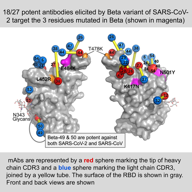
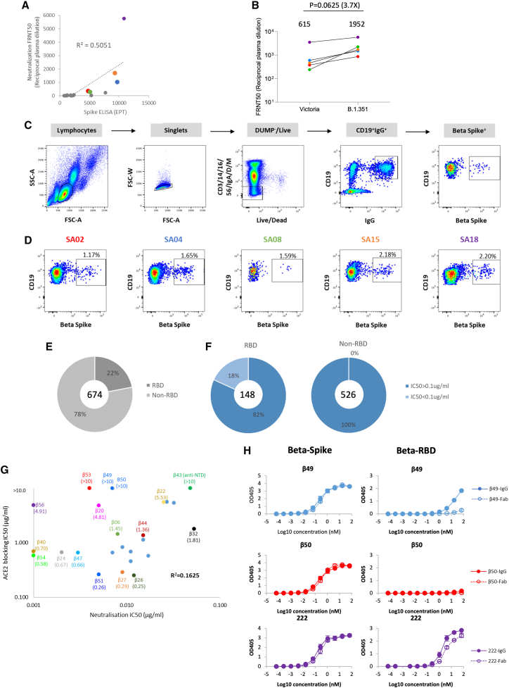
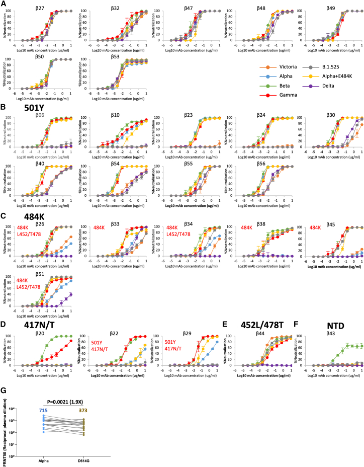
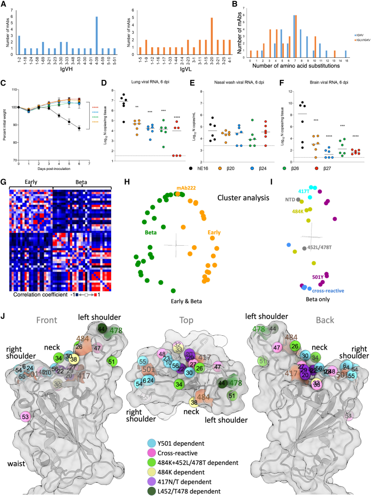
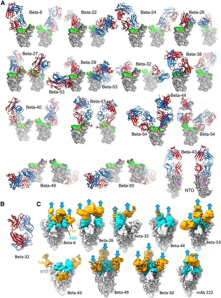
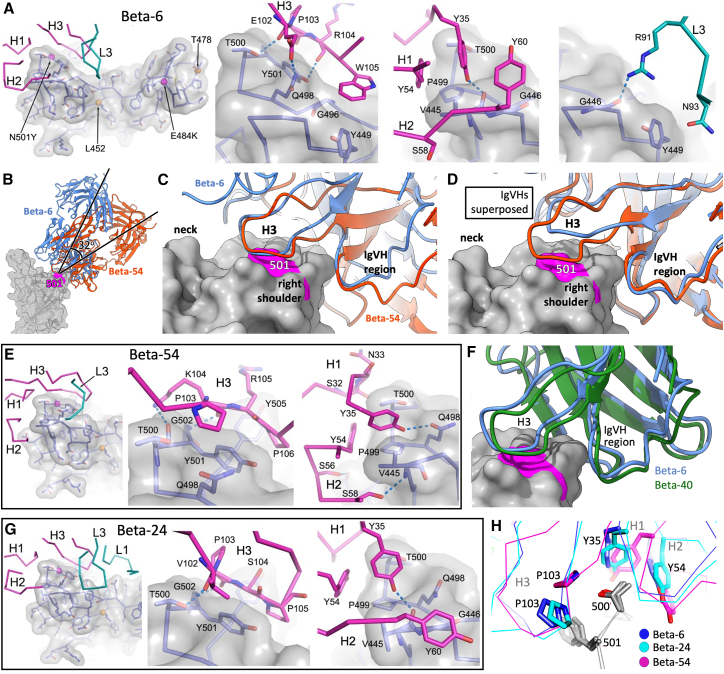
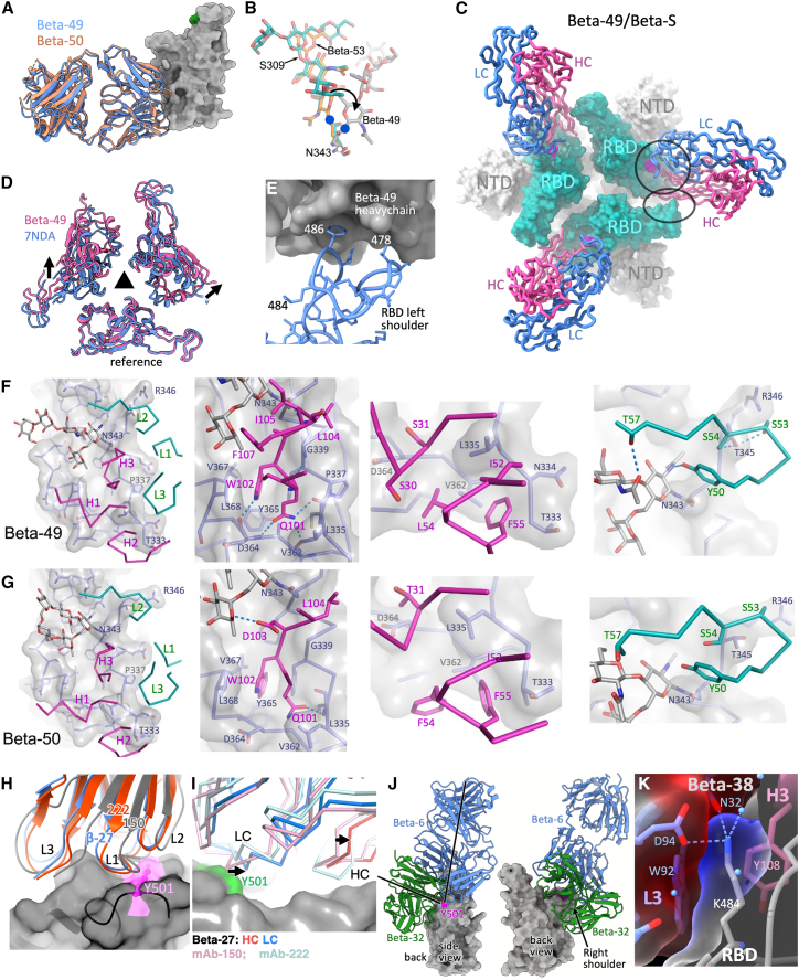
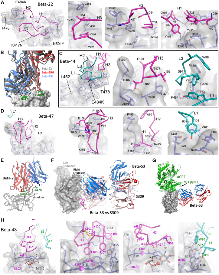
Alpha-B.1.1.7, Beta-B.1.351, Gamma-P.1, and Delta-B.1.617.2 variants of SARS-CoV-2 express multiple mutations in the spike protein (S). These may alter the antigenic structure of S, causing escape from natural or vaccine-induced immunity. Beta is particularly difficult to neutralize using serum induced by early pandemic SARS-CoV-2 strains and is most antigenically separated from Delta. To understand this, we generated 674 mAbs from Beta-infected individuals and performed a detailed structure-function analysis of the 27 most potent mAbs: one binding the spike N-terminal domain (NTD), the rest the receptor-binding domain (RBD). Two of these RBD-binding mAbs recognize a neutralizing epitope conserved between SARS-CoV-1 and -2, while 18 target mutated residues in Beta: K417N, E484K, and N501Y. There is a major response to N501Y, including a public IgVH4-39 sequence, with E484K and K417N also targeted. Recognition of these key residues underscores why serum from Beta cases poorly neutralizes early pandemic and Delta viruses.

Keywords: Beta variant; COVID-19; SARS-CoV-2; antibody; immune responses; neutralization; receptor-binding domain; spike protein; structure; vaccine.


Conflict of interest statement

Declaration of interests M.S.D. is a consultant for Inbios, Vir Biotechnology, NGM Biopharmaceuticals, Carnival Corporation, and on the Scientific Advisory Boards of Moderna and Immunome. The M.S.D. laboratory has received unrelated funding support in sponsored research agreements from Moderna, Vir Biotechnology, and Emergent BioSolutions. G.R.S. sits on the GSK Vaccines Scientific Advisory Board and is a founder member of RQ Biotechnology. The University of Oxford has protected intellectual property disclosed in this publication.

Figures

None
Graphical abstract
Figure 1
Figure 1
Isolation and characterization of Beta SARS-CoV-2-specific mAbs (A) Comparison of Beta SARS-CoV-2 neutralization and S binding ELISA by convalescent plasma from confirmed Beta SARS-CoV-2 infected donors. Plasma samples with FRNT50 >1:250 are highlighted and correspond to the cases shown in (D). (B) Neutralization titers against SARS-CoV-2 strain Victoria and the Beta variant for the 5 selected plasma samples with potent neutralizing properties, analysis used the Wilcoxon matched-pairs signed rank test and two-tailed p values were calculated; geometric means are indicated above each column. (C) Schematic of the Beta SARS-CoV-2 mAb isolation strategy. (D) Antigen-specific single B cells were isolated using labeled recombinant S protein as bait. The frequency of S-reactive IgG+ B cells was measured by FACS. (E) Epitope mapping of Beta SARS-CoV-2 specific mAbs against S and RBD were evaluated by ELISA. (F) Neutralization potencies (IC50) between anti-S (non-RBD) and anti-RBD mAbs against authentic Beta SARS-CoV-2 using a FRNT50 test. (G) Comparison of IC50 values for ACE2 binding and FRNT50 titers for the 27 potent mAbs, those selected for further structural study are highlighted. (H) Binding of Beta-49 and -50 Fab and IgG1 to Beta S trimer or Beta RBD measured by ELISA, comparison is made with binding of mAb 222, data are shown as mean ± SEM. See also Table S1A.
Figure 2
Figure 2
Cross reactivity of Beta-specific mAbs (A–F) Neutralization assays performed against Victoria, Alpha (N501Y), Beta (K417N, E484K, and N501Y), Gamma (K417T, E484K, and N501Y), Delta (L452R and T478K), Alpha+E484K (E484K and N501Y), and B.1.525 (E484K) live viral isolates with 27 potent Beta-specific mAbs. Titration curves are shown and mAbs grouped depending on the patterns of cross reactivity between the viral variants, potential binding determinants are indicated for the mAbs that show differential neutralization between isolates. Data are shown as mean ± SEM (A) Fully cross-reactive mAbs, (B) N501Y-dependent mAbs, (C) E484K-dependent mAbs, (D) K417N/T-dependent mAbs, (E) L452R/T478K-dependent mAbs, and (F) a single NTD-binding mAb. FRNT50 values are reported in Table S1. (G) FRNT50 titers of 17 Alpha convalescent sera against Alpha and B.1 (D614G), analysis used the Wilcoxon matched-pairs signed rank sum test, and two-tailed p values were calculated; geometric means are indicated above each column. See also Figure S1.
Figure 3
Figure 3
Gene usage, therapeutic use in K18-hACE2 mice, and computational analysis of responses (A) IgVH and IgVL gene usage for the 27 potent mAbs. (B) Amino acid substitutions in IgVH and IgVL for the 27 potent mAbs. (C–F) 8-week-old female K18-hACE2 transgenic mice were administered 103 FFU of SARS-CoV-2 Beta strain by intranasal inoculation. One day later, mice received a single 10 mg/kg dose of the indicated mAb treatment by intraperitoneal injection. Tissues were collected at 6 dpi. (C) Weight change following infection with SARS-CoV-2 (mean ± SEM; n = 6 mice per group, two experiments; one-way ANOVA with Dunnett’s test of area under the curve: ∗∗∗∗p < 0.0001). Viral RNA levels in the lung (D), nasal wash (E), and brain (F) (line indicates median; n = 6 mice per group, two experiments; one-way ANOVA with Dunnett’s test with comparison to control mAb: ∗∗p < 0.01, ∗∗∗p = 0.001, ∗∗∗∗p < 0.0001. Dotted line indicates the limit of detection of the assay. (G) Cross-correlation matrix showing agreement of neutralization titers for mAbs against seven variants of SARS-CoV-2. Every antibody is associated with a vector containing the residual neutralization titer after subtracting the mean for each variant and normalizing to a standard deviation of 1. (H–J) Each point (I and J) in the matrix is colored according to the dot product between vectors for antibody (I and J). (H) Major modes of variation after singular value decomposition of the matrix in (G). (I) Major modes of variation after singular value decomposition of a matrix similar to (G) but calculated for Beta mAbs and colored according to their designation as a fully cross-reactive, 501Y-specific, 484K-specific, or 417T-specific antibody. (J) Mapping of the Beta mAbs based on BLI competition measurements (STAR Methods; Dejnirattisai et al., 2021a). The mean positions of the mAbs are shown as spheres. Numbers match the antibody definitions in Tables S1A and S1B (βs omitted for clarity), colored by one aspect of the serological properties e.g., Y501-dependent indicates potent neutralization is only observed for those viruses with Tyr-501. Anatomical terms relate to the torso analogy (Dejnirattisai et al., 2021a). The RBD is shown as a semi-transparent surface with cartoon embedded. The outer two are related by 180° rotation about the vertical axis, and the central view is related to the “front” view by a 90° rotation about the horizonal axis. See also Figure S2 and Table S1B.
Figure 4
Figure 4
Overall structures of Beta RBD/Beta S complexes with Beta mAb Fabs reported in this paper (A) Front and back views of Beta RBD/Beta Fab complexes. Fabs drawn as ribbons with HC red and LC blue, and RBDs as gray surfaces with ACE2 footprint in green, mutation sites of the Beta variant in magenta and Delta variant in orange. All structures were crystallographic except Beta-26 and -32, which were derived by cryo-EM. (B) Crystal structure of Beta-32 Fab with HC red and LC blue. (C) Cryo-EM maps of Beta S complexes with Beta-6, -26, -32, -44, -53, -43, -49, and -50 and early pandemic mAb-222 Fabs. The bound Fabs are orange, RBD domains cyan, and the rest of S gray. Arrows indicate the RBD orientations. See also Figure S3 and Tables S2 and S4.
Figure 5
Figure 5
Structural details of IgVH4-30 and IgVH4-39 Beta Fab complexes (A) Beta-6, Beta-RBD interactions. Left panel shows interacting CDRs (HC magenta, LC cyan) with the Beta-RBD (semi-transparent gray surface, side chains as blue sticks, mutation sites of Beta [magenta] and Delta [orange] variants shown as spheres). Interactions of H3, H2, H1, and L3 loops are shown in the adjacent panels. (B) Comparison of binding orientations for Beta-6 (blue) and Beta-54 (red). (C) Closeup of (B) showing engagement of CDR-H3s with Tyr-501 (magenta). (D) Same as (C) but IgVHs are overlapped instead of RBDs. (E) Interactions of Beta-54 with Beta RBD. (F) Comparison of binding modes of Beta-40 IgVH (green) and Beta-6 (Blue). (G) Interactions of Beta-24 with Beta RBD. (H) Common features of the engagement used by Beta-6 (blue), -24 (cyan), and -54 (magenta). Y35 of CDR-H1 and Y54 of CDR-H2 are conserved among the IgVH4-30 and IgVH4-39 Beta mAbs reported here. See also Tables S2 and S3.
Figure 6
Figure 6
Engagement of other Beta IgVH Fabs with the Beta RBD (A) Almost identical binding of Beta-49 (blue) and Beta-50 (salmon) to the RBD. (B) Overlay of N343 RBD glycan from the (green) (Pinto et al., 2020), Beta-53 (yellow) and Beta-49 (gray) complexes, the side chain rotated into an unfavorable conformation in the latter. (C) Top view of the Beta-49 Fab/Beta S complex. S is shown as a surface (RBD cyan, position of glycan attachment to residue 343 magenta) while Beta-49 HC (dark pink) and LC (blue) are shown as cartoons. The HC contacts two RBDs, forming a primary (circle) and secondary (ellipse) epitope. (D) Top view of the RBDs in all RBD down S (PDB 7NDA) and in the Beta-49 bound state. The 3-fold axis of S is shown. One RBD is superposed (reference), arrows show the movement in the other RBDs induced on binding Beta-49. (E) Close up of the secondary epitope with some RBD residues marked. (F) Close up of Beta-49/Beta S interaction. The RBD is shown as sticks and a surface (glycan at N343 as sticks only), and Fab as sticks colored by chain. (G) Similar to (F) but for Beta-50. (H and I) Comparison of the binding of Beta-27 with mAbs 150 and 222. (H) Residue 501 is highlighted on the RBD surface. (I) Side view of the right shoulder and neck of the RBD. Arrows show shifts due to repositioning the HC CDR3. (J) Comparison of the attachment of Beta-6 and -32 to the RBD with axes (left panel) showing difference in pose. (K) K484 is enclosed by the Beta-38 HC and LC CDR3s. See also Figure S4 and Tables S2 and S4.
Figure 7
Figure 7
Details of interactions of Beta-22, -29, -47, -26, -53, and -43 (A) Interactions of Beta-22 (as Figure 6A). N417 specificity is achieved indirectly. (B) Identical binding modes of Beta-22 (gray) and Beta-29 (HC red, LC blue) IgVH3-30 mAbs. (C) Beta-44/Beta RDB interactions. (D) Beta-47/Beta RDB interactions. (E) Beta-26 binds the left shoulder contacting K484 and T478 of the RBD. (F) Beta-53 (HC red, LC blue) binds the same epitope as S309 (HC salmon, LC pale blue; PDB: 7BEP). (G) Binding of Beta-53 relative to ACE2 receptor. (H) Beta-43 binding to the NTD (gray surface). See also Figure S5.

References

    1. Altschul S.F., Gish W., Miller W., Myers E.W., Lipman D.J. Basic local alignment search tool. J. Mol. Biol. 1990;215:403–410. - PubMed
    1. Aricescu A.R., Lu W., Jones E.Y. A time- and cost-efficient system for high-level protein production in mammalian cells Acta Crystallogr D. Biol. Crystallogr. 2006;62:1243–1250. - PubMed
    1. Barnes C.O., Jette C.A., Abernathy M.E., Dam K.-M.A., Esswein S.R., Gristick H.B., Malyutin A.G., Sharaf N.G., Huey-Tubman K.E., Lee Y.E., et al. SARS-CoV-2 neutralizing antibody structures inform therapeutic strategies. Nature. 2020;588:682–687. - PMC - PubMed
    1. Caly L., Druce J., Roberts J., Bond K., Tran T., Kostecki R., Yoga Y., Naughton W., Taiaroa G., Seemann T., et al. Isolation and rapid sharing of the 2019 novel coronavirus (SARS-CoV-2) from the first patient diagnosed with COVID-19 in Australia. Med. J. Aust. 2020;212:459–462. - PMC - PubMed
    1. Case J.B., Bailey A.L., Kim A.S., Chen R.E., Diamond M.S. Growth, detection, quantification, and inactivation of SARS-CoV-2. Virology. 2020;548:39–48. - PMC - PubMed

Publication types

Supplementary concepts