Review
. 2021 Dec 18;16(1):83.
doi: 10.1186/s13024-021-00501-z.

Neuropathology and molecular diagnosis of Synucleinopathies

Affiliations
Review

Neuropathology and molecular diagnosis of Synucleinopathies

Shunsuke Koga et al. Mol Neurodegener. .

Abstract

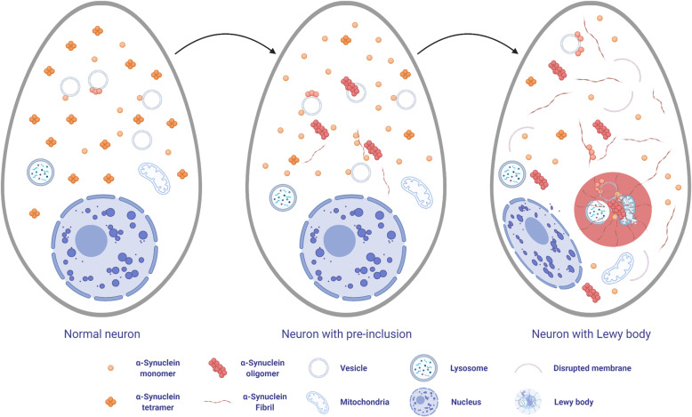
Synucleinopathies are clinically and pathologically heterogeneous disorders characterized by pathologic aggregates of α-synuclein in neurons and glia, in the form of Lewy bodies, Lewy neurites, neuronal cytoplasmic inclusions, and glial cytoplasmic inclusions. Synucleinopathies can be divided into two major disease entities: Lewy body disease and multiple system atrophy (MSA). Common clinical presentations of Lewy body disease are Parkinson's disease (PD), PD with dementia, and dementia with Lewy bodies (DLB), while MSA has two major clinical subtypes, MSA with predominant cerebellar ataxia and MSA with predominant parkinsonism. There are currently no disease-modifying therapies for the synucleinopathies, but information obtained from molecular genetics and models that explore mechanisms of α-synuclein conversion to pathologic oligomers and insoluble fibrils offer hope for eventual therapies. It remains unclear how α-synuclein can be associated with distinct cellular pathologies (e.g., Lewy bodies and glial cytoplasmic inclusions) and what factors determine neuroanatomical and cell type vulnerability. Accumulating evidence from in vitro and in vivo experiments suggests that α-synuclein species derived from Lewy body disease and MSA are distinct "strains" having different seeding properties. Recent advancements in in vitro seeding assays, such as real-time quaking-induced conversion (RT-QuIC) and protein misfolding cyclic amplification (PMCA), not only demonstrate distinct seeding activity in the synucleinopathies, but also offer exciting opportunities for molecular diagnosis using readily accessible peripheral tissue samples. Cryogenic electron microscopy (cryo-EM) structural studies of α-synuclein derived from recombinant or brain-derived filaments provide new insight into mechanisms of seeding in synucleinopathies. In this review, we describe clinical, genetic and neuropathologic features of synucleinopathies, including a discussion of the evolution of classification and staging of Lewy body disease. We also provide a brief discussion on proposed mechanisms of Lewy body formation, as well as evidence supporting the existence of distinct α-synuclein strains in Lewy body disease and MSA.

Keywords: AlphaFold; Biomarkers; Dementia with Lewy bodies; Lewy body disease; Multiple system atrophy; PMCA; Parkinson’s disease; RT-QuIC; cryo-EM structures.


Conflict of interest statement

The authors declare that they have no competing interests.

Figures

Fig. 1
Fig. 1
Structure of α-synuclein. Missense mutation sites linked to familial Parkinson’s disease and major post-translational modification sites are shown
Fig. 2
Fig. 2
Hypothetical scheme of Lewy body formation in neurons. α-Synuclein exists in equilibrium between monomers and tetramers in the cytoplasm. Under pathologic conditions, the tetramer-monomer ratio decreases, and monomers bind to vesicle membranes. On the surface of membrane, α-synuclein tend to oligomerize, and toxic oligomers can disrupt lipid membranes. Monomers and oligomers form insoluble fibrils in the cytoplasm. Disrupted membranes, organelles, α-synuclein oligomers and fibrils are involved in the Lewy body formation
Fig. 3
Fig. 3
Macroscopic findings of Lewy body disease brains. The midbrain and pons from Lewy body disease (A) and control (B) brains. Loss of neuromelanin pigment in the substantia nigra and locus coeruleus is observed in Lewy body disease. Abbreviations: LC, locus coeruleus; SN, substantia nigra
Fig. 4
Fig. 4
Representative images of histopathology of Lewy body disease. A, B, F-H hematoxylin and eosin staining, C-E immunohistochemistry for α-synuclein (NACP antibody). A, C Brainstem type Lewy body in the substantia nigra. B, D cortical type Lewy body (arrow) in the superior temporal cortex. E Lewy neurites in the CA2 sector of the hippocampus. F-G Lewy body disease shows neuronal loss with extracellular neuromelanin pigment in the substantia nigra (F), while it is minimum in a control case (G). H Spongiform change in the entorhinal cortex. Scale 100 μm in (A, B, and H); 50 μm in (C); 20 μm in (D-G, and I)
Fig. 5
Fig. 5
Macroscopic findings of multiple system atrophy (MSA). The brains from MSA (A, C, E, and G) and control (B, D, F, and H) cases. The pons and cerebellar white matter are atrophic in MSA (A). Loss of neuromelanin pigment in the locus coeruleus (C) and substantia nigra (E) is observed in MSA. The discoloration and atrophy of the lateral putamen in MSA (G). Abbreviations: CWM, cerebellar white matter; LC, locus coeruleus; SN, substantia nigra
Fig. 6
Fig. 6
Representative images of histopathology of MSA. A-D Hematoxylin and eosin staining. A Severe neuronal loss with gliosis in the lateral putamen. B Neurodegeneration is minimal in the same region in a case of minimal change MSA. C A typical MSA shows neuronal loss with extracellular neuromelanin pigment (arrows) in the substantia nigra, while it is well preserved in minimal change MSA (D). E-J Immunohistochemistry for α-synuclein (NACP antibody). Numerous GCIs in the putamen (E) and substantia nigra (F). Arrows indicate extracellular neuromelanin pigment (F). Neuronal cytoplasmic inclusions in the pontine nucleus (G, arrows) and inferior olivary nucleus (H, arrow). Abundant neuronal cytoplasmic inclusions in the dentate fascia in FTLD-synuclein (I) and hippocampal MSA (J). Scale bars: 50 μm in all images
Fig. 7
Fig. 7
The cryo-EM structures of recombinant α-synuclein fibrils. A Two polymorphs of filaments from wild type fibrils: rod (Protein Data Bank [PDB] ID: 6CU7) and twister (PDB ID: 6CU8). B E46K mutant fibrils (PDB ID: 6L43). C H50Q mutant fibrils (PDB ID: 6PES). D A53T mutant fibrils (PDB ID: 6LRQ). Dotted boxes indicate the inter-protofilament interfaces of each α-synuclein fibril
Fig. 8
Fig. 8
The cryo-EM structures of α-synuclein fibrils from MSA brains. Two distinct filaments: type I (PDB ID: 6XYO) and type II (PDB ID: 6XYP). Dotted boxes indicate the inter-protofilament interfaces. 43 K, 45 K, and 50 H are shown in red

References

    1. Hardy J, Gwinn-Hardy K. Genetic classification of primary neurodegenerative disease. Science. 1998;282:1075–1079. - PubMed
    1. Dickson DW, Lin W, Liu WK, Yen SH. Multiple system atrophy: a sporadic synucleinopathy. Brain Pathol. 1999;9:721–732. - PMC - PubMed
    1. Goedert M, Spillantini MG. Lewy body diseases and multiple system atrophy as alpha-synucleinopathies. Mol Psychiatry. 1998;3:462–465. - PubMed
    1. Kosaka K, Matsushita M, Oyanagi S, Mehraein P. A clinico-neuropathological study of “Lewy body disease.” Psy-chiatry Neurol Jpn. 1980;82:292–311. - PubMed
    1. Erskine D, Attems J. Insights into Lewy body disease from rare neurometabolic disorders. J Neural Transm (Vienna) 2021;128(10):1567–1575. - PMC - PubMed

Publication types

MeSH terms

Substances