. 2022 Jan;15(1):e008574.
doi: 10.1161/CIRCHEARTFAILURE.121.008574. Epub 2021 Dec 20.

Inflammatory Glycoprotein 130 Signaling Links Changes in Microtubules and Junctophilin-2 to Altered Mitochondrial Metabolism and Right Ventricular Contractility

Affiliations

Inflammatory Glycoprotein 130 Signaling Links Changes in Microtubules and Junctophilin-2 to Altered Mitochondrial Metabolism and Right Ventricular Contractility

Sasha Z Prisco et al. Circ Heart Fail. 2022 Jan.

Abstract

Background: Right ventricular dysfunction (RVD) is the leading cause of death in pulmonary arterial hypertension (PAH), but no RV-specific therapy exists. We showed microtubule-mediated junctophilin-2 dysregulation (MT-JPH2 pathway) causes t-tubule disruption and RVD in rodent PAH, but the druggable regulators of this critical pathway are unknown. GP130 (glycoprotein 130) activation induces cardiomyocyte microtubule remodeling in vitro; however, the effects of GP130 signaling on the MT-JPH2 pathway and RVD resulting from PAH are undefined.

Methods: Immunoblots quantified protein abundance, quantitative proteomics defined RV microtubule-interacting proteins (MT-interactome), metabolomics evaluated the RV metabolic signature, and transmission electron microscopy assessed RV cardiomyocyte mitochondrial morphology in control, monocrotaline, and monocrotaline-SC-144 (GP130 antagonist) rats. Echocardiography and pressure-volume loops defined the effects of SC-144 on RV-pulmonary artery coupling in monocrotaline rats (8-16 rats per group). In 73 patients with PAH, the relationship between interleukin-6, a GP130 ligand, and RVD was evaluated.

Results: SC-144 decreased GP130 activation, which normalized MT-JPH2 protein expression and t-tubule structure in the monocrotaline RV. Proteomics analysis revealed SC-144 restored RV MT-interactome regulation. Ingenuity pathway analysis of dysregulated MT-interacting proteins identified a link between microtubules and mitochondrial function. Specifically, SC-144 prevented dysregulation of electron transport chain, Krebs cycle, and the fatty acid oxidation pathway proteins. Metabolomics profiling suggested SC-144 reduced glycolytic dependence, glutaminolysis induction, and enhanced fatty acid metabolism. Transmission electron microscopy and immunoblots indicated increased mitochondrial fission in the monocrotaline RV, which SC-144 mitigated. GP130 antagonism reduced RV hypertrophy and fibrosis and augmented RV-pulmonary artery coupling without altering PAH severity. In patients with PAH, higher interleukin-6 levels were associated with more severe RVD (RV fractional area change 23±12% versus 30±10%, P=0.002).

Conclusions: GP130 antagonism reduces MT-JPH2 dysregulation, corrects metabolic derangements in the RV, and improves RVD in monocrotaline rats.

Keywords: glycoprotein; interleukin-6; monocrotaline; proteomics; pulmonary arterial hypertension.


Figures
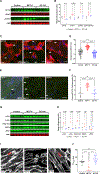
Figure 1:
Figure 1:. GP130 antagonism blunted RV STAT3 activation, normalized the MT-JPH2 pathway, and restored t-tubule architecture.
(A) Representative Western blots and (B) quantification of protein abundance in RV extracts from control, MCT-V, and SC-144 rats demonstrated GP130 inhibition normalized expression of GP130, STAT3, pSTAT3, and the ratio of pSTAT3/STAT3. Data shown as expression relative to control (n=4 per group). Representative confocal micrographs of RV free wall sections showed SC-144 reduced the amount of (C) GP130 receptors (white arrows) (green: GP130, blue: DAPI, red: wheat germ agglutinin) at the cell membrane as quantified in (D) as amount of GP130 expression per area (%) and amount of (E) pSTAT3 (white arrows) (green: pSTAT3, blue: DAPI) in cardiomyocyte nuclei as quantified in (F) as percent pSTAT3 positive nuclei per area. n=3–4 animals per group. (G) Representative Western blots and (H) protein quantification showed GP130 antagonism reduced expression of α- and β-tubulin, detyrosinated α-tubulin, and MAP4 and increased JPH2. Data shown as expression relative to control (n=4 per group). (I) Representative confocal images of RV free wall sections stained with Alexa Fluor-633 conjugated wheat germ agglutinin. SC-144 restored RV t-tubule architecture (red arrows). (J) Quantification of t-tubule architecture and organization by TTorg (arbitrary units are TTpower). n=3 animals per group, ≥7 cardiomyocytes were quantified per animal. Unpaired t-test was used to compare MCT-V vs. SC-144 in (B) and (H). ***p<0.001, ****p<0.0001, and (ns) not significantly different as assessed by Kruskal-Wallis ANOVA with Dunn post-hoc test in (D) and (J) and Brown-Forsythe and Welch ANOVA with Dunnett post-hoc analysis in (F).
Figure 2:
Figure 2:. Quantitative proteomic analysis of the MT-interactome in the RV revealed a link between microtubule remodeling and mitochondrial protein expression.
(A) Principal component analysis showed GP130 antagonism partially restored the global expression signature of the MT-interactome. (B) Hierarchical cluster analysis of MT-interacting proteins demonstrated the expression pattern of the MT-interactome in the RV of SC-144 rats more closely resembled control than MCT-V. (C) Ten most significantly enriched pathways identified using Ingenuity pathway analysis of dysregulated MT-interacting proteins in MCT-V RV when compared to control. The two most enriched pathways were mitochondrial dysfunction and oxidative phosphorylation. Hierarchical cluster analysis of proteins in complex I (D), complex II (E), complex III (F), complex IV (G), and complex V (H), the TCA cycle (I), and fatty acid oxidation (J). SC-144 corrected dysregulation of nearly all mitochondrial metabolic proteins.
Figure 3:
Figure 3:. SC-144 improved the RV metabolic signature, restored mitochondrial morphology, and corrected mitochondrial fission/fusion imbalance.
(A) Hierarchical cluster analysis demonstrated SC-144 normalized the global RV metabolic signature. (B) Random forest analysis highlighting the 15 metabolites that differentiate control, MCT-V, and SC-144. Hierarchical cluster analysis of (C) glycolysis, and (D) glutaminolysis identified a normalization of multiple metabolic pathways with SC-144 while most of the (E) acylcarnitine metabolites were increased by SC-144. (F) Representative electron micrographs of mitochondria. GP130 antagonism partially corrected the RV mitochondrial morphology as assessed by (G) mitochondrial area and (H) eccentricity index. GP130 antagonism reduced the amount of large, swollen mitochondria as quantified in (G) and restored the normal elongated mitochondrial shape as assessed in (H). n=3 RV per group, with >70 mitochondria measured per animal. (I) Representative Western blots and (J) quantification of protein abundance in RV extracts from control, MCT-V, and SC-144 demonstrated GP130 inhibition did not affect MFN1 expression and minimally changed MFN2 but normalized OPA1 (non-statistical change), FIS1, and DRP1 expression. *p<0.05, ****p<0.0001, and (ns) no statistical difference as determined by Brown-Forsythe ANOVA with Dunnett post-hoc test after transformation in (G) and (H) and unpaired t-test in (J).
Figure 4:
Figure 4:. Chemical stabilization of microtubules with paclitaxel in H9c2 cardiomyocytes altered mitochondrial metabolic function.
(A) Seahorse analysis of the oxygen consumption rate profile and (B) individual parameters of mitochondrial respiration. Con: control; OCR: oxygen consumption rate; Pac: paclitaxel. Data presented in (A) as mean ± standard deviation. *p<0.05, **p<0.01, ***p<0.001, ****p<0.0001, and (ns) no statistical difference as determined by t-test or Mann-Whitney U-test.
Figure 5:
Figure 5:. GP130 antagonism reduced RV hypertrophy and fibrosis.
SC-144 reduced RV hypertrophy as assessed by the Fulton index (RV/LV+S) (A) and RV weight normalized to body weight (B). n=10–26 animals per group. (C) SC-144 decreased cardiomyocyte area. (D) Representative images of cardiomyocytes in H&E stained RV free wall sections. n=2–3 RV per group, at least 21 cardiomyocytes measured per animal. (E) Representative Western blots and (F) quantification of collagen I and III protein abundance in RV extracts. n=4 per group. SC-144 decreased expression of collagen I and III protein expression compared to MCT-V, which corresponded with less RV fibrosis observed in trichrome staining of RV free wall as quantified in (G). n=2–3 RV per group, 3–4 areas sampled per animal. (H) Representative trichrome stained RV sections. White arrows highlight RV fibrosis. *p<0.05, **p<0.01, ***p<0.001, ****p<0.0001, and (ns) no statistical difference as determined by one-way ANOVA with Tukey post-hoc analysis in (A) and Kruskal-Wallis ANOVA with Dunn post-hoc test in (B), (C), and (G). Unpaired t-test between MCT-V and SC-144 was completed in (F).
Figure 6:
Figure 6:. SC-144 improved RV function.
(A) TAPSE, (B) percent RV free wall thickness change, (C) stroke volume, (D) cardiac output, and (E) cardiac output normalized to body weight were measured by echocardiography. n=9–14 rats per group. SC-144 improved RV function in all echocardiographic measures. Invasive hemodynamics and pressure-volume (PV) loops demonstrated that SC-144 augmented (F) RV ejection fraction (RVEF), (G) end-systolic elastance (Ees), and (H) RV-PA coupling (Ees/Ea). n=8–16 rats per group. *p<0.05, **p<0.01, ***p<0.001, ****p<0.0001, and (ns) no statistical difference as determined by one-way ANOVA with Tukey post-hoc analysis for (A), (B), (C), and (F), Kruskal-Wallis ANOVA with Dunn post-hoc test in (D), (E), and (H), and Brown-Forsythe ANOVA with Dunnett post-hoc analysis in (G).
Figure 7:
Figure 7:. Higher IL-6 levels in PAH patients were associated with worse RV function independent of changes in PAH severity.
Higher IL-6 levels are associated with higher NT-proBNP levels (A) and lower RVFAC (B). (C) Relationship between RVFAC and mPAP. Patients with higher IL-6 levels had lower RVFAC at each mPAP compared to patients with lower IL-6 levels (statistical difference in y-intercept, p=0.02; no difference in slope, p=0.84). (D) Relationship between RVFAC and PVR. PAH patients with higher IL-6 had reduced RVFAC at each PVR compared to patients with lower IL-6 (statistical difference in y-intercept, p=0.02; no difference in slope, p=0.56). n=73 total patients with the two groups stratified by median IL-6 level. There were no differences in (E) mPAP (p=0.35), (F) PVR (p=0.28), or (G) PAC (p=0.14) between patients with higher IL-6 levels compared to those with lower IL-6 levels. **p<0.01 and (ns) not significant as determined by Mann-Whitney U-test in (A), (B), (F), and (G) and unpaired t-test in (E). Linear regression evaluated differences between the lower and higher IL-6 curves in (C) and (D).
Figure 8:
Figure 8:. GP130-mediated RV dysfunction in PAH.
Heightened GP130 activation may induce STAT3-mediated gene transcription of microtubule proteins and pSTAT3 may interact with microtubules. GP130 activation causes microtubule remodeling, which leads to JPH2 dysregulation, t-tubule derangements, and mitochondrial dysfunction via imbalance of mitochondrial fission/fusion. These molecular changes ultimately manifest as RV dysfunction.

Comment in

References

    1. Thenappan T, Prins KW, Pritzker MR, Scandurra J, Volmers K, Weir EK. The critical role of pulmonary arterial compliance in pulmonary hypertension. Ann Am Thorac Soc. 2016;13:276–284 - PMC - PubMed
    1. Vonk Noordegraaf A, Chin KM, Haddad F, Hassoun PM, Hemnes AR, Hopkins SR, Kawut SM, Langleben D, Lumens J, Naeije R. Pathophysiology of the right ventricle and of the pulmonary circulation in pulmonary hypertension: An update. Eur Respir J. 2019;53:1801900. doi: 10.1183/13993003.01900-2018. - DOI - PMC - PubMed
    1. Prisco SZ, Thenappan T, Prins KW. Treatment targets for right ventricular dysfunction in pulmonary arterial hypertension. JACC Basic Transl Sci. 2020;5:1244–1260 - PMC - PubMed
    1. Prins KW, Tian L, Wu D, Thenappan T, Metzger JM, Archer SL. Colchicine depolymerizes microtubules, increases junctophilin-2, and improves right ventricular function in experimental pulmonary arterial hypertension. J Am Heart Assoc. 2017;6: e006195. doi: 10.1161/JAHA.117.006195. - DOI - PMC - PubMed
    1. Xie YP, Chen B, Sanders P, Guo A, Li Y, Zimmerman K, Wang LC, Weiss RM, Grumbach IM, Anderson ME, Song LS. Sildenafil prevents and reverses transverse-tubule remodeling and ca(2+) handling dysfunction in right ventricle failure induced by pulmonary artery hypertension. Hypertension. 2012;59:355–362 - PMC - PubMed

Publication types

MeSH terms