Review
. 2022 Apr;57(2):188-204.
doi: 10.1080/10409238.2021.1979461. Epub 2021 Dec 19.

Structure and function of ClpXP, a AAA+ proteolytic machine powered by probabilistic ATP hydrolysis

Affiliations
Review

Structure and function of ClpXP, a AAA+ proteolytic machine powered by probabilistic ATP hydrolysis

Robert T Sauer et al. Crit Rev Biochem Mol Biol. 2022 Apr.

Abstract

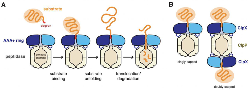
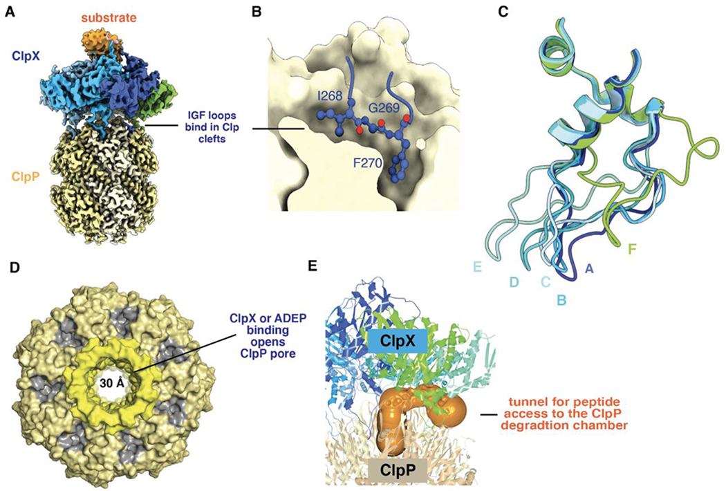
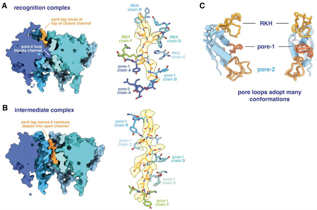
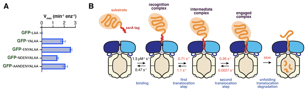
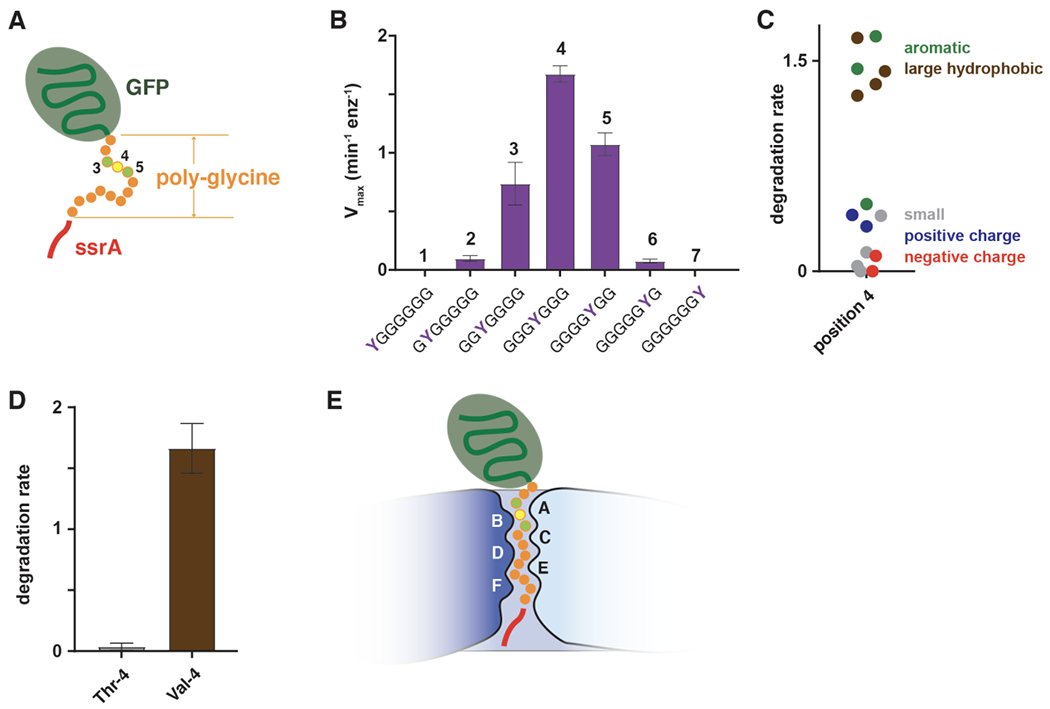
ClpXP is an archetypical AAA+ protease, consisting of ClpX and ClpP. ClpX is an ATP-dependent protein unfoldase and polypeptide translocase, whereas ClpP is a self-compartmentalized peptidase. ClpXP is currently the only AAA+ protease for which high-resolution structures exist, the molecular basis of recognition for a protein substrate is understood, extensive biochemical and genetic analysis have been performed, and single-molecule optical trapping has allowed direct visualization of the kinetics of substrate unfolding and translocation. In this review, we discuss our current understanding of ClpXP structure and function, evaluate competing sequential and probabilistic mechanisms of ATP hydrolysis, and highlight open questions for future exploration.

Keywords: AAA+ proteases; ATP hydrolysis; ClpP; ClpX; cryo-EM; molecular machine; protein degradation.


Conflict of interest statement

Disclosure statement

The authors declare no conflict of interest.

Figures

Figure 1.
Figure 1.
(A) AAA+ proteolytic mechanism. The AAA+ ring hexamer binds a peptide degron in a protein substrate, unfolds any native structure present, and then translocates the denatured polypeptide into a self-compartmentalized peptidase for degradation. (B). For the ClpXP protease, ClpX hexamers can bind to either heptameric ring of the ClpP peptidase.
Figure 2.
Figure 2.
(A) Cartoon depiction of the domain structure and important sequence motifs in a ClpX subunit. (B) ATP (transparent surface) is contacted by box-II, Walker-A, Walker-B, and sensor-II residues from one subunit (light blue) and by the arginine finger of the neighboring subunit (darker blue). (C) Substrate and ClpP binding loops in a large AAA+ domain of a ClpX subunit.
Figure 3.
Figure 3.
(A) Side view of ClpX hexamer (pdb code 6WRF) in cartoon representation. Substrate is colored orange and shown in sphere representation. (B) Top view of 6WRF ClpX hexamer. (C) Diagram showing the relative vertical positions of subunits in the 6WRF ClpX spiral.
Figure 4.
Figure 4.
(A) Nucleotides bound in different subunits of ClpX hexamers (T = ATP/ATPγS; D = ADP; apo = nucleotide free). Subunit names in structure 6VFS were changed to be consistent with the other structures. (B) Cartoon representation of ClpX subunits A and E from structure 6WRF after aligning their small AAA+ domains. A helix consisting of residues 83-100 is colored blue in subunit A and aquamarine in subunit E. (C) Contact map prepared using the https://www.molnac.unisa.it/BioTools/cocomaps/ website for ClpX subunits A & B (pdb code 6WRF). The positions of sequence motifs involved in nucleotide binding/hydrolysis and substrate binding are shown. (D) Top view of ClpX hexamer (pdb code 6WRF) in surface representation with positions of subunit-subunit contacts indicated by spheres in the same color as the subunit surface and positions of nucleotide shown as red spheres. (E) Same view as in panel D, except the surface is colored by rigid-body units (residues 62-314 of one subunit and residues 318-412 of the counter-clockwise subunit) and the hinges (residues 315-318) between the large and small AAA+ subunits of individual subunits are shown as dark spheres.
Figure 5.
Figure 5.
(A) Composite cryo-EM structure of ClpX bound to ClpP and protein substrate (pdb codes 6PPE and 6PP6). (B) An IGF sequence of ClpX binds deeply in a ClpP cleft. (C) IGF loops from aligned ClpX subunits (pdb code 6WRF) adopt a wide variety of conformations with respect to ClpP. This loop flexibility allows ClpX and ClpP to remain stably bound as ClpX adopts different conformations during its ATP-fueled mechanical cycle. (D) Axial view of ClpP (pdb code 6PPE) bound to ClpX (not shown) showing an open axial pore, the clefts that serve as docking sites for the IGF loops of ClpX or ADEPs (colored gray), and the collar of β hairpins that surround the axial pore (colored lighter yellow). (E) The orange surface shows a tunnel, calculated using CAVER (Pavelka et al., 2016), which may allow peptides to enter and leave the ClpP degradation chamber by passing between neighboring IGF loops of ClpX.
Figure 6.
Figure 6.
(A) The left panel shows a cutaway view of the ssrA tag in the recognition complex (pdb code 6WRF), emphasizing the blocked axial channel. The right panel shows the ssrA tag in this complex in stick representation with semi-transparent density, and emphasizes key contacts with specific pore-1, pore-2, or RKH loops of ClpX. (B) The left panel shows a cutaway view of the ssrA tag in the intermediate complex (pdb code 6WSG), highlighting the open ClpX channel and movement of the degron deeper into the axial channel. The right panel shows that five pore-1 loops of ClpX interact with the tag with a two-residue periodicity. (C) The pore loops of ClpX can adopt multiple conformations, facilitating flexible interactions with substrate peptides.
Figure 7.
Figure 7.
(A) Maximal rates of ClpXP degradation of GFP variants with degradation tags derived from the C-terminal 3, 5, 7, 9, or 11 amino acids of the ssrA tag (Fei et al., 2020b). (B) Multistep model for degradation of substrates with degrons of ~20 residues. The rate constants shown were determined for a slowly degraded titinI27 substrate (Saunders et al., 2020). Rate constants colored red become substantially smaller when ATPγS is substituted for ATP.
Figure 8.
Figure 8.
(A) GFP-g13senyalaa contains 13 glycines between the native barrel of GFP and a partial ssrA degron and is not degraded by ClpXP. (B) Effects of tyrosine substitutions at the N-terminal seven positions of the poly-glycine region of GFP-g13senyalaa on the maximal rate of ClpXP degradation (Bell et al., 2019). (C) Effects of position-4 side chain properties on ClpXP degradation (Bell et al., 2019). (D) Degradation of variants with threonine or valine at position 4 of the polyglycine region (Bell et al., 2019). (E) During unfolding, degron side chains 3-5 amino acids from the native domain of a substrate are the most important determinants of grip and are positioned to interact with the pore-1 loops of subunits A and B, near the top of the ClpX channel.

References

    1. Amor AJ, Schmitz KR, Baker TA, Sauer RT. 2019. Roles of the ClpX IGF loops in ClpP association, dissociation, and protein degradation. Protein Sci. 28(4):756–765. - PMC - PubMed
    1. Amor AJ, Schmitz KR, Sello JK, Baker TA, Sauer RT. 2016. Highly dynamic interactions maintain kinetic stability of the ClpXP protease during the ATP-fueled mechanical cycle. ACS Chem Biol. 11(6): 1552–1560. - PMC - PubMed
    1. Aubin-Tam ME, Olivares AO, Sauer RT, Baker TA, Lang MJ. 2011. Single-molecule protein unfolding and translocation by an ATP-fueled proteolytic machine. Cell. 145(2):257–267. - PMC - PubMed
    1. Baker TA, Sauer RT. 2006. ATP-dependent proteases of bacteria: recognition logic and operating principles. Trends Biochem Sci. 31(12):647–653. - PMC - PubMed
    1. Baker TA, Sauer RT. 2012. ClpXP, an ATP-powered unfolding and protein-degradation machine. Biochim Biophys Acta. 1823(1): 15–28. - PMC - PubMed

Publication types

MeSH terms

Substances

LinkOut - more resources