. 2022 Jan-Dec;14(1):2006123.
doi: 10.1080/19420862.2021.2006123.

Phenotypic whole-cell screening identifies a protective carbohydrate epitope on Klebsiella pneumoniae

Affiliations

Phenotypic whole-cell screening identifies a protective carbohydrate epitope on Klebsiella pneumoniae

Sophia K Berry et al. MAbs. 2022 Jan-Dec.

Abstract

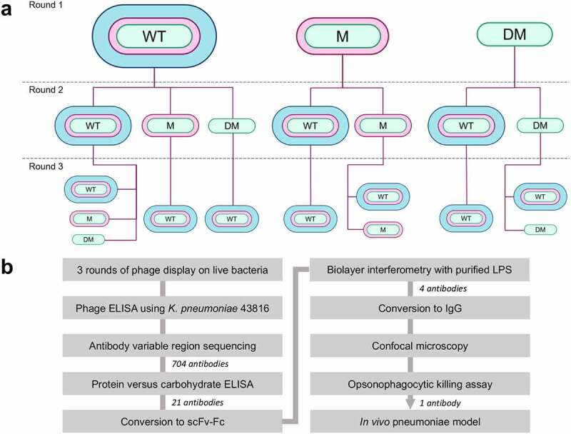
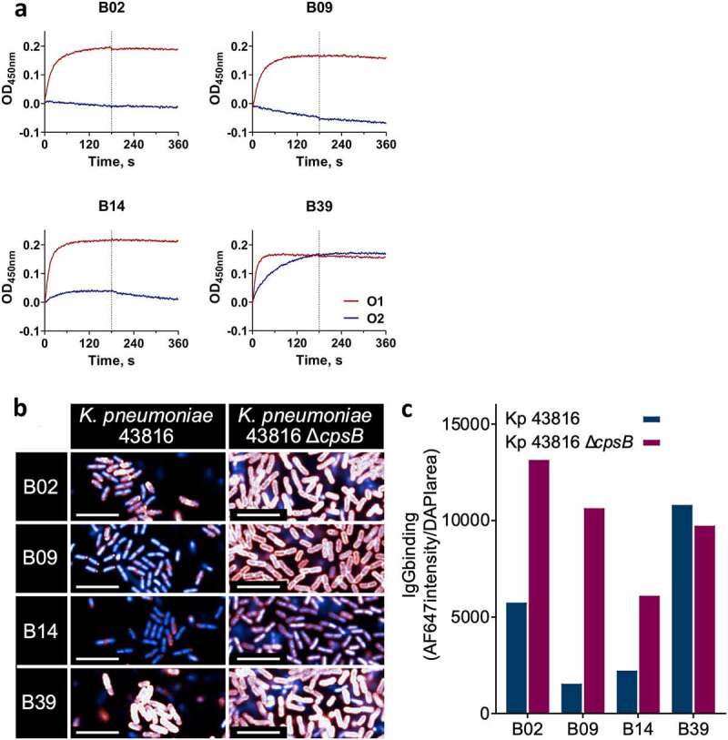
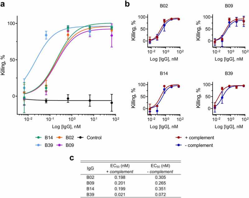
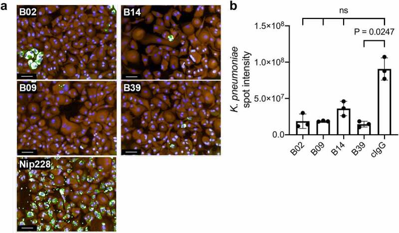
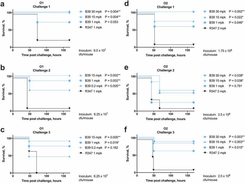
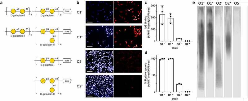
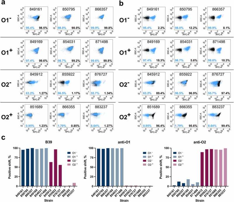
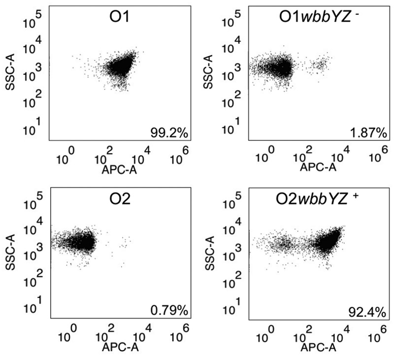
The increasing global occurrence of recalcitrant multi-drug resistant Klebsiella pneumoniae infections warrants the investigation of alternative therapy options, such as the use of monoclonal antibodies (mAbs). We used a target-agnostic phage display approach to K. pneumoniae bacteria lacking bulky, highly variable surface polysaccharides in order to isolate antibodies targeting conserved epitopes among clinically relevant strains. One antibody population contained a high proportion of unique carbohydrate binders, and biolayer interferometry revealed these antibodies bound to lipopolysaccharide (LPS). Antibodies that bound to O1 and O1/O2 LPS were identified. Antibodies were found to promote opsonophagocytic killing by human monocyte-derived macrophages and clearance of macrophage-associated bacteria when assessed using high-content imaging. One antibody, B39, was found to protect mice in a lethal model of K. pneumoniae pneumonia against both O1 and O2 strains when dosed therapeutically. High-content imaging, western blotting and fluorescence-activated cell sorting were used to determine binding to a collection of clinical K. pneumoniae O1 and O2 strains. The data suggests B39 binds to D-galactan-I and D-galactan-II of the LPS of O1 and O2 strains. Thus, we have discovered an mAb with novel binding and functional activity properties that is a promising candidate for development as a novel biotherapeutic for the treatment and prevention of K. pneumoniae infections.

Keywords: Klebsiella pneumoniae; Phage display; antimicrobial resistance; lipopolysaccharide; monoclonal antibody.


Conflict of interest statement

SR, RM, LI, CC, PW, and DT are current/former employees of the AstraZeneca Group and may have/had stock options in AstraZeneca.

Figures

Figure 1.
Figure 1.
Overview of the antibody discovery campaign. a) Flow diagram showing phage display selection campaign. The bacterial strain used at each round of selection is shown. WT (wild type) = K. pneumoniae 43816, M (mutant) = K. pneumoniae 43816 ΔcpsB, DM (double mutant) = K. pneumoniae 43816 ΔcpsBwaaL. b) Flow diagram summarizing the antibody discovery campaign. The number of antibodies triaged at each stage is indicated
Figure 2.
Figure 2.
In vitro characterization of carbohydrate binding antibodies. a) Binding of scFv-Fc to LPS purified from K. pneumoniae 43816 ΔcpsB (O1, red) and K. pneumoniae 8570 ΔcpsB (O2, blue) strains using BLI. Association and dissociation steps were performed for 180 seconds. b) and c) Binding of IgGs to K. pneumoniae 43816 and K. pneumoniae 43816 ΔcpsB. Fixed bacteria were treated with IgGs, then stained with nuclear stain DAPI (blue) and AF647 anti-human IgG (red). At high intensity, IgG binding signal appears white. Images were acquired using an Opera Phenix system (PerkinElmer) at 63x magnification and analyzed in Columbus (PerkinElmer). b. Representative images of IgGs binding to K. pneumoniae 43816 and K. pneumoniae 43816 ΔcpsB. Scale bar represents 10 μm. c IgG binding intensity was calculated by dividing AF647 intensity sum by DAPI area
Figure 3.
Figure 3.
OPK of K. pneumoniae by macrophages in the presence of carbohydrate binding antibodies, measured by release of luciferase. a) Killing of K. pneumoniae 43816 ΔcpsB lux by primary human macrophages in the presence of IgGs. Bacteria, IgGs and complement were added to plates containing macrophages and incubated for 5 hours. Luminescence was measured using an Envision multilabel plate reader (PerkinElmer). Control = negative isotype control. Killing by test IgG or control IgG was calculated as a percentage of wells containing no IgG using the following calculation: (IgG treatment/no IgG)*100. Error bars represent 1 SD. N = 3 individual macrophage donors. b) Killing of K. pneumoniae 43816 ΔcpsB lux by primary human macrophages in the presence of IgGs, with (red) and without (blue) complement. c) EC50 values for IgG treatment in the presence and absence of complement
Figure 4.
Figure 4.
OPK of K. pneumoniae by macrophages in the presence of carbohydrate binding antibodies by HCI. a) Representative images of bacterial clearance after a 7-hour incubation. Fixed and permeabilised cells were treated with rabbit polyclonal anti-K. pneumoniae 43816 and stained with the nuclear stain Hoechst (blue), macrophage stain cell mask Orange (Orange), and AF488 anti-rabbit IgG (green). Images from 15 fields were acquired using an Opera system (PerkinElmer) at 20 x magnification. Scale bar represents 50 μm. b) Quantification of AF488 intensity sum of K. pneumoniae spots in the macrophage cytoplasmic region. Kruskal-Wallis with Dunn’s correction for multiple comparisons test was used; Kruskal-Wallis statistic: 10.63. ns = not significant. cIgG = negative isotype control
Figure 5.
Figure 5.
Therapeutic activity of B39 antibody in a murine model of pneumonia. C57BL/6 mice were infected intranasally with the O1 strain K. pneumoniae 1131115 (a – c) or O2 strain K. pneumoniae 961842 (d – f) at an estimated inoculum size of 6.0 × 107 or 8.0 × 108 colony forming units (cfu) per mouse respectively. IgGs were administered one hour post challenge at various doses as indicated. Actual inoculum size, as indicated on each graph, was determined post challenge by plating serial dilutions. Data from each of three challenge experiments are shown separately. R347 = negative isotype control. A Mantel-Cox test was performed to compare each IgG treatment with R347. P-values were corrected for multiple comparisons using the Benjamini-Hochberg method. P-values: * = P ≤ 0.05; ** = P ≤ 0.005. N = 8 mice per group
Figure 6.
Figure 6.
Characterization of binding to O1 and O2 strains with/without gmlABC locus by HCI and western blot. a) Structures of the O1 and O2 gmlABC ± O-antigens. b) Representative images of B39 binding to each strain. Fixed bacteria were treated with B39, then stained with nuclear stain DAPI (blue, left panel) and AF647 anti-human IgG (red, right panel). 20 fields per well were acquired using an Opera system (PerkinElmer) at 60 x magnification and analyzed in Columbus. Scale bar represents 10 μm. N = 3. c) Quantification of IgG binding intensity. IgG binding intensity was calculated by dividing AF647 intensity sum by DAPI area. d) Quantification of % positive bacteria. % positive bacteria was calculated by dividing total AF647 positive area by DAPI area. e) Western blot analysis of B39 binding to a carbohydrate preparation from each strain. Bacterial whole cell lysates digested with proteinase K were immunoblotted and probed with B39 antibody. A strain with mannose-based O-antigen (O5) was included as a negative control. Strains used were as follows: O1 gmlABC – (O1): K. pneumoniae 43816; O1 gmlABC + (O1+): K. pneumoniae 1131115; O2 gmlABC – (O2): K. pneumoniae 845912; O2 gmlABC + (O2+): K. pneumoniae 961842; O5: K. pneumoniae 9181
Figure 7.
Figure 7.
Characterization of binding to K. pneumoniae O1 and O2 strains with/without the gmlABC locus by FACS. a) Binding of B39 (blue) and an anti-O1 IgG (black) by FACS. b) Binding of B39 (blue) and an anti-O2 IgG (black) by FACS. The % positive-shift in binding for each antibody is labeled by color. The numbers in the top-center of each box are the K. pneumoniae strain name. c) Summary of binding to K. pneumoniae O1 (blue) and O2 (magenta) strains by FACS. O1 = K. pneumoniae O1 gmlABC ; O1+ = K. pneumoniae O1 gmlABC +; O2 = K. pneumoniae O2 gmlABC ; O2 + = K. pneumoniae O2 gmlABC +.
Figure 8.
Figure 8.
Characterization of binding of B39 to K. pneumoniae O1/O2 isogenic mutants by FACS. Binding of B39 to K. pneumoniae 1131115 (O1), K. pneumoniae 961842 (O2), and the isogenic mutants K. pneumoniae 1131115 wbbYZ – (O1 wbbYZ –) and K. pneumoniae 961842 wbbYZ + (O2 wbbYZ +). The % positive-shift in binding for each strain is labeled

Similar articles

Cited by

References

    1. Friedlaender C. Ueber die Schizomyceten bei der acuten fibrösen Pneumonie. Arch für Pathol Anat und Physiol und für Klin Med. 1882;87:319–13.
    1. Podschun R, Ullmann U. Klebsiella spp. as nosocomial pathogens: epidemiology, taxonomy, typing methods, and pathogenicity factors. Clin Microbiol Rev. 1998;11(4):589–603. doi:10.1128/CMR.11.4.589. - DOI - PMC - PubMed
    1. Rock C, Thom KA, Masnick M, Johnson JK, Harris AD, Morgan DJ. Frequency of Klebsiella pneumoniae carbapenemase (KPC)–producing and non-KPC-producing Klebsiella species contamination of healthcare workers and the environment. Infect Control Hosp Epidemiol Off J Soc Hosp Epidemiol Am. 2014;35(4):426. doi:10.1086/675598. - DOI - PMC - PubMed
    1. Russo TA, Marr CM. Hypervirulent Klebsiella pneumoniae. Clin Microbiol Rev [Internet]. 2019;32(3):e00001–19. doi:10.1128/CMR.00001-19. - DOI - PMC - PubMed
    1. Lee C-R, Lee JH, Park KS, Jeon JH, Kim YB, Cha C-J, Jeong BC, Lee SH. Antimicrobial resistance of hypervirulent Klebsiella pneumoniae: epidemiology, hypervirulence-associated determinants, and resistance mechanisms. Front Cell Infect Microbiol. 2017;7:483. doi:10.3389/fcimb.2017.00483. - DOI - PMC - PubMed

Publication types

MeSH terms

LinkOut - more resources