. 2022 May;41(5):641-653.
doi: 10.1016/j.healun.2021.11.012. Epub 2021 Nov 25.

Lung transplant recipients with idiopathic pulmonary fibrosis have impaired alloreactive immune responses

Affiliations

Lung transplant recipients with idiopathic pulmonary fibrosis have impaired alloreactive immune responses

Ping Wang et al. J Heart Lung Transplant. 2022 May.

Abstract

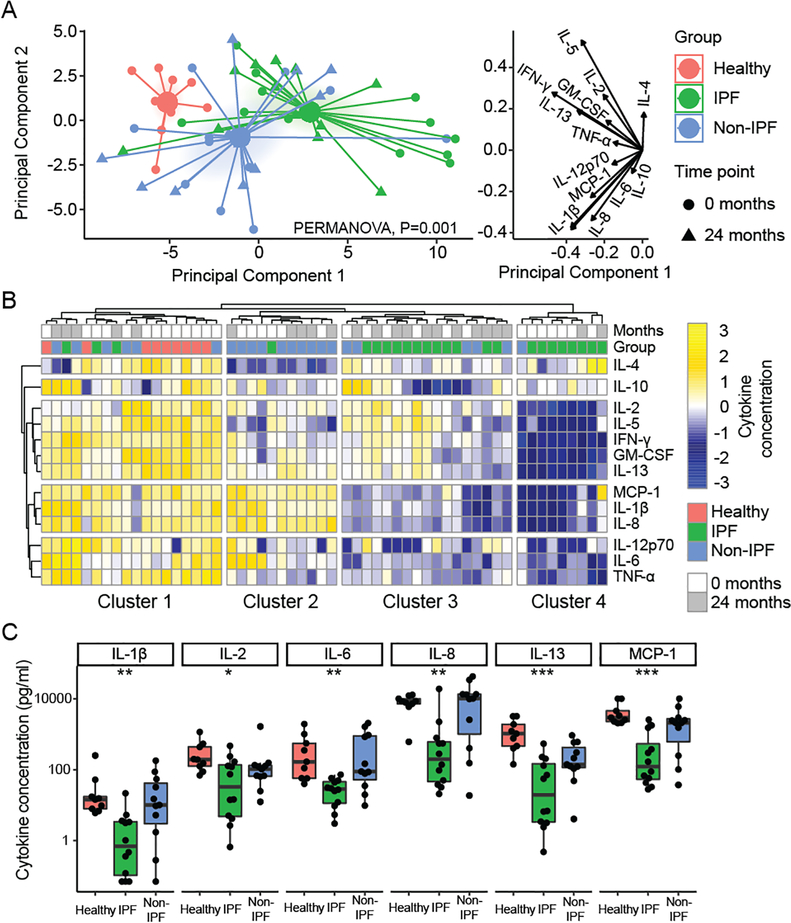
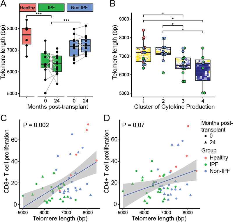
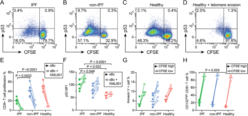
Background: Telomere dysfunction is associated with idiopathic pulmonary fibrosis (IPF) and worse outcomes following lung transplantation. Telomere dysfunction may impair immunity by upregulating p53 and arresting proliferation, but its influence on allograft-specific immune responses is unknown. We hypothesized that subjects undergoing lung transplantation for IPF would have impaired T cell proliferation to donor antigens.

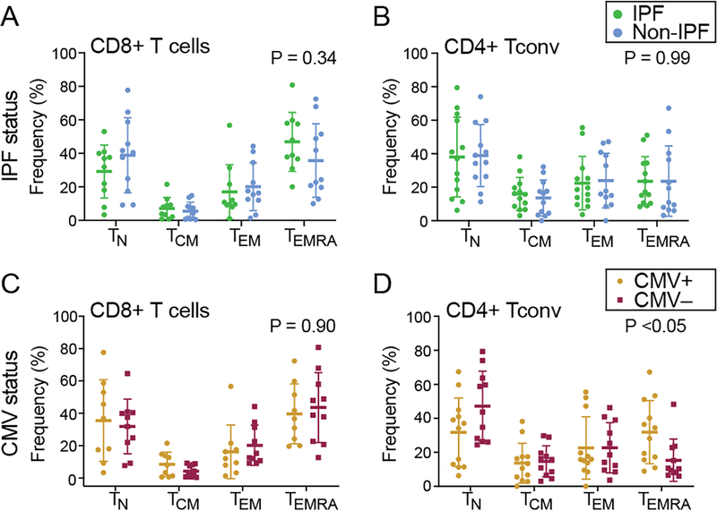
Methods: We analyzed peripheral blood mononuclear cells (PBMC) from 14 IPF lung transplant recipients and 12 age-matched non-IPF subjects, before and 2 years after transplantation, as well as PBMC from 9 non-transplant controls. We quantified T cell proliferation and cytokine secretion to donor antigens. Associations between PBMC telomere length, measured by quantitative PCR, and T cell proliferation to alloantigens were evaluated with generalized estimating equation models.

Results: IPF subjects demonstrated impaired CD8+ T cell proliferation to donor antigens pre-transplant (p < 0.05). IL-2, IL-7, and IL-15 cytokine stimulation restored T cell proliferation, while p53 upregulation blocked proliferation. IPF subjects had shorter PBMC telomere lengths than non-IPF subjects (p < 0.001), and short PBMC telomere length was associated with impaired CD8+ T cell proliferation to alloantigens (p = 0.002).

Conclusions: IPF as an indication for lung transplant is associated with short PBMC telomere length and impaired T cell responses to donor antigens. However, the rescue of proliferation following cytokine exposure suggests that alloimmune anergy could be overcome. Telomere length may inform immunosuppression strategies for IPF recipients.

Keywords: alloimmune response; idiopathic pulmonary fibrosis; immunosenescence; lung transplantation; telomeres.


Figures

Figure 1:
Figure 1:. Idiopathic pulmonary fibrosis (IPF) subjects demonstrate impaired CD8+ T proliferation to alloantigen pre-transplant.
Proliferation to alloantigens was measured by mixed lymphocyte reaction for IPF and non-IPF lung transplant recipients. Responder PBMC were cocultured with matched donor or pooled stimulated B cells (sBc) for 4 days and harvested for flow cytometric analysis and percent proliferation was defined based on the number of cells with ≥2-fold decreased in CFSE staining intensity. (A) Typical flow plots of proliferated CD8+ T cells for IPF and non-IPF lung transplant recipients. The percentage of proliferating (B) CD8+ Tconv and (C) CD4+ T in response to alloantigen were shown. P-values were calculated by two-tailed unpaired t-test. PBMC = peripheral blood mononuclear cells; sBc = stimulated B cells; Tconv = conventional T cells; pooled sBc = pooled sBc mixture from 6–8 donors. The data were shown as mean ± S.D.
Figure 2.
Figure 2.. Proliferation to donor-specific and pooled HLA at 2 years post-transplant.
PBMC from IPF and non-IPF lung transplant recipients 2 years post-transplant were stimulated with donor-derived matched sBc or pooled sBc from multiple donors in a mixed lymphocyte reaction. Proliferation of (A) CD8+ T cells and (B) CD4+ conventional T cells. Differences between IPF and non-IPF groups were assessed using unpaired Student’s t-test. Changes in (C) CD8+ T cells and (D) CD4+ Tconv are shown over time for the two groups in response to stimulation with matched donor-derived sBc. The dashed line highlights a subject who developed CLAD at the 2-year time point. The percentage of proliferating cells pre- and post-transplant were compared by two-tailed paired Student’s t-test.
Figure 3.
Figure 3.. Cytokine stimulation augments alloreactive T cell responses in both groups.
(A) Typical flow plots showed the portion of proliferated CD4+ Tconv and CD8+ T cells following coculture with alloantigens in the presence or absence of IL-2, IL-7 and IL-15. IPF and non-IPF PBMC pre-transplant (B, C) or 2 years post-transplant (D, E) were stimulated with matched donor antigens or pooled sBc in the presence or absence of cytokines. The data are shown as mean with 95% confidential interval (CI), such that confidence interval not crossing 0 implies a statistically significant increase in proliferation with the addition of cytokines. P-values are shown for comparisons between groups with a P < 0.10 by two-tailed Student’s t-test.
Figure 4.
Figure 4.. Alloreactive T cell cytokine profiling demonstrates decreased cytokine production in IPF subjects.
Cytokines were quantified in supernatant following coculture of pre-transplant or 2-year post-transplant recipient T cells with donor lymphocytes, or non-transplant referents with allogenic lymphocytes. (A) Principal component analysis assessed global differences in cytokine production across samples and showed that the groups were distinct across the first component, with IPF being farthest from normal, particularly for the pre-transplant time point. The groups were distinct as assessed by PERMANOVA (P = 0.001). The cytokine concentration value loadings for these principal components are shown on the right. (B) Four clusters were identified by unbiased hierarchical cluster analysis, within which there was distinct segregation of subjects (Fisher exact test, P < 0.001). Cluster 1 included all non-transplant referents and had robust production of most cytokines, while most IPF subjects were in cluster 3 and 4 and showed decreases in production of multiple cytokine groups. (C) Selected cytokines are shown in at the pre-transplant time-point compared across the three groups by Kruskal-Wallis test (*, P < 0.05; **, P < 0.01; ***, P < 0.001).
Figure 5.
Figure 5.. Similar T cell memory phenotype distributions between IPF and non-IPF subjects.
The memory phenotypes distribution for pre-transplant PBMC in the absence of alloantigen stimulation were analyzed by flow cytometry. The expression of CD45RO and CCR7 were analyzed for IPF and non-IPF PBMC (A-B) and CMV positive or negative recipients PBMC (C-D). Results for naïve T cells (TN: CCR7+CD45RO−), central memory (TCM: CCR7+CD45RO+), effector memory (TEM: CCR7-CD45RO+), and terminally differentiated effector memory (TEMRA: CCR7-CD45RO-) subsets were quantified. P-values shown are for the interaction between cell type and IPF status or CMV status by two-way ANOVA. The data are shown as mean ± S.D.
Figure 6.
Figure 6.. Short peripheral blood telomere length is associated with impaired alloimmune responses.
Telomere length was measured in PBMC DNA using quantitative PCR and shown for IPF, non-IPF, and non-transplant referents (healthy) both pre-transplant and at 2 years post-transplant (A). Telomere length was shorter in IPF subjects compared with non-IPF and non-transplant referents as assessed by unadjusted GEE models (P < 0.001). For Non-IPF versus healthy, we observed P = 0.09. (B) Telomere length measurements are shown stratified by cytokine profile cluster, as identified in Figure 4. After multiple comparison adjustments clusters 3 and 4, with impaired cytokine production, were found to have shorter telomere lengths than the clusters 1 and 2. Percent proliferation to matched or single donor alloantigens is shown versus telomere length for CD8+ T cells (C) and CD4+ Tconv (D). P-values represent statistical significance for the association of proliferation with telomere length as assessed by unadjusted GEE models.
Figure 7.
Figure 7.. P53 upregulation is associated with impaired CD8+ T cell proliferation to alloantigens in IPF.
Mixed lymphocyte reactions were performed to detect p53 expression in CD8+ T cells following stimulation with pooled alloantigens. Representative flow plots show p53 expression versus proliferation as measured by CFSE dilution in cells from subjects at 18 months post-transplant for (A) IPF or (B) other indications. P53 thresholds were set based on isotype control staining in the CFSElow population. Responses in healthy referents are shown in (C), as well as responses from healthy subject PBMC after treatment with KML001, a reagent that binds and erodes telomeres (D). (E) CD8+ T cell proliferation was less in IPF subjects than in healthy controls (P <0.0001) or non-IPF subjects (P = 0.0002). KML001 treatment resulted in reduced proliferation for all groups (P <0.0001). (F) Median fluorescence intensity (MFI) for P53 after subtraction of isotype control MFI is shown for non-responsive (CFSEhigh) CD8+ T cells with and without KML001. KML001 treatment resulted in increased p53 levels (P <0.0001), and cells from IPF subjects had increased p53 levels versus non-IPF (P=0.049) and healthy (P=0.002) groups. (G) Annexin V staining, a marker of apoptosis, was increased in proliferated CD8+ T cells versus unstimulated cells (P <0.0001) but not different between groups. (H) The common γ-chain receptor (CD132) was present on 99% of cells, but upregulated in following alloantigen stimulation (P <0.0001). CD132high cells were more common in non-responding (CFSElow) CD8+ T cells from IPF subjects (P = 0.005) as compared with healthy referents. Statistical comparisons were performed using 2-way ANOVA with Dunnett’s post-test.

References

    1. Lederer DJ, Martinez FJ. Idiopathic Pulmonary Fibrosis. N Engl J Med 2018;378:1811–23. - PubMed
    1. Richeldi L, Collard HR, Jones MG. Idiopathic pulmonary fibrosis. Lancet 2017;389:1941–52. - PubMed
    1. George PM, Patterson CM, Reed AK, Thillai M. Lung transplantation for idiopathic pulmonary fibrosis. Lancet Respir Med 2019;7:271–82. - PubMed
    1. Blackburn EH, Epel ES, Lin J. Human telomere biology: A contributory and interactive factor in aging, disease risks, and protection. Science 2015;350:1193–8. - PubMed
    1. Srinivas N, Rachakonda S, Kumar R. Telomeres and Telomere Length: A General Overview. Cancers (Basel) 2020;12. - PMC - PubMed

Publication types