. 2021 Dec 11:14:6799-6812.
doi: 10.2147/JIR.S329020. eCollection 2021.

Melatonin Alleviates Age-Associated Endothelial Injury of Atherosclerosis via Regulating Telomere Function

Affiliations

Melatonin Alleviates Age-Associated Endothelial Injury of Atherosclerosis via Regulating Telomere Function

Yinghua Xie et al. J Inflamm Res. .

Abstract

Background: Atherosclerosis is an aging-related disease, partly attributed to telomerase dysfunction. This study aims to investigate whether telomere dysfunction-related vascular aging is involved in the protection mechanism of melatonin (MLT) in atherosclerosis.

Methods: Young and aged ApoE-/- mice were used to establish atherosclerotic mice model. H&E staining and immunofluorescence assay were performed to detect endothelial cell injury and apoptosis. Inflammatory cytokines and oxidative stress-related factors were determined using corresponding commercial assay kits. Telomerase activity was detected by TRAP assay, and SA-β-gal staining was conducted to evaluate cellular senescence. HUVECs were treated with H2O2 for 1 h to induce senescence. Western blot was performed to measure protein expression.

Results: An obvious vascular endothelial injury, reflected by excessive production of inflammatory cytokines, elevated ROS, MDA and SOD levels, and more apoptotic endothelial cells, was found in atherosclerotic mice, especially in aged mice, which were then greatly suppressed by MLT. In addition, telomere dysfunction and senescence occurred in atherosclerosis, especially in aged mice, while MLT significantly alleviated the conditions. CYP1A1, one of the targeted genes of MLT, was verified to be upregulated in atherosclerotic mice but downregulated by MLT. Furthermore, H2O2 induced a senescence model in HUVECs, which was accompanied with a remarkably increased cell viability loss and apoptosis rate, and a downregulated telomerase activity of HUVECs, and this phenomenon was strengthened by RHPS4, an inhibitor of telomerase activity. However, MLT could partly abolish these changes in H2O2- and RHPS4-treated HUVECs, demonstrating that MLT alleviated vascular endothelial injury by regulating senescence and telomerase activity.

Conclusions: Collectively, this study provided evidence for the protective role of MLT in atherosclerosis through regulating telomere dysfunction-related vascular aging.

Keywords: atherosclerosis; melatonin; senescence; telomere; vascular aging.


Conflict of interest statement

The authors declare that they have no known competing financial interests or personal relationships that could have appeared to influence the work reported in this paper.

Figures

Figure 1
Figure 1
MLT alleviates vascular aging-related endothelial injury in ApoE−/− mice. Young-male ApoE−/− mice and aged-male ApoE−/− mice were received high fat diet (HFD) to induce atherosclerosis and intraperitoneally injected with melatonin (MLT; 20 mg/kg/d) for treatment. (A) H&E staining of intima of aorta, magnification×200. (B) TUNEL staining of intima of aorta, magnification×200. (C) Western blot and statistical analysis of protein expression of Bax, Bcl-2 and cleaved caspase3. N=3. *, **, ***p<0.05, 0.01, 0.001.
Figure 2
Figure 2
MLT alleviates vascular aging-related inflammatory response and oxidative stress in ApoE−/− mice. Young-male ApoE−/− mice and aged-male ApoE−/− mice were received high fat diet (HFD) to induce atherosclerosis and intraperitoneally injected with melatonin (MLT; 20 mg/kg/d) for treatment. (A) ELISA assay on the pro-inflammatory cytokines, including IL-6, IL-1β and TNF-α in blood. (B) ELISA analysis on the pro-inflammatory cytokines, including IL-6, IL-1β and TNF-α in blood vessel tissues. (C and D) The level of ROS in blood vessel tissues was detected using its assay kit. (E and F) The level of MDA and SOD in blood vessel tissues was detected using their commercial kits. N=3. *, **, ***p<0.05, 0.01, 0.001.
Figure 3
Figure 3
MLT attenuates senescence and regulates telomere-telomerase function in ApoE−/− mice. Young-male ApoE−/− mice and aged-male ApoE−/− mice were received high fat diet (HFD) to induce atherosclerosis and intraperitoneally injected with melatonin (MLT; 20 mg/kg/d) for treatment. (A) CYP1A1 activity was detected using P450-GloTM CYP1A1 Assay kit. (B) ELISA analysis on the level of 15-HETE. (C) Telomere length measurements were performed by Southern blot analysis of the terminal restriction fragments. (D) Telomerase activity was detected by TRAP assay. (E and F) SA-β-gal Staining Kit was used to detect SA-β-gal level. (G) Western blot and statistical analysis of protein expression of cyclin D1, CDK2, p16 and p21. N=3. *, **, ***p<0.05, 0.01, 0.001.
Figure 4
Figure 4
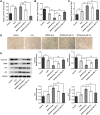
MLT alleviates cell apoptosis in HUVECs treated by H2O2 and RHPS4 HUVECs were treated with RHPS4, an inhibitor of telomerase activity, along with or without MLT (100 μM (MLT-L) and 500 μM (MLT-H)), 30 min prior to H2O2 induction. (A and B) cell viability was detected using MTT assay. (C) Flow cytometry assay and statistical assay on cell apoptosis. (D) Western blot and statistical analysis of protein expression of Bax, Bcl-2 and cleaved caspase3. N=3. **, ***p<0.01, 0.001.
Figure 5
Figure 5
MLT inhibits senescence and improves telomerase activity in HUVECs treated by H2O2 and RHPS4 HUVECs were treated with RHPS4, an inhibitor of telomerase activity, along with or without MLT (100 μM (MLT-L) and 500 μM (MLT-H)), 30 min prior to H2O2 induction. (A) CYP1A1 activity was detected using P450-GloTM CYP1A1 Assay kit. (B) Telomerase activity was detected by TRAP assay. (C and D) SA-β-gal Staining Kit was used to detect SA-β-gal level. (E) Western blot and statistical analysis of protein expression of cyclin D1, CDK2, p16 and p21. N=3. *, **, ***p<0.05, 0.01, 0.001.

References

    1. Bentzon JF, Otsuka F, Virmani R, Falk E. Mechanisms of plaque formation and rupture. Circ Res. 2014;114(12):1852–1866. doi:10.1161/CIRCRESAHA.114.302721 - DOI - PubMed
    1. Liu Y, Chen KJ. Atherosclerosis, vascular aging and therapeutic strategies. Chin J Integr Med. 2012;18(2):83–87. doi:10.1007/s11655-012-0996-z - DOI - PubMed
    1. Perk J, De Backer G, Gohlke H, et al. European Guidelines on cardiovascular disease prevention in clinical practice (version 2012). The Fifth Joint Task Force of the European Society of Cardiology and Other Societies on Cardiovascular Disease Prevention in Clinical Practice (constituted by representatives of nine societies and by invited experts). Eur Heart J. 2012;33(13):1635–1701. doi:10.1093/eurheartj/ehs092 - DOI - PubMed
    1. Benjamin EJ, Virani SS, Callaway CW, et al. Heart disease and stroke statistics-2018 update: a report from the American Heart Association. Circulation. 2018;137(12):e67–e492. doi:10.1161/CIR.0000000000000558 - DOI - PubMed
    1. Sturlaugsdottir R, Aspelund T, Bjornsdottir G, et al. Prevalence and determinants of carotid plaque in the cross-sectional REFINE-Reykjavik study. BMJ Open. 2016;6(11):e012457. doi:10.1136/bmjopen-2016-012457 - DOI - PMC - PubMed