. 2021 Dec 1:12:761922.
doi: 10.3389/fphar.2021.761922. eCollection 2021.

Physalin A Inhibits MAPK and NF-κB Signal Transduction Through Integrin αVβ3 and Exerts Chondroprotective Effect

Affiliations

Physalin A Inhibits MAPK and NF-κB Signal Transduction Through Integrin αVβ3 and Exerts Chondroprotective Effect

Rui Lu et al. Front Pharmacol. .

Abstract

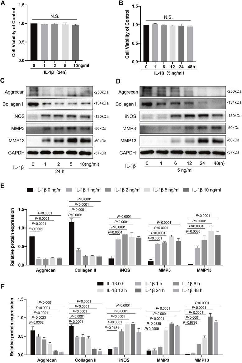
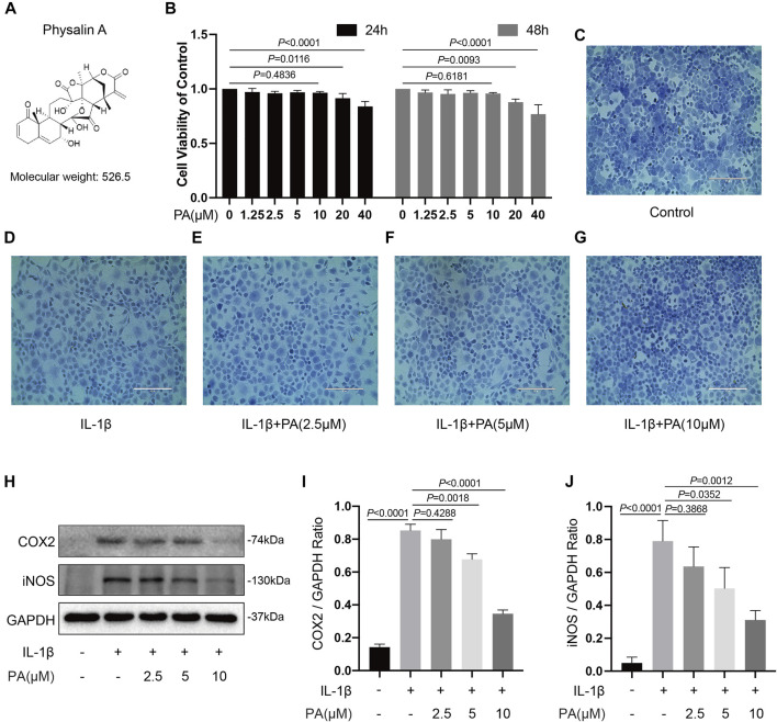
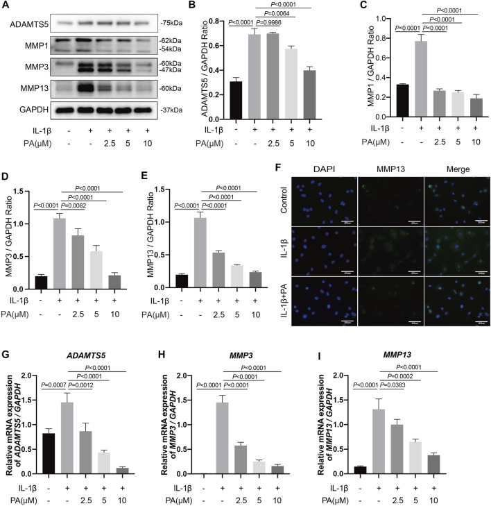
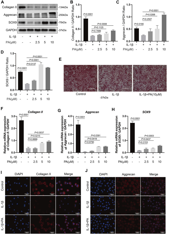
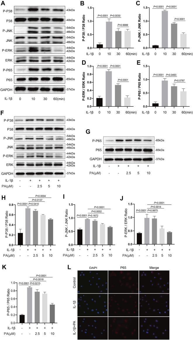
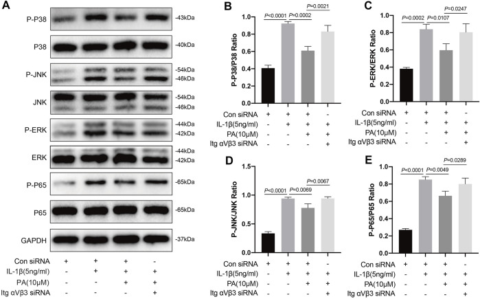
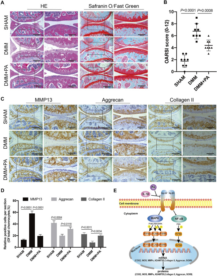
Osteoarthritis (OA) is a common articular ailment presented with cartilage loss and destruction that is common observed in the elderly population. Physalin A (PA), a natural bioactive withanolide, exerts anti-inflammatory residences in more than a few diseases; however, little is known about its efficacy for OA treatment. Here, we explored the therapeutic effects and potential mechanism of PA in mouse OA. After the in vitro administration of PA, the expression of inflammation indicators including inducible nitric oxide synthase and cyclooxygenase-2 was low, indicating that PA could alleviate the IL-1β-induced chondrocyte inflammation response. Moreover, PA reduced IL-1β-induced destruction of the extracellular matrix by upregulating the gene expression of anabolism factors, including collagen II, aggrecan, and sry-box transcription factor 9, and downregulating the gene expression of catabolic factors, including thrombospondin motif 5 and matrix metalloproteinases. In addition, the chondroprotective effect of PA was credited to the inhibition of mitogen-activated protein kinase (MAPK) and nuclear factor-κB (NF-κB) signaling pathways. Furthermore, in vivo experiments showed that intra-articular injection of PA could alleviate cartilage destruction in a mouse OA model. However, the anti-inflammatory, anabolism enhancing, catabolism inhibiting, and MAPK and NF-κB signaling pathway inhibiting properties of PA on IL-1β-induced chondrocytes could be reversed when integrin αVβ3 is knocked down by siRNA. In conclusion, our work demonstrates that PA exhibits a chondroprotective effect that may be mediated by integrin αVβ3. Thus, PA or integrin αVβ3 might be a promising agent or molecular target for the treatment of OA.

Keywords: MAPK; NF-κB; integrin; osteoarthritis; physalin A; αvβ3.


Conflict of interest statement

The authors declare that the research was conducted in the absence of any commercial or financial relationships that could be construed as a potential conflict of interest.

Figures

FIGURE 1
FIGURE 1
Effects of IL-1β on knee chondrocytes in mice. (A) Mice chondrocytes were treated with various concentration gradients of IL-1β (0, 1, 2, 5, and 10 ng/ml) for 24 h, (B) or with IL-1β at the concentration of 5 ng/ml for different time points (0, 1, 6, 12, 24, and 48 h), and cell viability was detected with a CCK-8 kit. (C) Western blots and (E) quantitative analysis of aggrecan, collagen II, iNOS, MMP3, and MMP13 for chondrocytes exposed to different concentrations of IL-1β (0, 1, 2, 5, and 10 ng/ml) for 24 h. (D) Western blotting results and (F) quantitative analysis of aggrecan, collagen II, iNOS, MMP3, and MMP13 for chondrocytes stimulated with IL-1β (5 ng/ml) at several time points (0, 1, 6, 12, 24, and 48 h). GAPDH was used as an internal reference. Data are presented as means ± SD (n = 3). The exact p value was marked in the corresponding figure, N.S. indicated no significance, and p < 0.05 was considered statistically significant.
FIGURE 2
FIGURE 2
Effects of PA on cell viability and PA suppressed inflammatory responses induced by IL-1β. (A) Molecular structure of PA. (B) Mice chondrocytes were treated with PA (0, 1.25, 2.5, 5, 10, 20, and 40 μM) for 24 and 48 h, and cell viability was detected with a CCK-8 kit. (C–G) Toluidine blue staining of chondrocytes treated with IL-1β (5 ng/ml) and PA for 24 h (scale bar 200 μm). (H) Western blotting results and (I,J) quantitative analysis of COX2 and iNOS in IL-1β–induced chondrocytes treated with PA. GAPDH was used as an internal reference. Data are presented as means ± SD (n = 3). The exact p value was marked in the corresponding figure and p < 0.05 was considered statistically significant.
FIGURE 3
FIGURE 3
PA suppressed excess expression of the catabolic indicators of chondrocytes induced by IL-1β, including ADAMTS5, MMP1, MMP3, and MMP13. Mice chondrocytes were treated with 5 ng/ml of IL-1β, alone or with PA (2.5, 5, and 10 μM) for 24 h (A) Western blotting results and (B–E) quantitative analysis of ADAMTS5, MMP1, MMP3, and MMP13. (F) MMP13 expression was observed by immunofluorescence staining when chondrocytes were treated with 5 ng/ml of IL-1β, alone or with 10 μM of PA (scale bar 200 μm). (G–I) Relative mRNA levels of ADAMTS5, MMP3, and MMP13 in chondrocytes stimulated with 5 ng/ml of IL-1β, alone or with PA (2.5, 5, and 10 μM) for 24 h. GAPDH was used as an internal reference. Data are presented as means ± SD (n = 3). The exact p value was marked in the corresponding figure and p < 0.05 was considered statistically significant.
FIGURE 4
FIGURE 4
PA upregulated anabolic indicators including collagen II, aggrecan, and SOX9 expression in IL-1β-induced chondrocytes. (A) Western blotting results and (B–D) quantitative analysis of collagen II, aggrecan, and SOX9. (E) Safranin O staining reflected the content of proteoglycan among the control, IL-1β (5 ng/ml), and IL-1β (5 ng/ml) + PA (10 μM) groups (scale bar 200 μm). (F–H) Relative mRNA levels of collagen II, aggrecan, and SOX9 in chondrocytes stimulated with 5 ng/ml of IL-1β, alone or with PA (2.5, 5, and 10 μM) for 24 h. GAPDH was used as an internal reference. Data are presented as means ± SD (n = 3). The exact p value was marked in the corresponding figure and p < 0.05 was considered statistically significant. (I,J) Collagen II and aggrecan expression observed by immunofluorescence staining when chondrocytes were treated with 5 ng/ml of IL-1β, alone or with 10 μM of PA (scale bar 200 μm).
FIGURE 5
FIGURE 5
PA inhibited the IL-1β-induced activation of MAPK and NF-κB signaling pathways. (A) Western blots and (B–E) quantitative analysis of MAPK pathway related proteins (P-P38, P38, P-JNK, JNK, P-ERK, and ERK) and NF-κB pathway related proteins (P-P65, P65) in IL-1β-induced chondrocytes at the time points (0, 10, 30, and 60 min). (F,G) Western blotting results and (H–K) quantification analysis of MAPK pathway related proteins (P-P38, P38, P-JNK, JNK, P-ERK, and ERK) and NF-κB pathway related proteins (P-P65, P65) in chondrocytes pretreated with the administration of PA (2.5, 5, and 10 μM) for 24 h and followed the stimulation of 5 ng/ml of IL-1β for 10 min. (L) Nuclear translocation of P65 was detected by immunofluorescence staining after chondrocytes pretreated with the administration of PA (10 μM) for 24 h and followed the stimulation of 5 ng/ml of IL-1β for 10 min (scale bar 200 μm). Non-phosphorylated protein (P38, JNK, ERK, and P65) was used as an internal control. Data are presented as means ± SD (n = 3) and the exact p value was marked in the corresponding figure and p < 0.05 was considered statistically significant.
FIGURE 6
FIGURE 6
Knockdown of integrin αVβ3 weakened the anti-inflammatory, anabolism enhancing, and catabolism inhibiting effect of PA on IL-1β-induced chondrocytes. (A,B) Relative mRNA levels of integrin αV (Itg αV) and integrin β3 (Itg β3) in chondrocytes stimulated with 5 ng/ml of IL-1β, alone or with PA (2.5, 5, and 10 μM) for 24 h (C,D) Itg αV and Itg β3 were knocked down by siRNA transfection, and the knockdown efficiency was verified by RT-PCR. (E,F) Inflammatory markers (COX2, iNOS) were detected by western blotting and the band density of protein levels were quantified after mice chondrocytes were added with or without 5 ng/ml of IL-1β, 10 μM of PA, and Itg αVβ3 siRNA. (G–I) Western blotting was applied to measure the anabolic (aggrecan, collagen II) and catabolic markers (MMP1, MMP3, MMP13, and ADAMTS5) in the Itg αVβ3-deficiency mice chondrocytes along with or without the administration of 5 ng/ml of IL-1β and 10 μM of PA, and the band density of these protein levels were quantified in the histogram. GAPDH was used as an internal reference. Data are presented as means ± SD (n = 3). The exact p value was marked in the corresponding figure and p < 0.05 was considered statistically significant.
FIGURE 7
FIGURE 7
Knockdown of integrin αVβ3 reversed the inhibition of MAPK and NF-κB signaling pathways by PA. (A) Western blotting results and (B–E) quantitative analysis of MAPK pathway related proteins (P-P38, P38, P-JNK, JNK, P-ERK, and ERK) and NF-κB pathway related proteins (P-P65, P65) from the Itg αVβ3-deficiency mice chondrocytes along with or without the administration of 5 ng/ml of IL-1β and 10 μM of PA. The non-phosphorylated protein (P38, JNK, ERK, and P65) was used as an internal control. Data are presented as means ± SD (n = 3) The exact p value was marked in the corresponding figure and p < 0.05 was considered statistically significant.
FIGURE 8
FIGURE 8
PA attenuated cartilage destruction in the in vivo mouse OA model. (A) HE, Safranin O, and Fast Green staining and (B) OARSI scores of mice knee joints from the sham, DMM, and DMM + PA groups at 8 weeks after the corresponding treatment (scale bars, 200 and 400 μm). (C) Immunohistochemistry staining and (D) quantification of the expression of MMP13, aggrecan, collagen II were measured among the three groups (scale bars 200 and 400 μm). Data are presented as means ± SD (n = 6). The exact p value was marked in the corresponding figure and p < 0.05 was considered statistically significant. (E) The potential molecular mechanism of PA’s chondroprotective effect. PA could alleviate the inflammatory response and cartilage degradation of IL-1β-induced chondrocytes by inhibting the MAPK and NF-κB signaling pathways, and the beneficial effect of PA on OA may be mediated through integrin αVβ3.

References

    1. Appleton C. T. (2018). Osteoarthritis Year in Review 2017: Biology. Osteoarthritis Cartilage 26 (3), 296–303. 10.1016/j.joca.2017.10.008 - DOI - PubMed
    1. Attur M. G., Dave M. N., Clancy R. M., Patel I. R., Abramson S. B., Amin A. R. (2000). Functional Genomic Analysis in Arthritis-Affected Cartilage: Yin-Yang Regulation of Inflammatory Mediators by Alpha 5 Beta 1 and Alpha V Beta 3 Integrins. J. Immunol. 164 (5), 2684–2691. 10.4049/jimmunol.164.5.2684 - DOI - PubMed
    1. Buckwalter J. A., Mankin H. J., Grodzinsky A. J. (2005). Articular Cartilage and Osteoarthritis. Instr. Course Lect 54, 465–480. - PubMed
    1. Chen D. H., Zheng G., Zhong X. Y., Lin Z. H., Yang S. W., Liu H. X., et al. (2021). Oroxylin A Attenuates Osteoarthritis Progression by Dual Inhibition of Cell Inflammation and Hypertrophy. Food Funct. 12 (1), 328–339. 10.1039/d0fo02159h - DOI - PubMed
    1. Cheng C., Tian J., Zhang F., Deng Z., Tu M., Li L., et al. (2021). WISP1 Protects against Chondrocyte Senescence and Apoptosis by Regulating αvβ3 and PI3K/Akt Pathway in Osteoarthritis. DNA Cel Biol 40 (4), 629–637. 10.1089/dna.2020.5926 - DOI - PubMed