Randomized Controlled Trial
. 2021 Dec 1:12:772555.
doi: 10.3389/fimmu.2021.772555. eCollection 2021.

Transcutaneous Vagal Nerve Stimulation Alone or in Combination With Radiotherapy Stimulates Lung Tumor Infiltrating Lymphocytes But Fails to Suppress Tumor Growth

Affiliations
Randomized Controlled Trial

Transcutaneous Vagal Nerve Stimulation Alone or in Combination With Radiotherapy Stimulates Lung Tumor Infiltrating Lymphocytes But Fails to Suppress Tumor Growth

Eva Reijmen et al. Front Immunol. .

Abstract

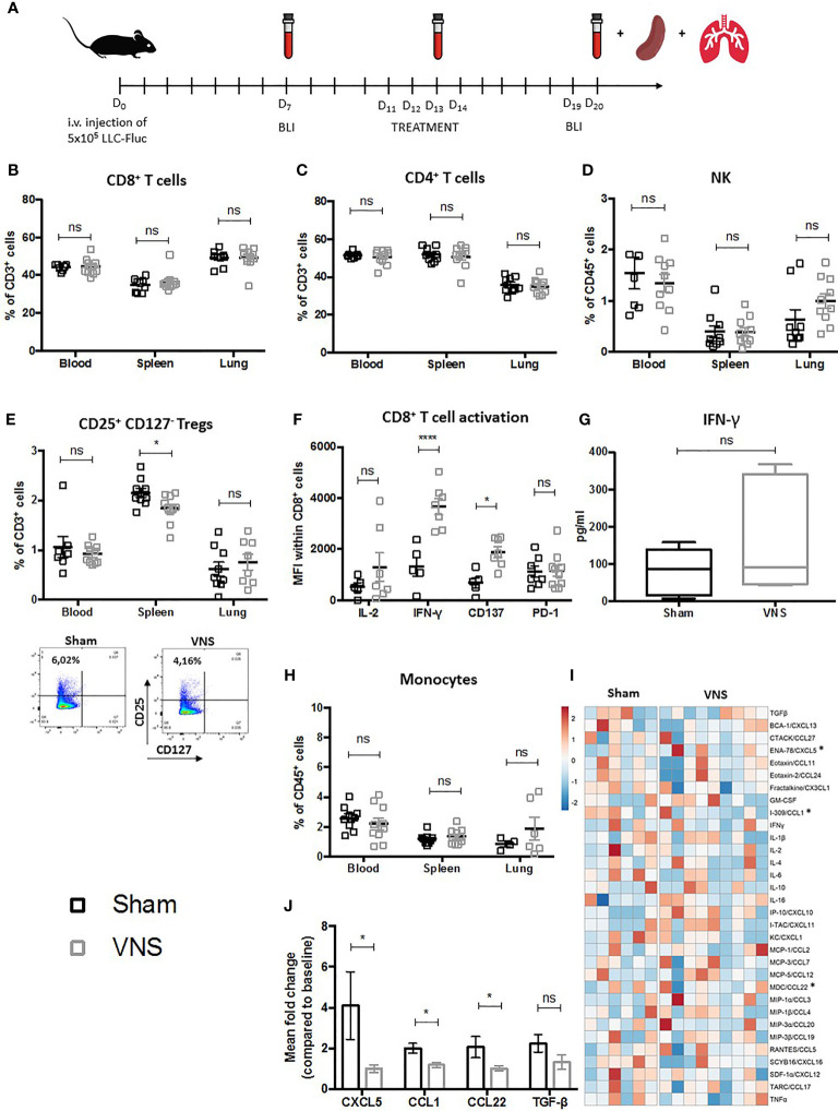
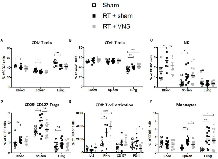
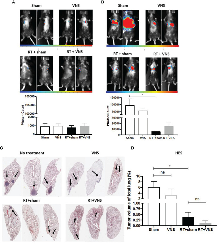
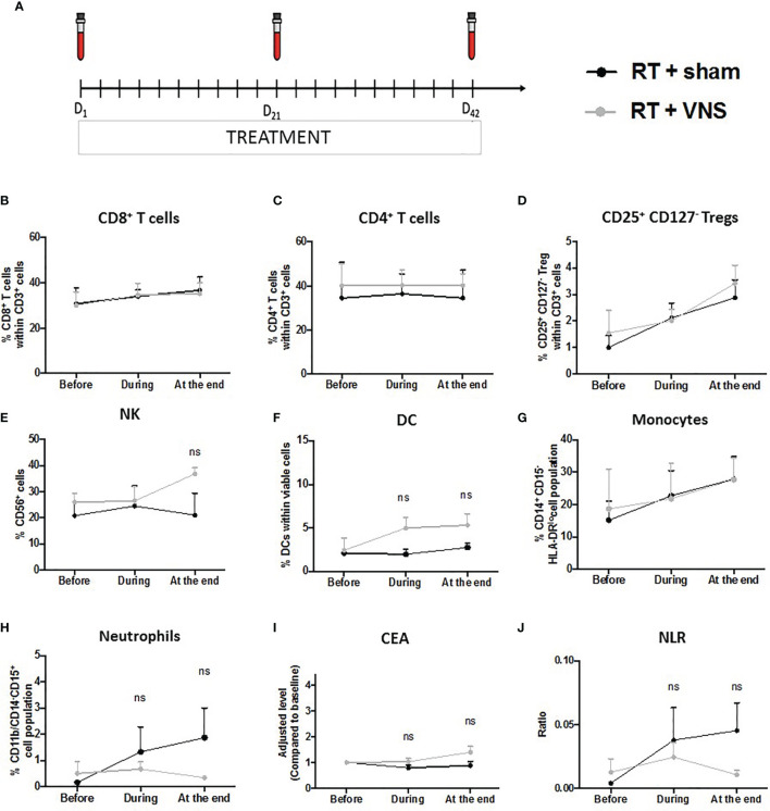
The combination of radiotherapy (RT) with immunotherapy represents a promising treatment modality for non-small cell lung cancer (NSCLC) patients. As only a minority of patients shows a persistent response today, a spacious optimization window remains to be explored. Previously we showed that fractionated RT can induce a local immunosuppressive profile. Based on the evolving concept of an immunomodulatory role for vagal nerve stimulation (VNS), we tested its therapeutic and immunological effects alone and in combination with fractionated RT in a preclinical-translational study. Lewis lung carcinoma-bearing C57Bl/6 mice were treated with VNS, fractionated RT or the combination while a patient cohort with locally advanced NSCLC receiving concurrent radiochemotherapy (ccRTCT) was enrolled in a clinical trial to receive either sham or effective VNS daily during their 6 weeks of ccRTCT treatment. Preclinically, VNS alone or with RT showed no therapeutic effect yet VNS alone significantly enhanced the activation profile of intratumoral CD8+ T cells by upregulating their IFN-γ and CD137 expression. In the periphery, VNS reduced the RT-mediated rise of splenic, but not blood-derived, regulatory T cells (Treg) and monocytes. In accordance, the serological levels of protumoral CXCL5 next to two Treg-attracting chemokines CCL1 and CCL22 were reduced upon VNS monotherapy. In line with our preclinical findings on the lack of immunological changes in blood circulating immune cells upon VNS, immune monitoring of the peripheral blood of VNS treated NSCLC patients (n=7) did not show any significant changes compared to ccRTCT alone. As our preclinical data do suggest that VNS intensifies the stimulatory profile of the tumor infiltrated CD8+ T cells, this favors further research into non-invasive VNS to optimize current response rates to RT-immunotherapy in lung cancer patients.

Keywords: immunosuppressive tumor microenvironment (TME); lung cancer; neuromodulation; radiotherapy; transcutaneous vagal nerve stimulation; tumor infiltrating lymphocytes.


Conflict of interest statement

The authors declare that the research was conducted in the absence of any commercial or financial relationships that could be construed as a potential conflict of interest.

Figures

Figure 1
Figure 1
Schematic representation of preclinical experimental setup and evaluation of immune cell populations in lung tumor bearing mice treated with VNS. (A) C57BL/6 mice were i.v. injected with 5x10⁵ Fluc expressing LLC tumor cells. Seven days later in vivo BLI was performed to randomize mice with similar photon counts to the VNS treated, RT treated, RT+VNS treated or sham treated control group. Mice were daily treated with VNS (25Hz) and/or RT (3,2 Gy) for four consecutive days starting at day eleven after tumor injection. Blood samples were collected at day 7 (baseline), day 13 and day 20. On day 20 after tumor injection, mice were euthanized to collect perfused lungs and spleens. (B–E, H) Flow cytometric analysis of (B) CD8+ T cells, (C) CD4+ T cells, (D) CD56+ NK cells, (E) CD4+CD25+CD127- Tregs with on the right a representative dot plot from splenic CD4+CD25+CD127- Tregs from a sham or VNS treated animal and (H) CD11b+Ly6C+ monocytes in blood, spleen and lung TME. (F, G) Cell suspensions derived from murine lung tissue were co-cultured with LLC cells at a 10:1 ratio for 24 hours. Next, expression of IL-2, IFN-γ, CD137 (4-1BB) and PD-1 was assessed on the CD8+ T cells specifically using flow cytometry (F) and IFN-γ secretion was evaluated via a mouse IFN-γ ELISA kit (G). (I, J) Data obtained from a 31-plex cytokine array on plasma samples obtained from VNS or sham treated mice. (I) Heatmap of the 31 different cytokines, in which the meanfold change of each cytokine concentration compared to baseline is visually represented with red, white, and blue to indicate high, median, and low, respectively. (J) Box plots show the significant fold changes of ENA-78 (CXCL5), I-309 (CCL1), MDC (CCL22) next to the non-significant decrease of TGF-β in the same plasma samples. Data represent three pooled experiments six days after the last treatment. Data are shown as mean ± SD of n>9 (VNS) and n>6 (sham). *P < 0,0332; ****0,0001 by two-tailed unpaired t-tests. ns, non significant.
Figure 2
Figure 2
Evaluation of immune cell populations in lung tumor bearing mice treated with RT+VNS. Flow cytometric analysis of lymphoid (A) CD8+ T cells, (B) CD4+ T cells, (C) CD56+ NK cells, (D) CD4+CD25+CD127- Tregs in blood spleen and lung TME. (E) Cell suspensions from murine lung tissue were co-cultured with LLC cells at ratio 10:1 in vitro. After 24 hours, CD8+ TIL expression of the function markers IL-2, IFN-γ, CD137 (4-1BB) and PD-1 was assessed via flow cytometry. (F) Flow cytometric analysis of myeloid CD11b+Ly6C+ monocytes in blood spleen and lung TME. Of note, the sham group in this figure represents the same group as in . Data represent three pooled experiments six days after the last treatment. Data are shown as mean ± SD of n=13 (RT) and n=14 (VNS+RT). *P < 0,0332; **0,0021; ***0,0002; ****0,0001 by two-tailed unpaired t-tests. ns, non significant.
Figure 3
Figure 3
Therapeutic impact of VNS mono- or combitreatment with RT. (A, B) On day 7 and 19, lung tumors were evaluated using in vivo BLI. Images of two animals from 4 different treatment groups on (A) day 7 and (B) day 19 with the integrated light signal of 7 minutes at peak activity and photon counts as a measure of tumor size according to ROI. (C) Lung histopathology of formalin-fixed, paraffin-embedded lung tissue stained with HES. (D) Histology measured nodule volume shown as percentage (%) of total lung volume. Data are shown as mean ± SD of n=4 (sham), n=5 (VNS), n=5 (RT) and n=5 (VNS+RT). *P < 0,0332 by two-tailed unpaired t-tests. ns, non significant.
Figure 4
Figure 4
Schematic representation of the clinical trial and impact on lymphoid and myeloid cell populations in NSCLC patients. (A) Subjects’ visit schedule. Bloods were drawn before the first VNS treatment was performed (baseline), after 3 weeks of ccRTCT (D21) and at the end of ccRTCT (D42). Flow cytometric analysis comparing the levels of (B) CD8+ T cells, (C) CD4+ T cells, (D) CD25+CD127- Tregs, (E) NK cells, (F) DC, (G) CD14+CD15HLA-DRlo/– monocytes and (H) CD11b+CD14CD15+ neutrophils in both cohorts. Adjusted serum levels (normalized against baseline levels) of the prognostic markers (I) CEA and (J) NLR. ns, non significant.

References

    1. Siegel RL, Miller KD, Fuchs HE, Jemal A. Cancer Statistics, 2021. CA Cancer J Clin (2021) 71(1):7–33. doi: 10.3322/caac.21654 - DOI - PubMed
    1. Memmott RM, Wolfe AR, Carbone DP, Williams TM. Predictors of Response, Progression-Free Survival, and Overall Survival in Patients With Lung Cancer Treated With Immune Checkpoint Inhibitors. J Thorac Oncol (2021) 16:1086–98. doi: 10.1016/j.jtho.2021.03.017 - DOI - PubMed
    1. Liu L, Bai H, Wang C, Seery S, Wang Z, Duan J, et al. . Efficacy and Safety of First-Line Immunotherapy Combinations for Advanced NSCLC: A Systematic Review and Network Meta-Analysis. J Thorac Oncol (2021) 16(7):1099–117. doi: 10.1016/j.jtho.2021.03.016 - DOI - PubMed
    1. Hanahan D, Weinberg RA. Hallmarks of Cancer: The Next Generation. Cell (2011) 144(5):646–74. doi: 10.1016/j.cell.2011.02.013 - DOI - PubMed
    1. Mittal D, Gubin MM, Schreiber RD, Smyth MJ. New Insights Into Cancer Immunoediting and Its Three Component Phases-Elimination, Equilibrium and Escape. Curr Opin Immunol (2014) 27:16–25. doi: 10.1016/j.coi.2014.01.004 - DOI - PMC - PubMed

Publication types

MeSH terms