

A high resolution and high detection efficiency depth-encoding detector for brain positron emission tomography based on a 0.75 mm pitch scintillator array
- PMID: 34925535
- PMCID: PMC8681625
- DOI: 10.1088/1748-0221/16/05/p05015
A high resolution and high detection efficiency depth-encoding detector for brain positron emission tomography based on a 0.75 mm pitch scintillator array
Abstract
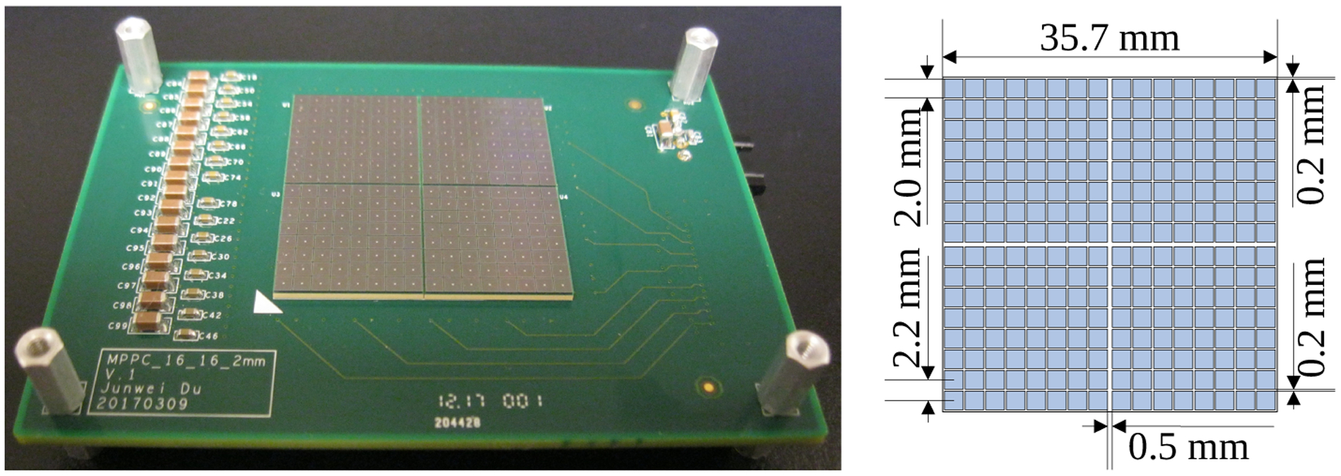
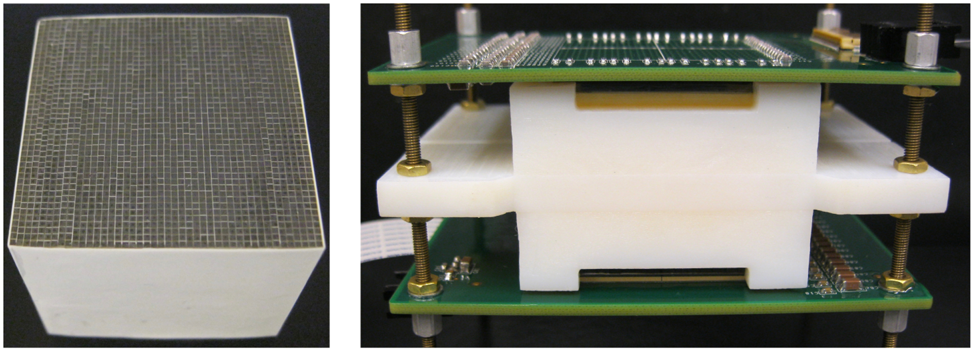
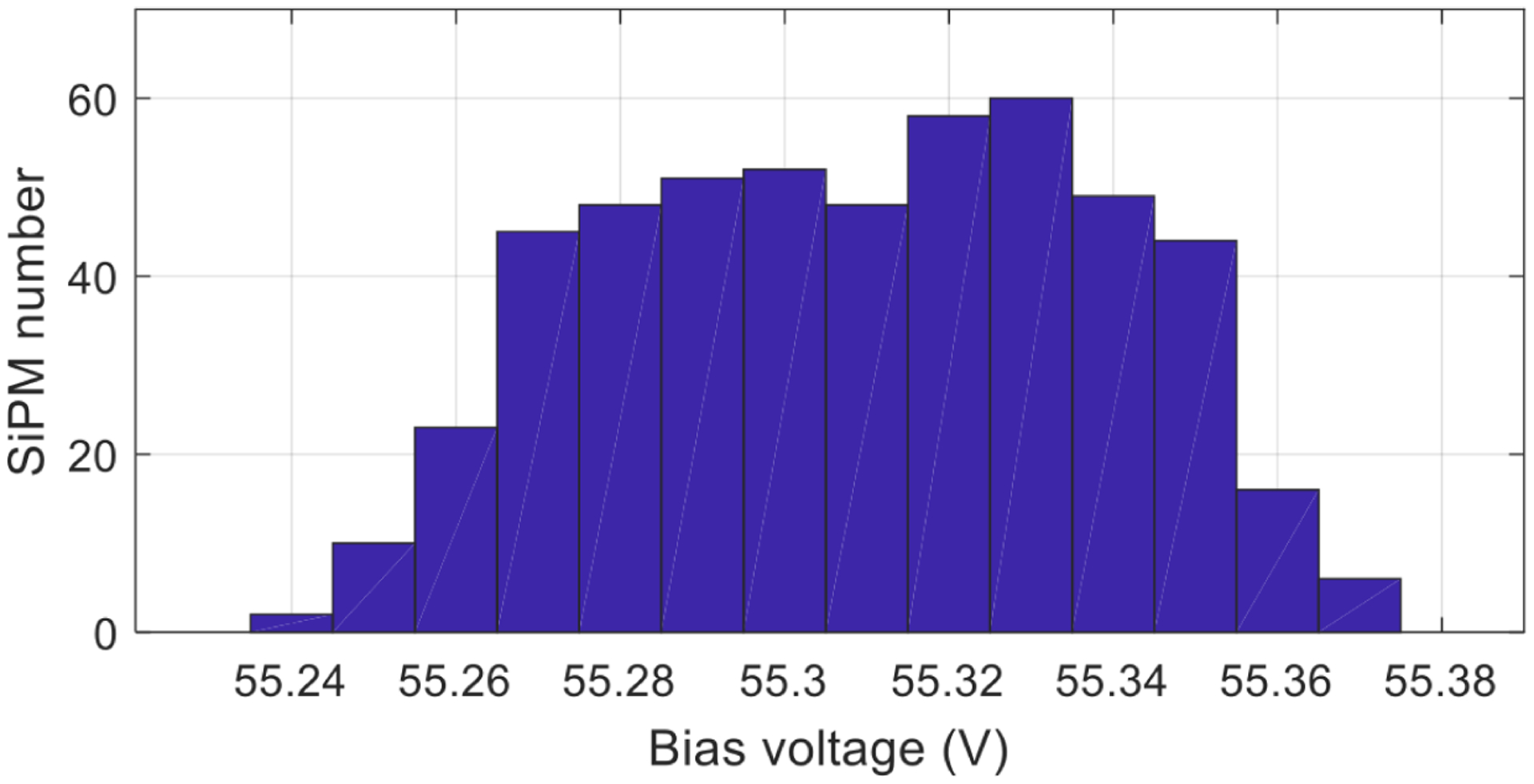
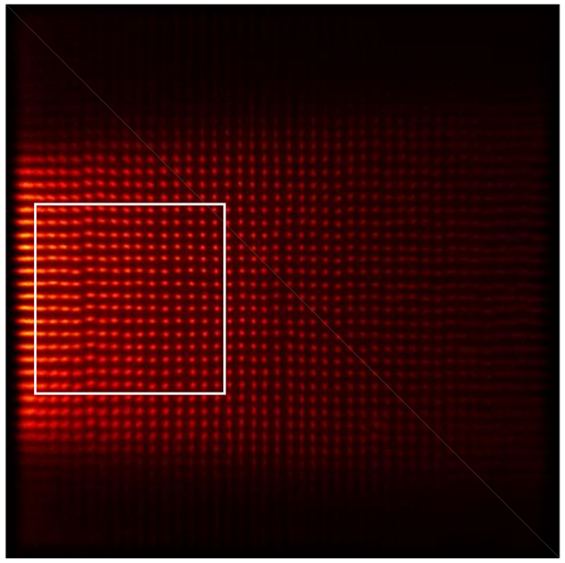
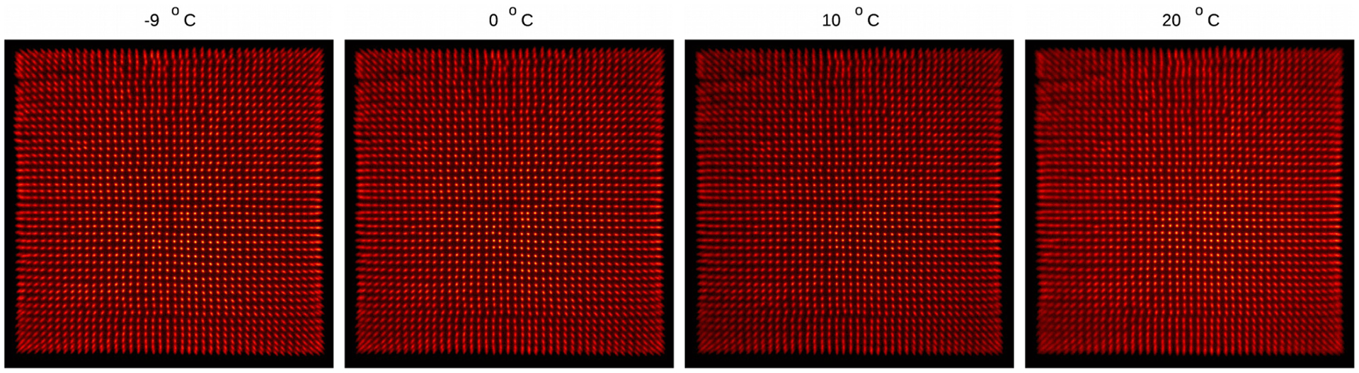
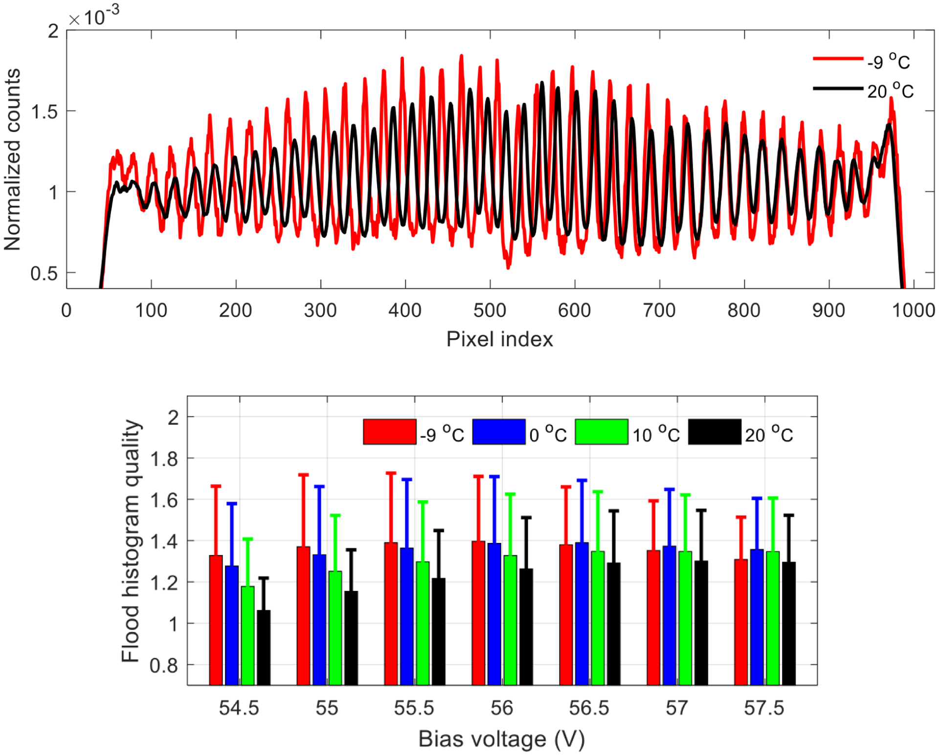
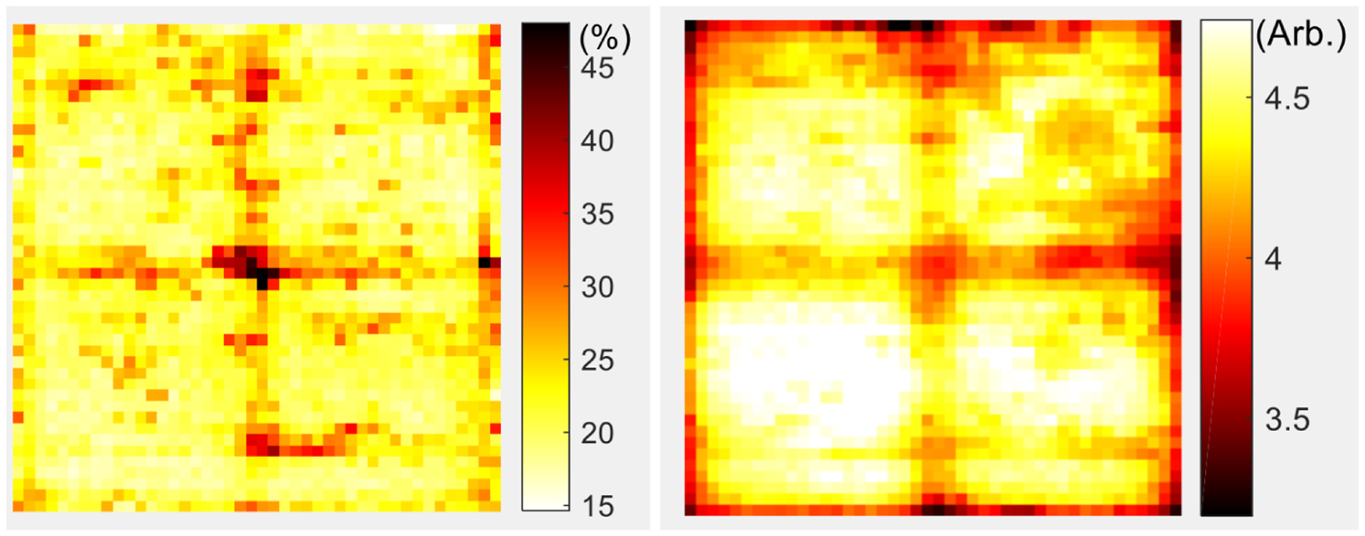
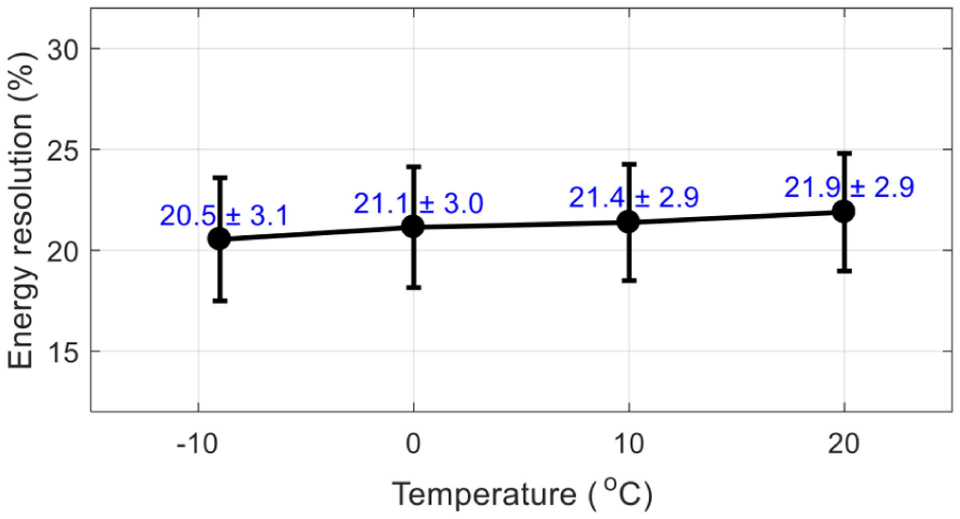
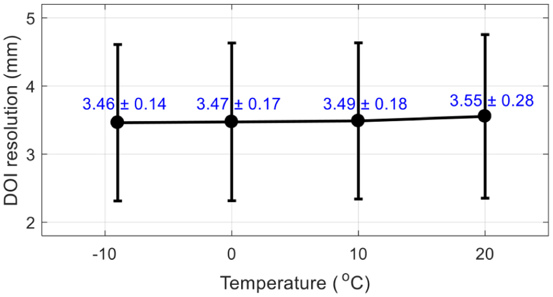
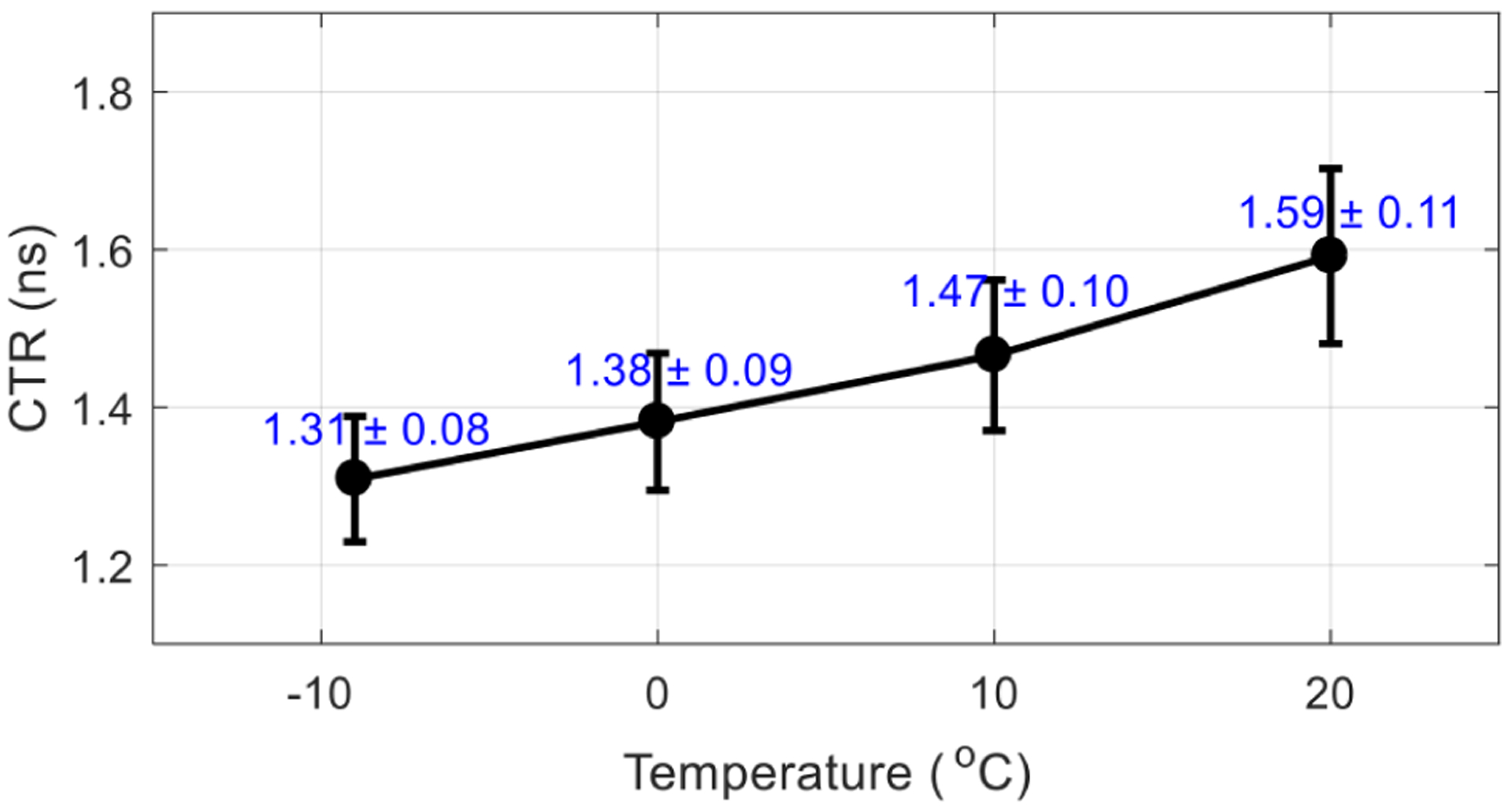
The quantitative accuracy and precision of brain positron emission tomography (PET) studies can be considerably improved using dedicated brain PET scanners with a uniform high resolution and a high sensitivity across the brain volume. One approach to building such a system is to construct the PET scanner using depth-of-interaction (DOI) encoding detectors with finely segmented and thick crystal arrays. In this paper, the performance of a DOI PET detector based on two 16 × 16 arrays of 2 × 2 mm2 SiPMs coupled to both ends of a 44 × 44 array of 0.69 × 0.69 × 30 mm3 polished LYSO crystals was evaluated at different temperatures (-9°C, 0°C, 10°C, and 20°C) for brain PET applications. The pitch size of the LYSO array is 0.75 mm. The flood histograms show that all the crystal elements in the LYSO array can be resolved except some edge crystals, due to the limited light sharing. The average energy resolution, average DOI resolution, and average timing resolution across crystal elements are 21.1 ± 3.0%, 3.47 ± 0.17 mm, and 1.38 ± 0.09 ns, respectively, which were obtained at a bias voltage of 56.5 V and a temperature of 0°C.
Keywords: Detector design and construction technologies and materials; Gamma camera; Gamma detectors (scintillators, CZT, HPGe, HgI etc); PET PET/CT; SPECT; coronary CT angiography (CTA).
Figures
Similar articles
-
Development of depth encoding small animal PET detectors using dual-ended readout of pixelated scintillator arrays with SiPMs.Med Phys. 2018 Feb;45(2):613-621. doi: 10.1002/mp.12722. Epub 2017 Dec 30. Med Phys. 2018. PMID: 29222959
-
High-resolution TOF-DOI PET detectors through the implementation of dual-ended readout with SiPM arrays of different pixel sizes on the two ends.Med Phys. 2025 Feb;52(2):867-879. doi: 10.1002/mp.17544. Epub 2024 Nov 28. Med Phys. 2025. PMID: 39607086
-
Performance of long rectangular semi-monolithic scintillator PET detectors.Med Phys. 2019 Apr;46(4):1608-1619. doi: 10.1002/mp.13432. Epub 2019 Feb 20. Med Phys. 2019. PMID: 30723932
-
High spatial resolution PET detectors based on 10 mm × 10 mm linearly-graded SiPMs and 0.5 mm pitch LYSO arrays.Phys Med Biol. 2025 Jan 17;70(2):10.1088/1361-6560/ada084. doi: 10.1088/1361-6560/ada084. Phys Med Biol. 2025. PMID: 39689427
-
A depth-of-interaction encoding PET detector module with dual-ended readout using large-area silicon photomultiplier arrays.Phys Med Biol. 2018 Dec 14;63(24):245019. doi: 10.1088/1361-6560/aaee32. Phys Med Biol. 2018. PMID: 30523925 Free PMC article.
Cited by
-
Performance Comparison of DOI-Encoding PET Detectors Based on 1.1-mm Pitch BGO Arrays With Different Reflectors.IEEE Trans Radiat Plasma Med Sci. 2024 Mar;8(3):257-262. doi: 10.1109/trpms.2024.3361891. Epub 2024 Feb 5. IEEE Trans Radiat Plasma Med Sci. 2024. PMID: 39279872 Free PMC article.
References
-
- Watanabe M et al., Performance evaluation of a high-resolution brain PET scanner using four-layer MPPC DOI detectors, Phys. Med. Biol 62 (2017) 7148. - PubMed
Grants and funding
LinkOut - more resources
Full Text Sources
Miscellaneous