Case Reports
. 2021 Nov 8;13(11):e19370.
doi: 10.7759/cureus.19370. eCollection 2021 Nov.

A Rare Case of an Intra-nasal Ectopic Tooth in a Young Woman

Affiliations
Case Reports

A Rare Case of an Intra-nasal Ectopic Tooth in a Young Woman

Rajamani Anand et al. Cureus. .

Abstract

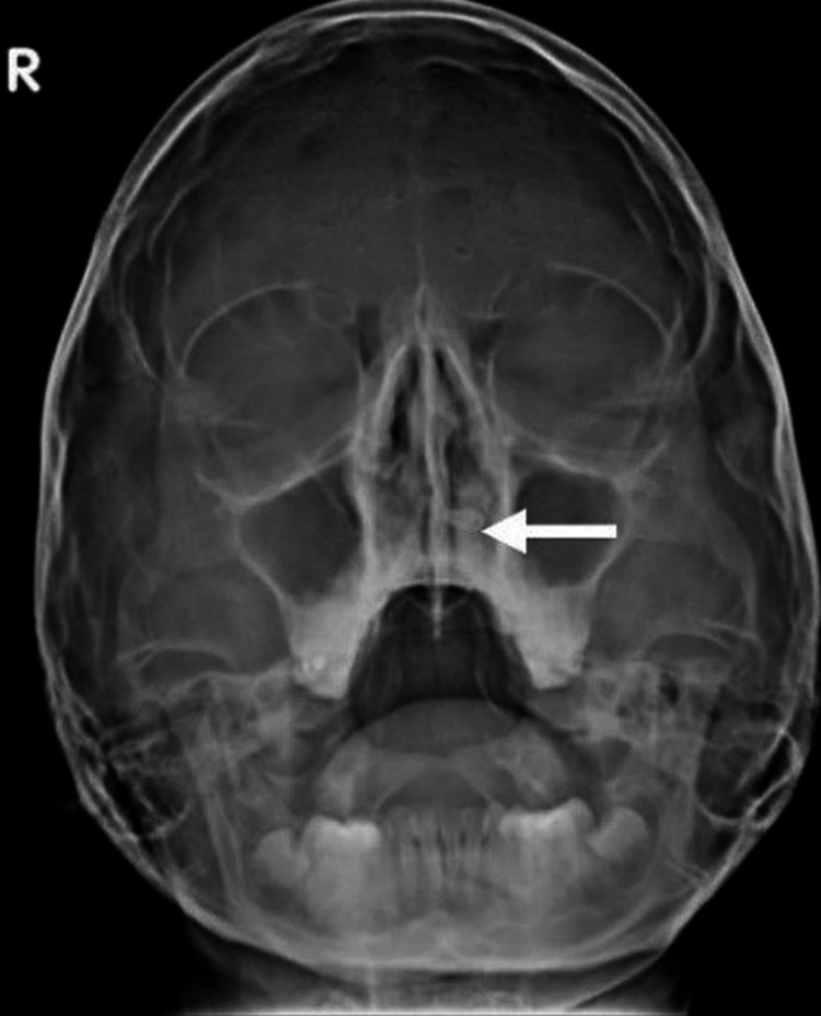
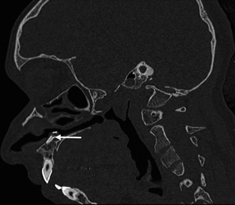
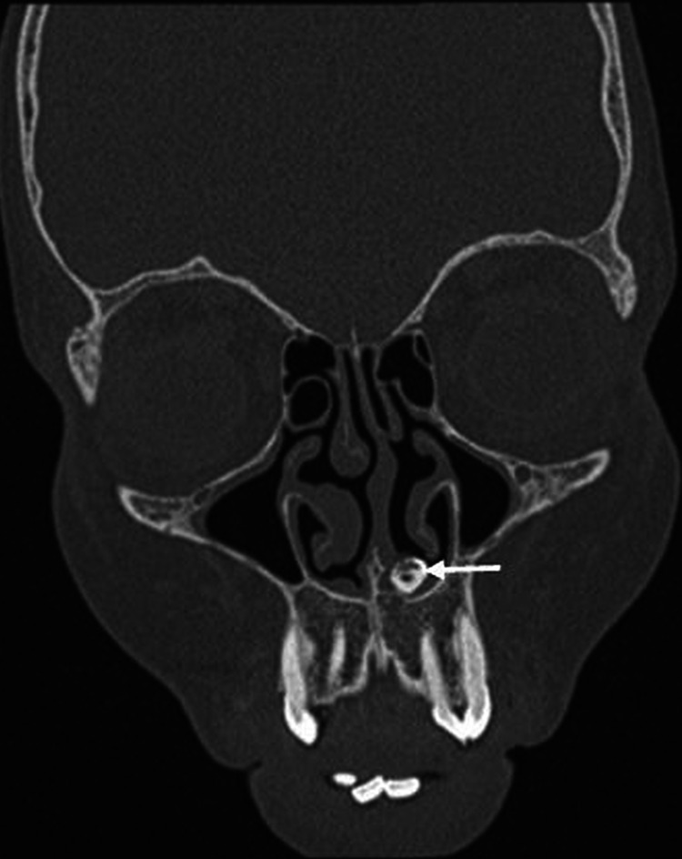
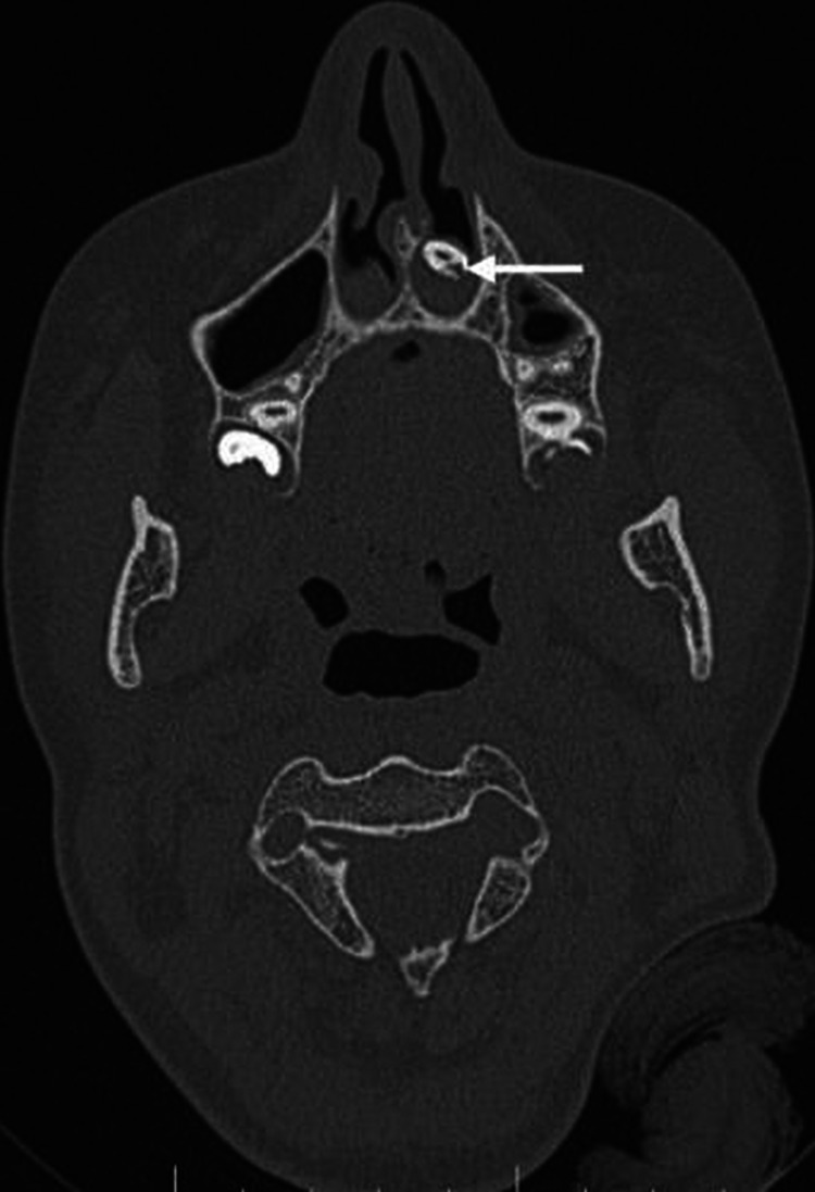
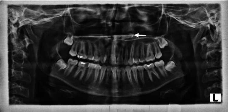
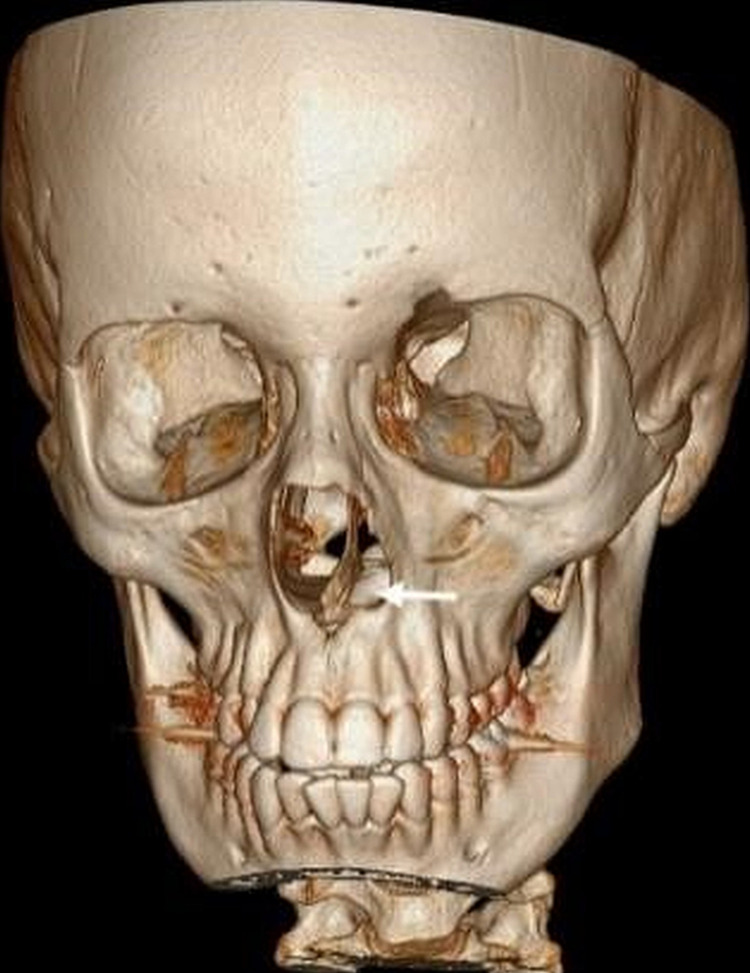
Teeth in non-dentate areas including the intra-sinus and intranasal teeth are rarely encountered in clinical practice. Although the majority of patients remain asymptomatic, the usual presenting complaints are nasal obstruction, epistaxis, hyposmia and headache. In this article, we present a case of an intranasal tooth in a 15-year-old female who presented with complaints of hyposmia and nasal obstruction. Computed tomography (CT) of the paranasal sinuses and nasal cavity showed a tooth-like structure in the left inferior nasal cavity extending from the hard palate. The mainstay of treatment is the surgical removal of the ectopic tooth under anaesthesia. Even in asymptomatic patients, surgical removal of the nasal tooth is advised to prevent complications. Along with a clinician's understanding of the condition, imaging aids in the diagnosis of an ectopic tooth. Imaging, particularly with CT, also helps plan the surgical approach to treatment.

Keywords: ectopic tooth; hyposmia; mesiodens; nasal tooth; supernumerary tooth.


Conflict of interest statement

The authors have declared that no competing interests exist.

Figures

Figure 1
Figure 1. Plain x-ray shows the radiopaque structure in the left nasal cavity, embedded in the hard palate (white arrow).
Figure 2
Figure 2. Sagittal image showing complete extension of an intranasal tooth (white arrow), which is embedded in the hard palate.
Figure 3
Figure 3. Coronal image showing a bone-like structure (white arrow) embedded in the hard palate extending into the left nasal cavity.
Figure 4
Figure 4. Axial CT scan showing an ectopic tooth in the left nasal cavity surrounded by soft tissue (white arrow).
Figure 5
Figure 5. Orthopantomogram showing tooth-like radiopaque structure (white arrow); a shape resembling a canine with a relatively smaller size. The radiodensity of this radiopaque structure resembles enamel, dentin, the pulp chamber and the pulp canal in relation to the left nasal floor.
Figure 6
Figure 6. Reconstructed 3D CT image showing tooth-like structure in the left nasal cavity (white arrow) embedded in the hard palate.
3D CT - Three-dimensional computed tomography scan

References

    1. Intranasal ectopic tooth in adult and pediatric patients: a report of two cases. Bergamaschi IP, Olsson B, Sebastiani AM, et al. Case Rep Surg. 2019;2019:8351825. - PMC - PubMed
    1. A rhinolith turning out to be an intranasal tooth. Ong HY, Ng JJ, Ong HJ, Wong SJ, Gopalan S. Cureus. 2021;13:0. - PMC - PubMed
    1. Intranasal tooth: ectopic eruption 1 year after maxillofacial trauma. Agrawal M, Khan TS, Gupta T, Khanna S. BMJ Case Rep. 2014;2014:204432. - PMC - PubMed
    1. Supernumerary teeth--an overview of classification, diagnosis and management. Garvey MT, Barry HJ, Blake M, et al. http://www.cda-adc.ca/jcda/vol-65/issue-11/612.html. J Can Dent Assoc. 1999;65:612–616. - PubMed
    1. Dental number anomalies and their prevalence according to gender and jaw in school children 7 To 14 years. Sejdini M, Çerkezi S. Open Access Maced J Med Sci. 2018;6:867–873. - PMC - PubMed

Publication types

LinkOut - more resources