. 2021 Nov 29:8:1899-1908.
doi: 10.1016/j.toxrep.2021.11.021. eCollection 2021.

Dunalialla salina microalgea and its isolated zeaxanthin mitigate age-related dementia in rats: Modulation of neurotransmission and amyloid-β protein

Affiliations

Dunalialla salina microalgea and its isolated zeaxanthin mitigate age-related dementia in rats: Modulation of neurotransmission and amyloid-β protein

Farouk K El-Baz et al. Toxicol Rep. .

Abstract

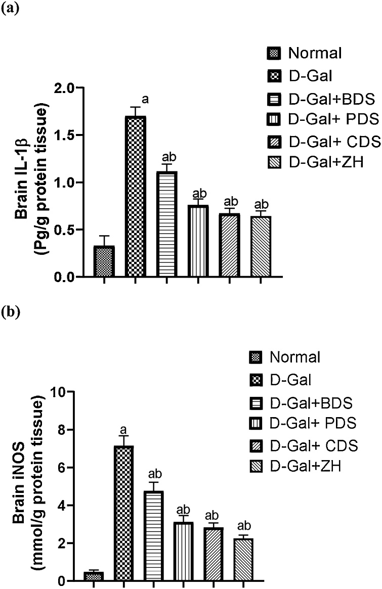
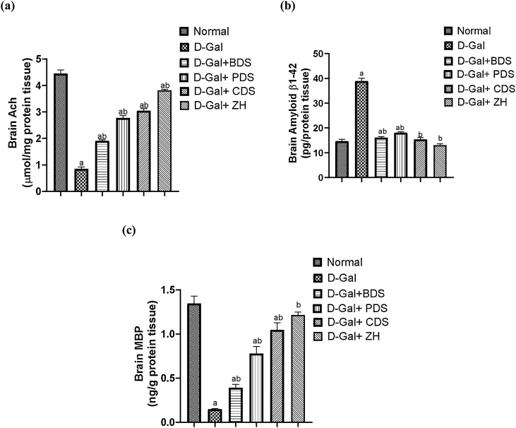
Age-related deterioration of sensorimotor and cognitive abilities suggests that the brain undergoes regressive alterations with aging that compromise its function. Thus, the present study was designed to assess the efficacy of Dunaliella salina in counteracting D-galactose (D-gal)-induced dementia brain aging and its modulatory role in attenuating amyloid β (Aβ) protein and neurotransmitters. Aging associated dementia was generated by injection of D-gal (200 mg/kg; i.p) of rats for 8 weeks. D. salina biomass (250 mg/kg), polar (30 mg/kg), its carotenoid (30 mg/kg) fractions as well as the isolated zeaxanthin (250 μg/kg) were given orally simultaneously with D-gal for additional two weeks. Twenty-four hours after the last treatment dose; behavioral, biochemical and histopathological assessment were performed. Results showed that oral treatment of motor deficit rats with D. salina biomass and its isolated polar and carotenoid fractions showed amelioration in the motor coordination assessed by the rotarod test and in the memory and learning capabilities evaluated by Morris water maze test. D. salina also showed a reduction in brain levels of inflammatory indicators viz. interlekin-1β and inducible nitric oxide synthetase as well as brain contents of Aβ protein and myelin base protein. Likewise, oral treatment with D. salina biomass and its isolated polar and carotenoid fractions exhibited an increase in the rats' brain neurotransmitters and their metabolites. Furthermore, histopathological investigations have confirmed all of these results. Our findings suggest that D. salina overcomes brain aging and thereby repairs age-related dementia, both for its modulating function in attenuating the Aβ protein and neurotransmitters.

Keywords: Aging; Amyloid-β protein; Dementia; Dunalialla salina; Neurotransmitters.


Conflict of interest statement

The authors have declared no conflict of interest.

Figures

None
Graphical abstract
Fig. 1
Fig. 1
Virtual 2-dimensional (A) and 3-dimentional (B) interaction of β-carotene and 2-dimensional (C) and 3-dimentional (D) interaction of zeaxanthin with the active site of TauTubulin Kinase 1 (TTK1).
Fig. 2
Fig. 2
Virtual 2-dimensional (A) and 3-dimentional (B) interaction of β-carotene and 2-dimensional (C) and 3-dimentional (D) interaction of zeaxanthin with the active site of Acetylcholinesterase.
Fig. 3
Fig. 3
Effect of D. salina biomass and its isolated polar and carotenoid fractions as well as the separated zeaxanthin on locomotor activity in dementia-induced rats. Data was expressed as mean ± SEM, n= 6 rats/group. a Significantly different from the normal control. b Significantly different from the AD group at P < 0.05.
Fig. 4
Fig. 4
Effect of D. salina biomass and its isolated polar and carotenoid fractions as well as the separated zeaxanthin on mean time spent in the quadrant in dementia-induced rats. Data was expressed as mean ± SEM, n= 6 rats/group. a Significantly different from the normal control. b Significantly different from the AD group at P < 0.05.
Fig. 5
Fig. 5
Effect of D. salina biomass and its isolated polar and carotenoid fractions as well as the separated zeaxanthin on brain IL-1β (a) and iNOS (b) levels in dementia-induced rats. Data was expressed as mean ± SEM, n= 6 rats/group. a Significantly different from the normal control. b Significantly different from the AD group at P < 0.05.
Fig. 6
Fig. 6
Effect of D. salina biomass and its isolated polar and carotenoid fractions as well as the separated zeaxanthin on brain Ach (a), Aβ (b) and MBP (c) contents in dementia-induced rats. Data was expressed as mean ± SEM, n= 6 rats/group. a Significantly different from the normal control. b Significantly different from the AD group at P < 0.05.
Fig. 7
Fig. 7
Effect of D. salina biomass and its isolated polar and carotenoid fractions on brain histopathological alterations associated with dementia induced in rats. Brain tissue of, (a) normal rats showing normal cerebral cortical neurons with large cell bodies and nuclei with single prominent nuclei normal (black arrows), (b,c,d,e) D-gal group showing decreased number of normal neurons and increased number of degenerated neurons, which appeared with shrunken cell bodies and intensely stained eosinophilic cytoplasm as well as a small pyknotic nuclei (red arrows) in addition to activation of microglia (arrow head) (b), diffuse gliosis and presence of reactive astrocytes with more distinct eosinophilic cytoplasm and large eccentric nuclei (black arrow) (c), deposition of amyloid plaques with eosinophilic core surrounded by astrocytes and microglia (black arrow) (d) and cerebral amyloid angiopathy (black arrow) (e), (f) D-gal-BDS group showing degeneration of cerebral cortical neurons (black arrow) associated with neuronophagia (red arrow), (g) D-gal-PDS group showing decreased number of degenerated neurons associated with mild gliosis (arrow), (h) D-gal-CDS group showing pronounced decrease of degenerated neurons (black arrows), and (i) D-gal-ZH group showing sparse degenerated neurons (black arrow). (Stain H&E; Scale bar=100μm).
Fig. 8
Fig. 8
Effect of D. salina biomass and its isolated polar and carotenoid fractions on hippocampus histopathological alterations associated with dementia induced in rats. Hippocampus of, (a, b) normal rats showing normal neurons (black arrows), (c, d) D-gal group showing increased number of degenerated neurons (black arrows), (e, f) D-gal-BDS group showing degeneration of hippocampal pyramidal neurons (black arrow) associated with activation of astrocytes (red arrow), (g, h) D-gal-PDS group showing decreased number of degenerated neurons associated with mild gliosis (black arrow for degenerated neurons and red arrow for astrocyte), (I, j) D-gal-CDS group showing pronounced decrease of degenerated neurons (black arrow), and (k, l) D-gal-ZH group showing sparse degenerated neurons (black arrow). (Stain H&E; Scale bar=100μm).

References

    1. Aloizou A.-M., Siokas V., Vogiatzi C., Peristeri E., Docea A.O., Petrakis D., et al. Pesticides, cognitive functions and dementia: a review. Toxicol. Lett. 2020;326:31–51. - PubMed
    1. Duong S., Patel T., Chang F. Dementia: what pharmacists need to know. Can. Pharm. J. (Ott) 2017;150(2):118–129. - PMC - PubMed
    1. Singh S.K., Srivastav S., Yadav A.K., Srikrishna S., Perry G. Overview of alzheimer’s disease and some therapeutic approaches targeting aβ by using several synthetic and herbal compounds. Oxid. Med. Cell. Longev. 2016;2016 7361613-7361613. - PMC - PubMed
    1. Razgonova M.P., Veselov V.V., Zakharenko A.M., Golokhvast K.S., Nosyrev A.E., Cravotto G., et al. Panax ginseng components and the pathogenesis of Alzheimer’s disease (Review) Mol. Med. Rep. 2019;19(4):2975–2998. - PMC - PubMed
    1. Gubandru M., Margina D., Tsitsimpikou C., Goutzourelas N., Tsarouhas K., Ilie M., et al. Alzheimer’s disease treated patients showed different patterns for oxidative stress and inflammation markers. Food and Chem. Toxicol.: an Int. J. Publ. Br. Ind. Biol. Res. Assoc. 2013;61:209–214. - PubMed

LinkOut - more resources