. 2021 Dec 3:8:100390.
doi: 10.1016/j.ejro.2021.100390. eCollection 2021.

Diagnostic value of dynamic magnetic resonance imaging of temporomandibular joint dysfunction

Affiliations

Diagnostic value of dynamic magnetic resonance imaging of temporomandibular joint dysfunction

Thomas J Vogl et al. Eur J Radiol Open. .

Abstract

Background: To estimate the diagnostic value of dynamic magnetic resonance imaging (MRI) for the assessment of the temporomandibular joint (TMJ) compared to standard static MRI sequences in patients with TMJ dysfunction (TMD).

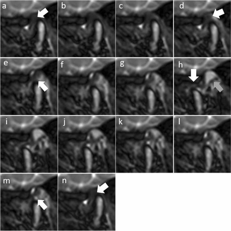
Methods and materials: This retrospective study included 71 patients with clinical diagnose of TMD. We acquired 5 static T1- and T2-weighted sequences in parasagittal and paracoronal views and one dynamic sequence (trueFISP) in parasagittal view for each TMJ. Image analysis included evaluation of morphology and function of intra-articular structures and rating of the dynamic images as more, equally, or less informative compared to static MRI sequences.

Results: Mean age was 35.0 ± 14.7 years and 50/71 (70.4%) were female. 127/142 (89.4%) TMJs were of diagnostic quality. 42/127 (33.1%) TMJs showed no disc displacement (DD), 56 (44.1%) had DD with disc reduction (DDwR), and 29 (22.8%) had DD without disc reduction (DDwoR). In 38/127 (29.9%) TMJs, dynamic images were rated "more informative", in 84/127 (66.2%) "equally informative", and in 5/127 (3.9%) "less informative" compared to solely static images. Overall, 27/71 (38.0%) patients benefited from additional dynamic sequences compared to solely static images. Dynamic images were "more informative" in TMJs with DDwR (23/56 [41.1%], p < 0.001) and in TMJs with DDwoR (13/29 [44.8%], p = 0.007), while it had no beneficial value for TMJ without DD. For evaluation of joint effusion, static T2-weighted images were rated better in 102/127 (80.3%) TMJs compared to dynamic images (<0.001).

Conclusion: Dynamic MRI sequences are beneficial for the evaluation of morphology and function of the TMJ compared to static sequences, especially in patients with temporomandibular disc displacement.

Keywords: DDwR, disc displacement with reduction; DDwoR, disc displacement without reduction; Dynamic MRI; FLASH, Fast Low-Angle Shot; HASTE, Half-Fourier Acquisition Single-shot Turbo spin Echo; ID, Internal Derangement; Static MRI; TMD, Temporomandibular joint dysfunction; TMJ, Temporomandibular joint; Temporomandibular joint; Temporomandibular joint dysfunction; trueFISP, true fast imaging with steady state precession.


Figures

Fig. 1
Fig. 1
T1-weighted static images of the left temporomandibular joint of a 52-year-old male patient with anterior displaced disc (arrows). Anteriorly (11`o clock) positioned disc with contact to the condyle in closed mouth position (a). At open mouth position the condyle is located on the disc (b). The translation of the condyle and disc is reduced.
Fig. 2
Fig. 2
Dynamic MRI sequences of the patient from Fig. 1. The dynamic sequence starts in closed mouth position and slightly anterior positioned disc (a, white arrow). During mouth opening (a-g) the condyle translates to its physiological position below the disc (b and e, white arrows) until complete open mouth position is reached with the condyle being anterior of the tuberculum (g). The posterior ligament (gray arrow in g) of the disc is attached to the temporal bone, stretched at maximum mouth opening and prevents the disc (white arrow in g) from gliding further anterior. During mouth closing the condyle translates below the disc (white arrows in j and l) back into the fossa (h-m) until closed mouth position is reached and the disc is slightly anterior positioned (n, white arrow).
Fig. 3
Fig. 3
Left temporomandibular joint of a 21-year-old male patient with disc displacement without reduction (disc adhesion). In closed mouth position (a), the disc (white arrow in a) is displaced anteriorly the degenerated condyle (gray arrow in a) between mandibular fossa and tuberculum mandibulae (arrow-head in a). During mouth opening (b-g), the condyle glides from posterior below the disc (arrow in b), deforms the disc (c), and glides anterior of the disc (arrow in e) until maximal mouth opening position is reached (h). Reversed movement can be observed during mouth closing (arrows in i to n). The disc does not move during mouth opening and closing and stucks on the temporal bone (disc adhesion).
Fig. 4
Fig. 4
Right temporomandibular joint of a 31-year-old female patient with disc displacement with reduction. In closed mouth position, the disc is displaced anterior the condyle (arrow in a). During the initial phase of mouth opening, rotation but no translation is observed (b and c). Subsequently, the condyle glides below the disc (arrow in d) until the condyle is repositioned on the disc (arrow in e). Condyle and disc translate (e-h) on the anterior until maximum mouth opening (white arrow in h). Mouth opening is naturally limited by the posterior ligament of the disc (gray arrow in h). Dynamic sequences provide detailed visualization of condyle and disc movements during mouth closing (i to m). In the last part of mouth closing, the condyle glides down from the disc (arrow in m to n).
Fig. 5
Fig. 5
Scheme of the physiologic anatomy in closed mouth position: The posterior border of the disc is located at 11‘o clock. Positions of the posterior border of the disc between 11 and 12 o′clock were seen as physiological, while positions between 9 and 11 o′clock are defined as slightly anterior, below 9′ o clock as total anterior, and above 12 o′clock as posterior disc displacement.

References

    1. Westesson P.L. Reliability and validity of imaging diagnosis of temporomandibular joint disorder. Adv. Dent. Res. 1993;7:137–151. doi: 10.1177/08959374930070020401. - DOI - PubMed
    1. Petersson A. What you can and cannot see in TMJ imaging - an overview related to the RDC/TMD diagnostic system. J. Oral Rehabil. 2010;37:771–778. doi: 10.1111/j.1365-2842.2010.02108.x. - DOI - PubMed
    1. Greess H., Anders K. Indikation und Aussagekraft von Computertomographie und Magnetresonanztomographie des Kiefergelenks. Rontgenpraxis. 2005;56:1–11. doi: 10.1016/j.rontge.2005.05.001. - DOI - PubMed
    1. Vogl T.J., Lauer H.-C., Lehnert T., Naguib N.N.N., Ottl P., Filmann N., Soekamto H., Nour-Eldin N.-E.A. The value of MRI in patients with temporomandibular joint dysfunction: correlation of MRI and clinical findings. Eur. J. Radiol. 2016;85:714–719. doi: 10.1016/j.ejrad.2016.02.001. - DOI - PubMed
    1. Jung Y.-W., Park S.-H., On S.-W., Song S.-I. Correlation between clinical symptoms and magnetic resonance imaging findings in patients with temporomandibular joint internal derangement. J. Korean Assoc. Oral Maxillofac. Surg. 2015;41:125–132. doi: 10.5125/jkaoms.2015.41.3.125. - DOI - PMC - PubMed

LinkOut - more resources