. 2022 Jun;240(6):1020-1033.
doi: 10.1111/joa.13615. Epub 2021 Dec 19.

The hyoid as a sound conducting apparatus in laryngeally echolocating bats

Affiliations

The hyoid as a sound conducting apparatus in laryngeally echolocating bats

Chelsie C G Snipes et al. J Anat. 2022 Jun.

Abstract

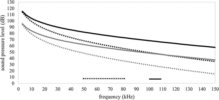
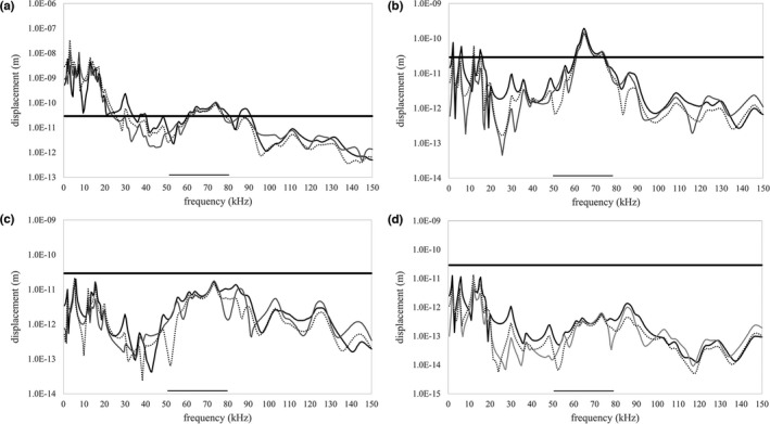
The morphology of the stylohyal-tympanic bone articulation found in laryngeally echolocating bats is highly indicative of a function associated with signal production. One untested hypothesis is that this morphology allows the transfer of a sound signal from the larynx to the tympanic bones (auditory bulla) via the hyoid apparatus during signal production by the larynx. We used µCT data and finite element analysis to model the propagation of sound through the hyoid chain into the tympanic bones to test this hypothesis. We modeled sound pressure (dB) wave propagation from the basihyal to the tympanic bones, vibratory behavior (m) of the stylohyal-tympanic bone unit, and the stylohyal and tympanic bones when the stylohyal bone is allowed to pivot on the tympanic bone. Sound pressure wave propagation was modeled using the harmonic acoustics solver in ANSYS and vibratory behavior was modeled using coupled modal and harmonic response analyses in ANSYS. For both analyses (harmonic acoustics and harmonic response), the input excitation on the basihyal and thyrohyals was modeled as the estimated pressure (Pa) imposed by the collision of the vibrating thyroid cartilage of the larynx against these bones during signal production. Our models support the hypothesis that this stereotypical hyoid morphology found in laryngeally echolocating bats can transfer sound to the auditory bullae at an amplitude that is likely heard for the species Artibeus jamaicensis and Rhinolophus pusillus.

Keywords: Chiroptera; bioacoustics; biomechanics; sonar.


Figures

FIGURE 1
FIGURE 1
Volume rendered µCT scans of Artibeus jamaicensis (b and d) and Rhinolophus pusillus (a and c) showing basihyal and fused thyrohyals (green), hypohyals (purple), ceratohyals (pink), stylohyals (blue), and tympanic (grey) bone. Intervening cartilaginous segments are white. The calcified larynx of the R. pusillus is indicated in orange, while the only part of the larynx that is calcified in A. jamaicensis is the cricoid (Carter 2020) and is not shown here. Points of view are from cranial (a), ventral (b), and lateral (c and d) aspects
FIGURE 2
FIGURE 2
(a) Artibeus jamaicensis and (b) Rhinolophus pusillus show the dorsal aspects of the FE model geometry with the laryngeal surface (area of sinusoidal excitation input) shaded in red. (c) A. jamaicensis and (d) R. pusillus show the medial aspects of the FE model geometry with the area where vibratory data were collected shaded in blue. The orange dot in (c) indicates the location of the facet where vibratory data were collected at the end of the stylohyal in A. jamaicensis. (e) and (f) show the lateral aspect of the FE model geometry for A. jamaicensis with the bonded area between the stylohyal and tympanic bones shaded in green. (e) Shows the scenario where the entire interface between the stylohyal and tympanic bones is bonded and (f) shows the scenario where there is a pivot‐like articulation between them
FIGURE 3
FIGURE 3
Sagittal slices through the tympanic bone, stylohyal bone, skull bones, and cochlea in Rhinolophus pusillus (a and b) and Artibeus jamaicensis (c and d). Blue circles indicate the locations from where sound pressure level (dB) data were collected in our acoustic models
FIGURE 4
FIGURE 4
Fixed support boundary conditions for the harmonic response analyses for Artibeus jamaicensis (a–c) and Rhinolophus pusillus (d–f). The green triangles (a and d) indicate the location of the fixed support attaching the tympanic bones the skull. The red triangles (b–f) indicate the location of the fixed support attaching the thryohyal bones to thyrohyal cartilage (which attaches to the thyroid cartilage of the larynx). The black triangle (c and f) indicates the location of the fixed point used to constrain the basihyal at one point. The black and yellow triangles indicate the locations of the fixed supports used to constrain the basihyal at three points. The black, yellow, and blue triangles indicate the locations of the fixed supports used to constrain the basihyal at five points. Elements of the volume rendered models are color coded as follows: basihyal and fused thyrohyals (green), hypohyals (purple), ceratohyals (pink), stylohyals (blue), tympanic bones (grey), cartilaginous segments (white)
FIGURE 5
FIGURE 5
Contour plots of sound pressure level (dB) for Rhinolophus pusillus (a) at 105 kHz and Artibeus jamaicensis (b) at 63 kHz. Input pressures on laryngeal surfaces of the hyoid apparatus are 63 Pa (130 dB) for Rhinolophus pusillus and 6.3 Pa (110 dB) for A. jamaicensis
FIGURE 6
FIGURE 6
Frequency response curves for sound pressure level (dB) measured from facets on the left and right tympanic bones where the cranial end of the stylohyal articulates. Data points represent the average sound pressure level measured between tympanic bones (left and right) at a particular frequency. Black curves represent data from models with a 63 Pa (130 dB) input excitation and gray curves represent data from models with a 6.3 Pa (110 dB) input excitation. Solid black and gray curves are from Rhinolophus pusillus and dotted black and grey lines are from Artibeus jamaicensis. The solid and dotted bars along the horizontal axis indicate echolocation frequency ranges for each species, A. jamaicensis (dotted) and R. pusillus (solid)
FIGURE 7
FIGURE 7
Frequency response curves in all three axes (x—solid black, y—solid gray, z—dotted black) for maximum tympanic ring displacement (m) in Artibeus jamaicensis with the stylohyal and tympanic bones fused together across the entire interface. The x‐axis (solid black line) is orthogonal to the tympanic ring and therefore orthogonal to the plane of the tympanic membrane; the y‐axis is along the occipito‐rostral plane and the z‐axis is along the dorso‐ventral plane. The 2.9e‐11 m displacement threshold is indicated by the thick horizontal black line and the echolocation frequency range is indicated by the black bar along the horizontal axis. Data are from an unconstrained basihyal (a), basihyal constrained at one point (b), basihyal constrained at three points (c), and basihyal constrained at five points (d)
FIGURE 8
FIGURE 8
Frequency response curves in all three axes (x—solid black, y—solid grey, z—dotted black) for maximum tympanic ring displacement (m) in Rhinolophus pusillus with the stylohyal and tympanic bones fused together across the entire interface. The x‐axis (solid black line) is orthogonal to the tympanic ring and therefore orthogonal to the plane of the tympanic membrane; the y‐axis is along the occipito‐rostral plane and the z‐axis is along the dorso‐ventral plane. The 2.9e‐11 mm displacement threshold is indicated by the thick horizontal black line and the echolocation frequency range is indicated by the black bar along the horizontal axis. Data are from an unconstrained basihyal (a), basihyal constrained at one point (b), basihyal constrained at three points (c), and basihyal constrained at five points (d)
FIGURE 9
FIGURE 9
Frequency response curves in all three axes (x—solid black, y—solid grey, z—dotted black) for maximum displacement (m) of the cranial end of the stylohyal in Artibeus jamaicensis with the stylohyal and tympanic bones articulated with a pivot‐like connection. The x‐axis (solid black line) is orthogonal to the tympanic ring and therefore orthogonal to the plane of the tympanic membrane; the y‐axis is along the occipito‐rostral plane and the z‐axis is along the dorso‐ventral plane. The 2.9e‐11 mm displacement threshold is indicated by the thick horizontal black line and the echolocation frequency range is indicated by the black bar along the horizontal axis. Data are from an unconstrained basihyal (a), basihyal constrained at one point (b), basihyal constrained at three points (c), and basihyal constrained at five points (d)
FIGURE 10
FIGURE 10
Frequency response curves in all three axes (x—solid black, y—solid grey, z—dotted black) for maximum tympanic ring displacement (m) in Artibeus jamaicensis with the stylohyal and tympanic bones articulated with a pivot‐like connection. The x‐axis (solid black line) is orthogonal to the tympanic ring and therefore orthogonal to the plane of the tympanic membrane; the y‐axis is along the occipito‐rostral plane and the z‐axis is along the dorso‐ventral plane. The 2.9e‐11 mm displacement threshold is indicated by the thick horizontal black line and the echolocation frequency range is indicated by the black bar along the horizontal axis. Data are from an unconstrained basihyal (a), basihyal constrained at one point (b), basihyal constrained at three points (c), and basihyal constrained at five points (d)

References

    1. Brinkløv, S. , Kalko, E.K. & Surlykke, A. (2009) Intense echolocation calls from two ‘whispering’ bats, Artibeus jamaicensis and Macrophyllum macrophyllum (Phyllostomidae). Journal of Experimental Biology, 212, 11–20. - PubMed
    1. Carter, R.T. (2020) Reinforcement of the larynx and trachea in echolocating and non‐echolocating bats. Journal of Anatomy, 237, 495–503. - PMC - PubMed
    1. Carter, R.T. & Adams, R.A. (2014) Ontogeny of the larynx and flight ability in Jamaican fruit bats (Phyllostomidae) with considerations for the evolution of echolocation. The Anatomical Record, 297, 1270–1277. - PubMed
    1. Carter, R.T. , Shaw, J.B. & Adams, R.A. (2014) Ontogeny of vocalization in Jamaican fruit bats with implications for the evolution of echolocation. Journal of Zoology, 293, 25–32.
    1. Currey, J.D. (2006) Bones: structure and mechanics. Princeton, NJ: Princeton University Press, pp. 124–129, 269.