Review
. 2022 Jul 1;102(3):1159-1210.
doi: 10.1152/physrev.00022.2021. Epub 2021 Dec 20.

Mechanisms and physiological implications of cooperative gating of clustered ion channels

Affiliations
Review

Mechanisms and physiological implications of cooperative gating of clustered ion channels

Rose E Dixon et al. Physiol Rev. .

Abstract

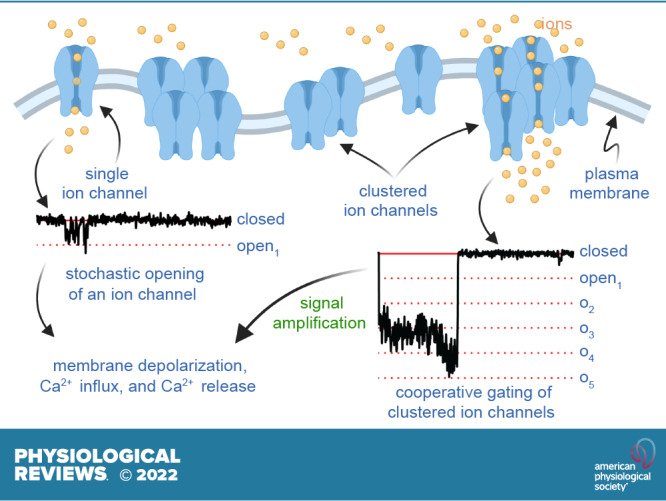
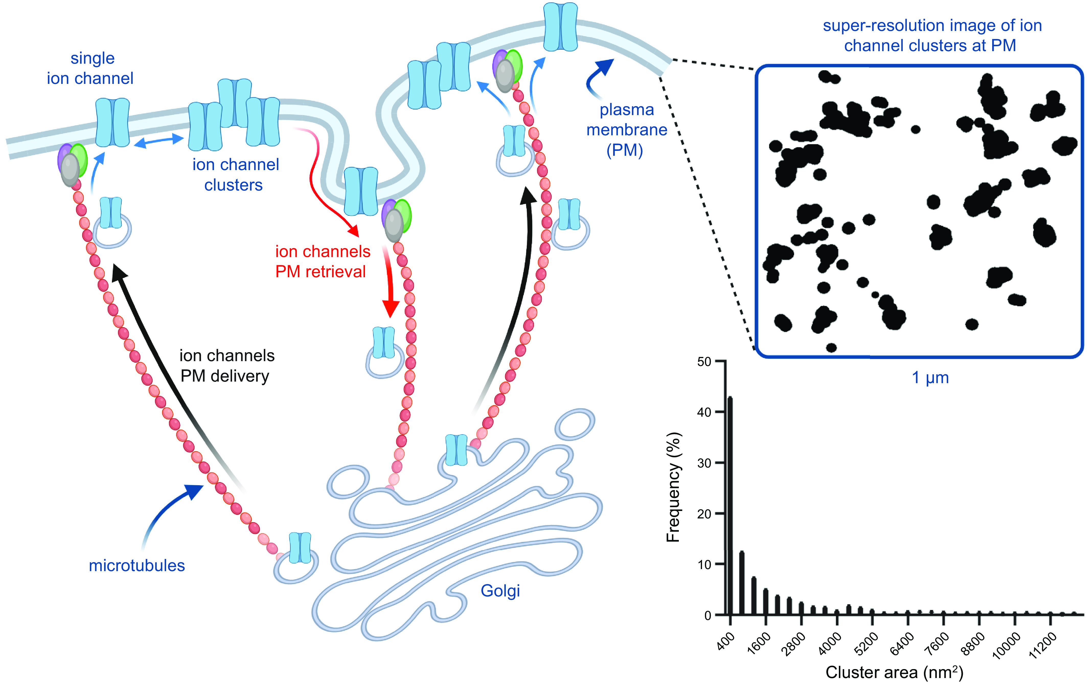
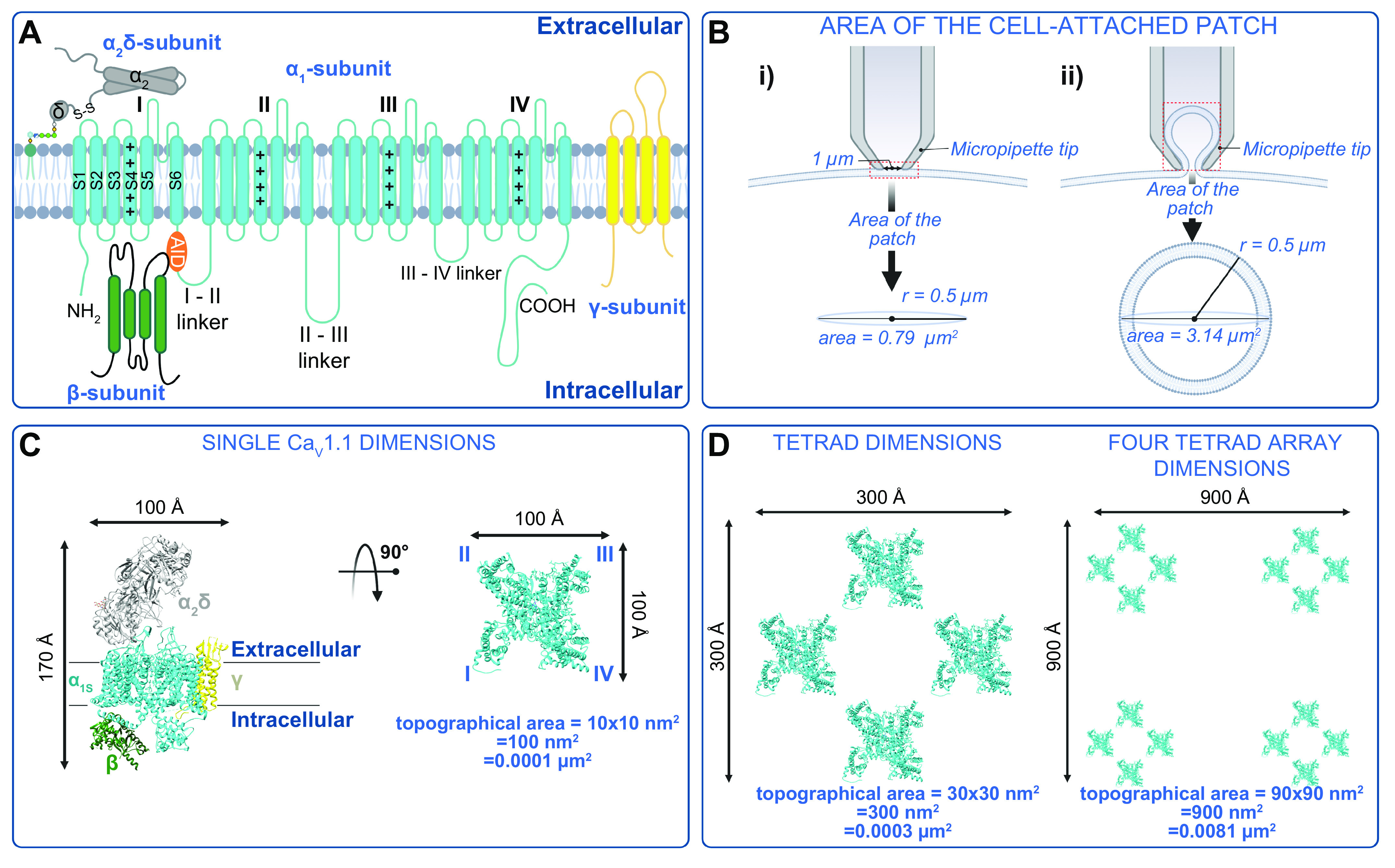
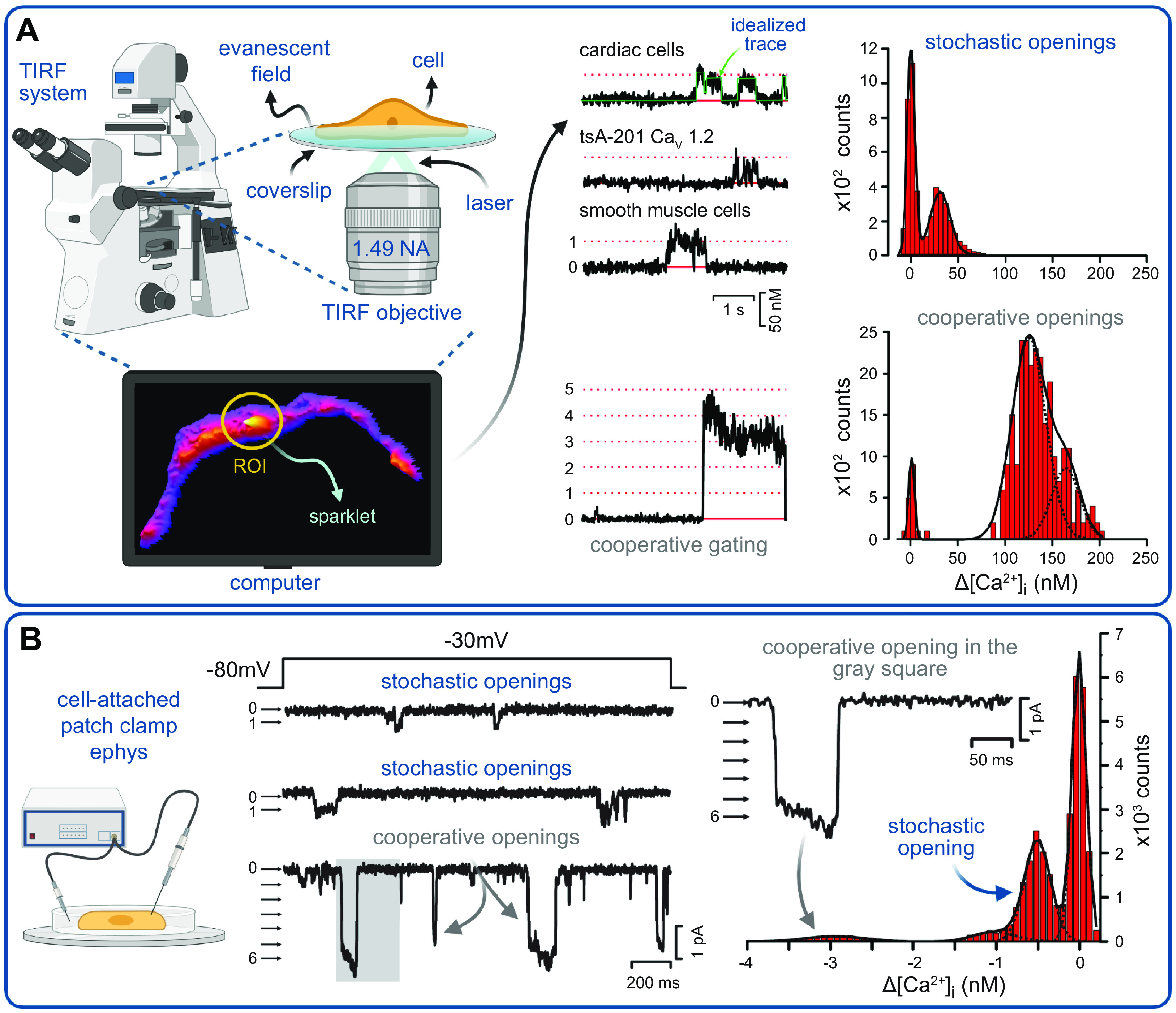
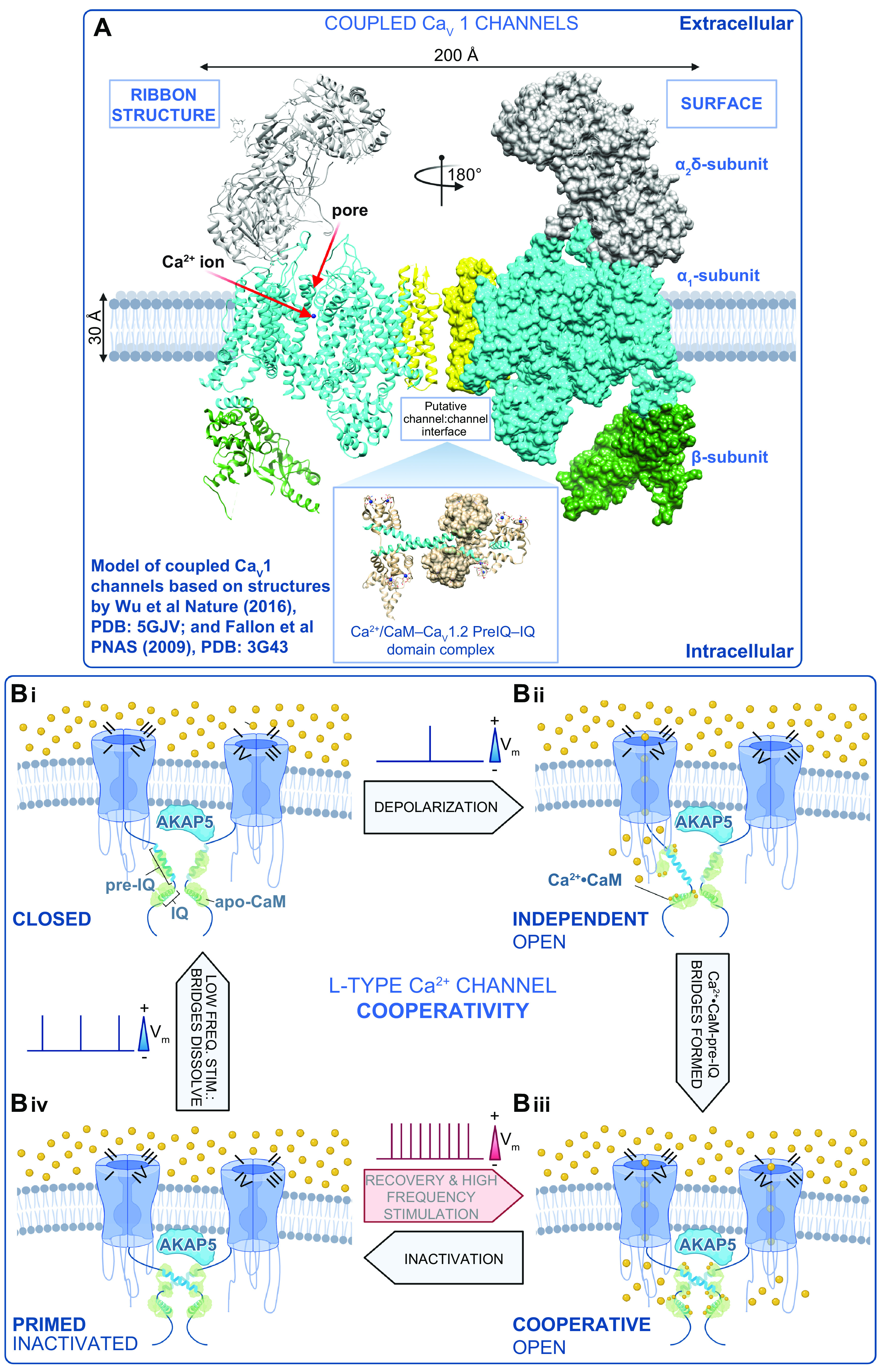
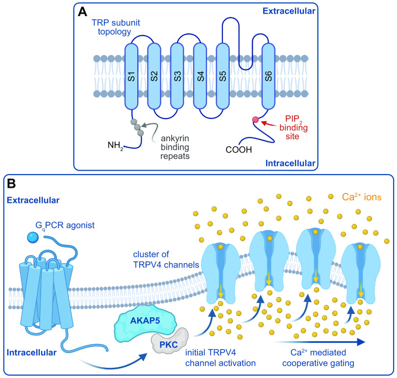
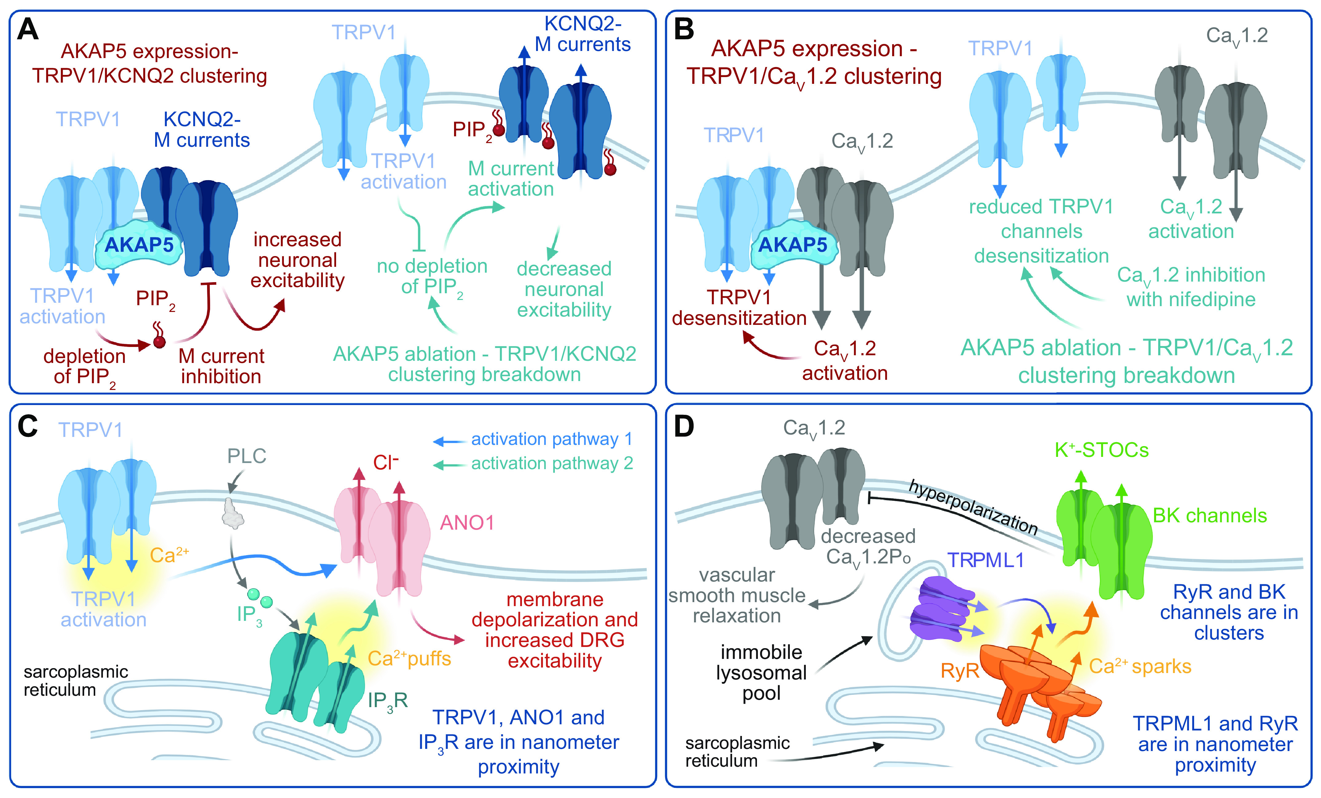
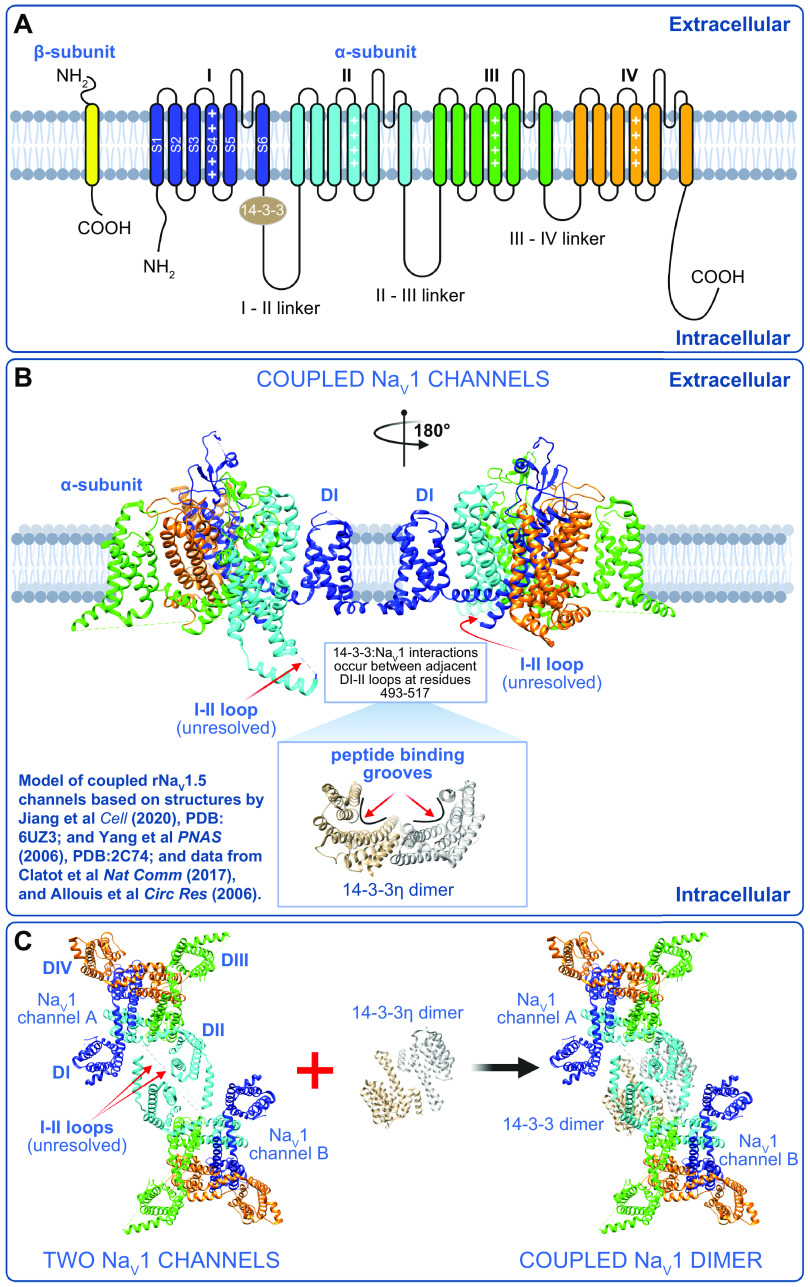
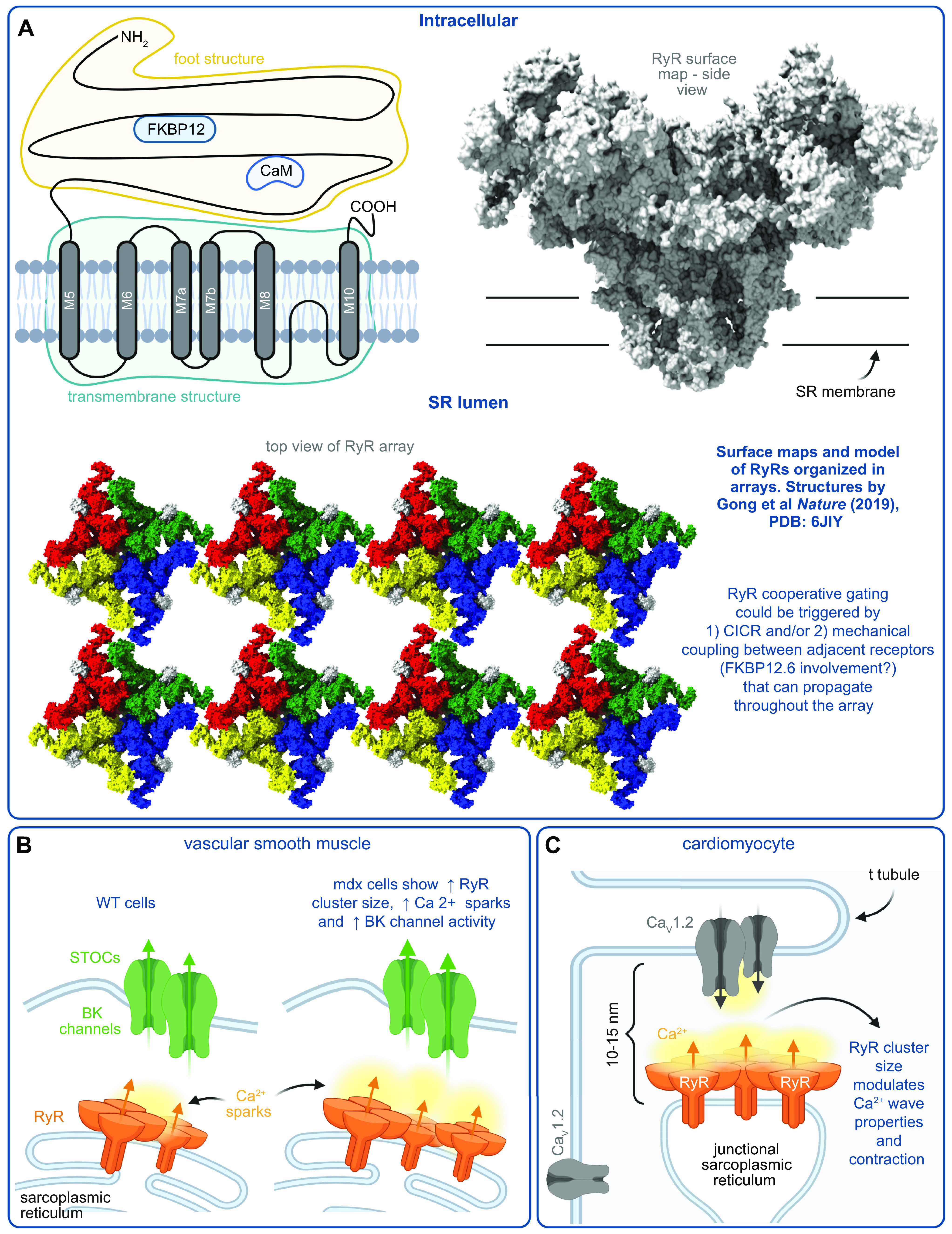
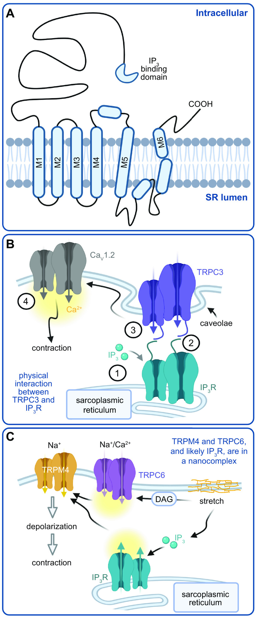
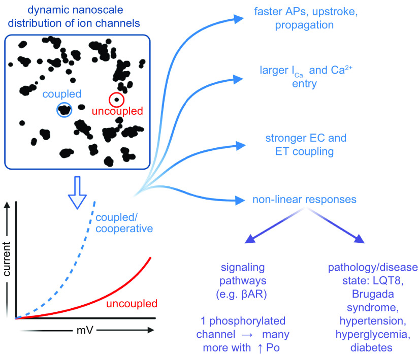
Ion channels play a central role in the regulation of nearly every cellular process. Dating back to the classic 1952 Hodgkin-Huxley model of the generation of the action potential, ion channels have always been thought of as independent agents. A myriad of recent experimental findings exploiting advances in electrophysiology, structural biology, and imaging techniques, however, have posed a serious challenge to this long-held axiom, as several classes of ion channels appear to open and close in a coordinated, cooperative manner. Ion channel cooperativity ranges from variable-sized oligomeric cooperative gating in voltage-gated, dihydropyridine-sensitive CaV1.2 and CaV1.3 channels to obligatory dimeric assembly and gating of voltage-gated NaV1.5 channels. Potassium channels, transient receptor potential channels, hyperpolarization cyclic nucleotide-activated channels, ryanodine receptors (RyRs), and inositol trisphosphate receptors (IP3Rs) have also been shown to gate cooperatively. The implications of cooperative gating of these ion channels range from fine-tuning excitation-contraction coupling in muscle cells to regulating cardiac function and vascular tone, to modulation of action potential and conduction velocity in neurons and cardiac cells, and to control of pacemaking activity in the heart. In this review, we discuss the mechanisms leading to cooperative gating of ion channels, their physiological consequences, and how alterations in cooperative gating of ion channels may induce a range of clinically significant pathologies.

Keywords: calcium signaling; channel clustering; cooperative gating; excitability; stochastic self-assembly.


Conflict of interest statement

No conflicts of interest, financial or otherwise, are declared by the authors.

Figures

None
Graphical abstract
FIGURE 1.
FIGURE 1.
Ion channel proteins translocate to the membrane of the rough endoplasmic reticulum (ER), traffic to the Golgi, and are transported to the surface membrane on microtubules, where they form clusters. Left: cartoon depicting the synthesis, membrane translocation, processing, and trafficking of ion channels to the surface membrane. Top right: superresolution image of ion channel clusters in the surface membrane. Bottom right: histogram of ion channel cluster areas from images like that above. The purple, green, and gray ovals at the tip of the microtubules cartoon represent microtubule-anchoring/binding proteins such as BIN1. Histogram was modified from Sato et al. (5) with permission. Cartoon created with Biorender.com.
FIGURE 2.
FIGURE 2.
Structure and dimensions of voltage-gated Ca2+ (CaV) channels. A: illustration of the pore-forming CaVα1 and auxiliary CaVβ, CaVα2δ, and CaVγ. AID, alpha-interacting domain. B: 2 illustrations of patch pipettes forming a gigaseal enabling voltage control and single-channel recordings from the underlying patch of membrane. The pipette may enclose either a circle (i) or a 3-dimensional Ω-shaped patch of membrane (ii) that may contain a large number of ion channels. C: dimensions of CaV1.1 (PDB: 5GJV) from Wu et al. (76) (left) and the topological view/footprint of a single α1S pore-forming subunit (right), illustrating that many channels can theoretically be housed in a single patch. D: the dimensions of a single CaV1.1 tetrad (left) and an array of 4 tetrads (right). Figure created with Biorender.com.
FIGURE 3.
FIGURE 3.
Recording and analysis of CaV1.2 cooperative gating events: total internal reflection fluorescence (TIRF) microscopy configuration (A) and cell-attached patch-clamp electrophysiology (ephys) system (B) for recording Ca2+ sparklets and single-channel currents, respectively. [Ca2+]i, intracellular Ca2+ concentration. Traces and histograms used to detect and analyze CaV1.2 cooperative openings in different cell types are modified from Navedo et al. (13) with permission. Figure created with Biorender.com.
FIGURE 4.
FIGURE 4.
Mechanism of L-type Ca2+ channel cooperative gating. A: model of the interaction between 2 CaV1 channels based on structures by Wu et al. (76) and Fallon et al. (189). B: schematic showing our proposed model of L-type Ca2+ channel cooperative gating based on the analysis of CaV1.2 and CaV1.3 channels (12, 40). Two channels are illustrated for simplicity and are drawn bridged by Ca2+·calmodulin (CaM) in the manner of the 2 published crystal structures of COOH-terminal fragment dimers (189, 190). Although this is speculative at present, we do know that the interactions depend on Ca2+·CaM (12, 42) and intact pre-IQ motifs (12, 42) and occur in AKAP scaffolded microdomains (at least for CaV1.2) (13, 173). For >2 channel multimers to gate coordinately, we postulate that there are at least 2 possibilities: 1) Ca2+·CaM may “daisy-chain” adjacent pre-IQ motifs, utilizing the 2 CaM-binding sites on that motif, or 2) one lobe of Ca2+·CaM may bind to pre-IQ and the other may bind to another of the established CaM binding sites on these channels (for review see Ref. 445). In our scheme, we begin at Bi, where the membrane is at its resting potential and channels are close to one another but not interacting in the closed state. Bii: with depolarization, a subset of the channels stochastically open and Ca2+ flows into the cell, binding to CaM. Biii: Ca2+·CaM facilitates the physical, functional interactions of adjacent channels that depend on binding to the pre-IQ motif. In this functionally cooperative state, the opening of 1 channel is allosterically communicated to the attached channel and they gate coordinately. Biv: CaV channels undergo voltage- and Ca2+-dependent inactivation but remain associated for a time after the Ca2+ signal has decayed, leaving them in a primed state as the cells repolarize. Thus, with high-frequency activation, e.g., in the heart during fight or flight, the channels can immediately transition to the cooperative open state, resulting in an immediate facilitation of Ca2+ influx. During low-frequency stimulation, e.g., during resting heart rate, the physical bridges between adjacent channels in a cluster are dissolved, and the resting confirmation is assumed as the cycle is resumed. Vm, membrane potential. Figure created with Biorender.com.
FIGURE 5.
FIGURE 5.
Model of cooperative gating of transient receptor potential (TRP) channels. A: TRP subunit topology. PIP2, phosphatidylinositol 4,5-bisphosphatase. B: model by which Gq agonists trigger AKAP5-dependent, PKC-mediated cooperative gating of TRPV4 channels. GqPCR, Gq protein-coupled receptor. Figure created with Biorender.com.
FIGURE 6.
FIGURE 6.
Functional coupling of transient receptor potential (TRP) channels with other ion channels. A: functional coupling between TRPV1 and KCNQ2 modulates neuronal excitability. PIP2, phosphatidylinositol 4,5-bisphosphatase. B: clustering of TRPV1 and CaV1.2 channels regulates TRPV1 desensitization in neurons. C: a tripartite clustering of TRPV1, inositol trisphosphate (IP3) receptor (IP3R), and ANO1 controls membrane depolarization and dorsal root ganglion (DRG) excitability. PLC, phospholipase C. D: mucolytic TRP (TRPML)1s located in immobile lysosomes near ryanodine receptor (RyR) channels control large-conductance (BK) channel activity to regulate vascular smooth muscle contractility. Po, open probability. STOCs, spontaneous transient outward currents. Figure created with Biorender.com.
FIGURE 7.
FIGURE 7.
Voltage-gated Na+ (NaV) channel structure and a model of coupled NaV channels. A: illustration of the pore-forming NaV and auxiliary NaVβ subunits. Location of the 14-3-3 protein is highlighted in the I–II linker. B and C: models of the interaction between 2 NaV channels based on structures by Jiang et al. (446) and Yang et al. (447) and data from Clatot et al. (10) and Allouis et al. (448). Figure created with Biorender.com.
FIGURE 8.
FIGURE 8.
Ryanodine receptor (RyR) structure, organization, and a model of RyR interactions with other ion channels. A: illustration of an RyR subunit and RyR surface map highlighting its 3-dimensional structure. RyRs organize into clusters or arrays that enable cooperative gating. Surface map and structure based on Gong et al. (449) and organization based on Yin et al. (450). CaM, calmodulin; SR, sarcoplasmic reticulum. B: model of RyR organization in wild-type (WT) and mdx cells and its impact on large-conductance (BK) channel activity in vascular smooth muscle. STOCs, spontaneous transient outward currents. C: model of the organization and communication between RyR and CaV1.2 in the t-tubules of cardiac myocytes. Figure created with Biorender.com.
FIGURE 9.
FIGURE 9.
Topology of inositol trisphosphate (IP3) receptors (IP3Rs) and models of IP3R interactions with other ion channels. A: structure of an IP3R subunit. SR, sarcoplasmic reticulum. B: model. IP3 (1) promotes (2) a physical interaction between IP3Rs and canonical transient receptor potential (TRP) (TPRC)3 channels that (3) activates TRPC3 and promotes membrane depolarization, resulting in (4) an increase in the open probability of CaV1.2 channels, an elevation in intracellular Ca2+, and contraction of vascular smooth muscle cells. C: stretch promotes IP3 production and activation of IP3Rs, as well as diacylglycerol (DAG) production leading to activation of TRPC6 channels. The activation of IP3Rs and TRPC6 channels increases local intracellular Ca2+ near melastatin TRP (TRPM)4 channels, which are then activated to cause membrane depolarization and vascular smooth muscle contraction. In this model, it is proposed that IP3Rs, TPRC6, and TRPM4 form a nanocomplex. Figures created with Biorender.com.
FIGURE 10.
FIGURE 10.
Proposed physiological implications of the cooperative gating of ion channels within membrane clusters. AP, action potential; EC, excitation-contraction; ET, excitation-transcription; βAR, β-adrenergic receptor. Figure created with Biorender.com.

References

    1. Hodgkin AL, Huxley AF. A quantitative description of membrane current and its application to conduction and excitation in nerve. J Physiol 117: 500–544, 1952. doi:10.1113/jphysiol.1952.sp004764. - DOI - PMC - PubMed
    1. Hille B. Ionic Channels of Excitable Membranes. Sunderland, MA: Sinauer Associates Inc., 2001, p. 814.
    1. Neher E, Sakmann B. Single-channel currents recorded from membrane of denervated frog muscle fibres. Nature 260: 799–802, 1976. doi:10.1038/260799a0. - DOI - PubMed
    1. Neher E, Sakmann B, Steinbach JH. The extracellular patch clamp: a method for resolving currents through individual open channels in biological membranes. Pflugers Arch 375: 219–228, 1978. doi:10.1007/BF00584247. - DOI - PubMed
    1. Sato D, Hernández-Hernández G, Matsumoto C, Tajada S, Moreno CM, Dixon RE, O’Dwyer S, Navedo MF, Trimmer JS, Clancy CE, Binder MD, Santana LF. A stochastic model of ion channel cluster formation in the plasma membrane. J Gen Physiol 151: 1116–1134, 2019. doi:10.1085/jgp.201912327. - DOI - PMC - PubMed

Publication types

Substances

LinkOut - more resources