. 2022 Feb 1;95(1130):20210687.
doi: 10.1259/bjr.20210687. Epub 2021 Dec 20.

Brenner tumors

Affiliations

Brenner tumors

Filipa de Sousa Costeira et al. Br J Radiol. .

Abstract

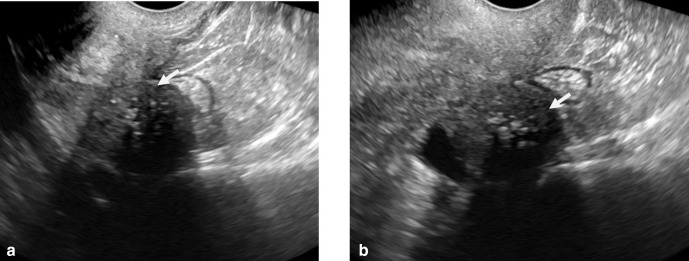
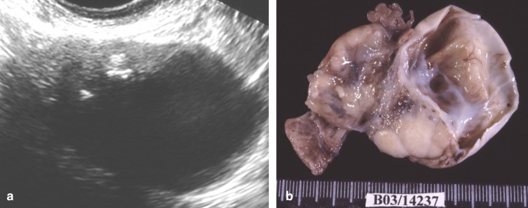
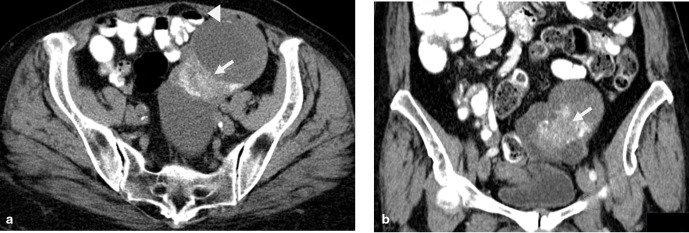
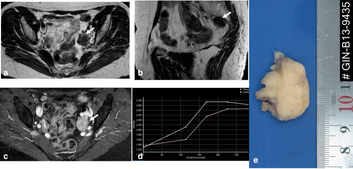
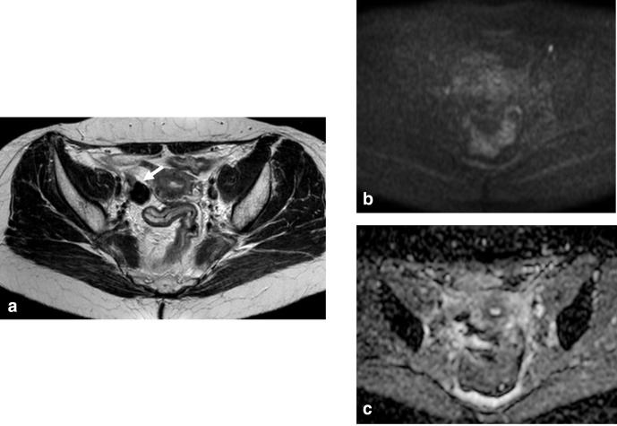
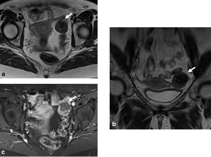
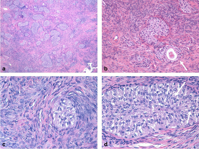
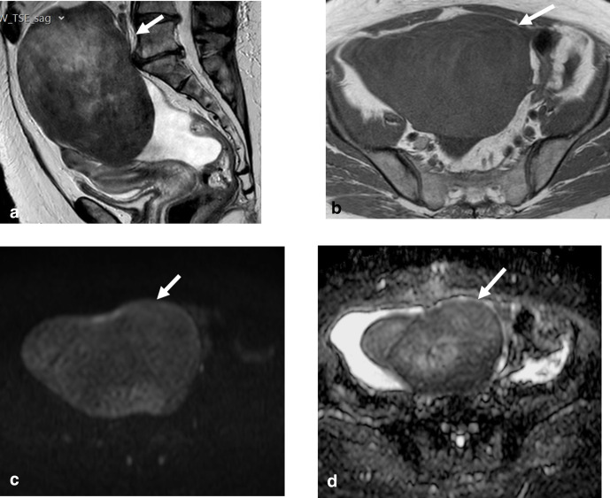
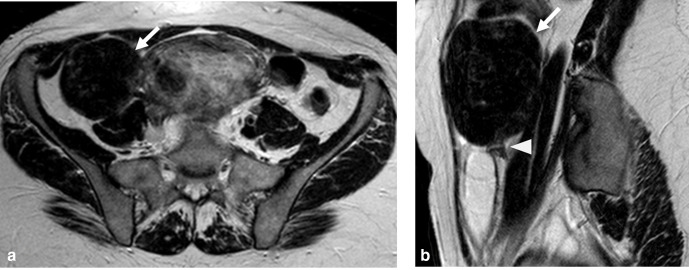
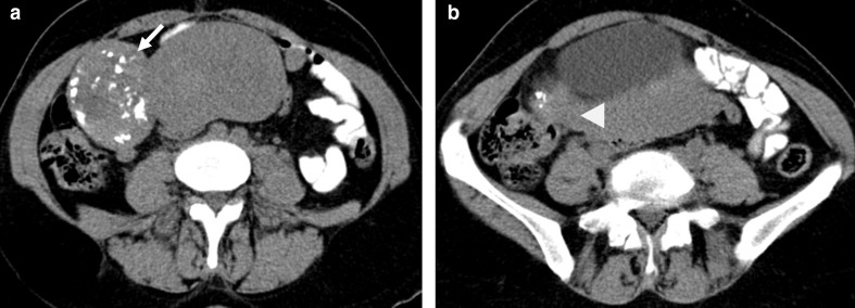
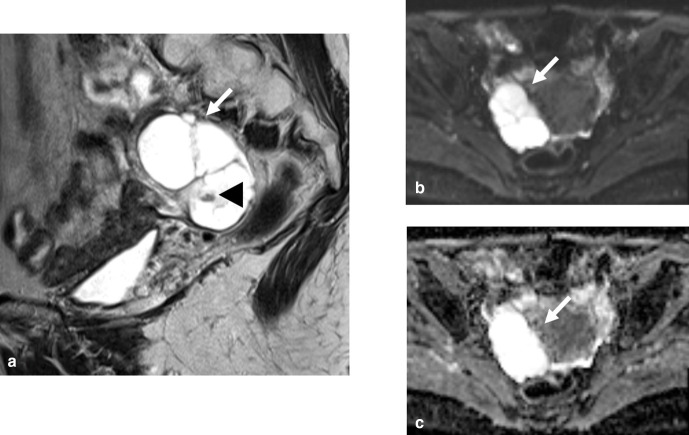
Brenner tumors are rare ovarian neoplasms composed of ovarian transition cells surrounded by dense fibrous tissue. Most of them are small tumors (<2 cm), detected incidentally in asymptomatic women. Its predominantly fibrous content results in relatively low signal on T2 weighted images, establishing differential diagnosis with ovarian fibroma and thecoma. Their imaging features are very similar, the differentiation is based on secondary characteristics, such as signs or symptoms of estrogen excess and the presence of a second ovarian neoplasm, which has been reported in up to 30% of patients with Brenner tumor. Although originally thought to be universally benign, there have been scattered reports in the past decades of borderline and malignant forms of Brenner tumors.


Figures

Figure 1.
Figure 1.
Solid Brenner tumor of the right ovary. Transvaginal ultrasound images show solid, hypoechoic ovarian tumor, with multiple milimetric calcifications (arrow), representing a benign Brenner tumor.
Figure 2.
Figure 2.
Benign mixed Brenner tumor and mucinous cystadenoma of the right ovary. (a) Transvaginal ultrasound images show mixed cystic and solid components, with calcifications in the solid portion (arrow); (b) Macroscopic evaluation revealed an ovary partially replaced by a solid and cystic tumor.
Figure 3.
Figure 3.
Benign mixed Brenner tumor and mucinous cystadenoma of the left ovary. (a) CT axial image shows mixed cystic (arrow head) and solid neoplasm, with milimetric calcifications (arrow), in which the solid part represents the Brenner tumor component; (b) CT coronal image better depicts the calcifications present in the solid component (arrow).
Figure 4.
Figure 4.
Solid Brenner tumor of the left ovary. (a) Axial T2WI shows a solid, hypointense left ovarian tumor (arrow); (b) Sagittal T2WI depicts a solid, hypointense tumor (arrow); (c) Tumor exhibits heterogeneous enhancement on T1WI FATSAT after contrast images (arrow); (d) The neoplasm presents a Type 2 curve, typical of benign tumors; (e) Patient underwent left oophorectomy. The ovary is almost entirely replaced by a pale yellow tumor, firm in consistency, with 30 mm in the long axis. FATSAT, fat saturation; WI, weighted image.
Figure 5.
Figure 5.
Borderline Brenner tumor of the right ovary. (a) Axial T2WI shows a solid, hypointense right ovarian tumor (arrow). (b,c) The neoplasm does not exhibit restriction to diffusion. Histopathological analysis revealed a Borderline Brenner tumor. WI, weighted image.
Figure 6.
Figure 6.
Malignant Brenner tumor of the left ovary. (a) Axial T2WI shows a solid, hypointense, heterogeneous, left ovarian tumor (arrow). (b) Coronal T2WI depicts a markedly hypointense central area, surrounded by a less hypointense component. (c) Tumor exhibits heterogeneous enhancement on T1WI FATSAT post-contrast images (arrow). Histopathological analysis revealed a Malignant Brenner tumor. FATSAT, fat saturation; WI, weighted image.
Figure 7.
Figure 7.
Brenner tumor (a) low power view of a dense ovarian tumor with dispersed nest of epithelial cells. (b) Small nests of bland urothelial type of epithelium with focal lumina (arrow); (c) dense fibromatous stroma with bland tumor nest; (d) nuclear grooving in some cells ”coffee beans” (arrows).
Figure 8.
Figure 8.
Meigs Syndrome – Ovarian fibroma and ascites. (a) Axial T2WI shows an ovarian neoplasm that is hypointense (arrow), as well as mild ascites (arrow head); (b) The tumor exhibits hypointensity on T1WI (c) and (d) The ovarian tumor shows no diffusion restriction.
Figure 9.
Figure 9.
Pedunculated leiomyoma. (a) Axial T2WI shows a solid, hypointense tumor (arrow); (b) Sagittal T2WI depicts that the tumor (arrow) is connected to the uterus by a stalk (arrow head). WI, weighted image.
Figure 10.
Figure 10.
Pedunculated leiomyoma. (a) CT axial image shows an hypodense, solid right ovarian neoplasm, with calcifications (arrow); (b) The tumor is connected to the uterus by means of a stalk (arrow head).
Figure 11.
Figure 11.
Cystadenofibroma of the left ovary. (a) Sagittal T2WI shows a cystic, multilocular, neoplasm (arrow), with an area of mixed low signal intensity with small cystic locules (arrow head). (b) Axial T2WI better depicts the solid area of mixed low signal intensity. (c, d) The tumor shows no diffusion restriction on b1000 images. WI, weighted image.

References

    1. Jung SE, Lee JM, Rha SE, Byun JY, Jung JI, Hahn ST. Ct and MR imaging of ovarian tumors with emphasis on differential diagnosis. Radiographics 2002; 22: 1305–25. doi: 10.1148/rg.226025033 - DOI - PubMed
    1. Roth LM, Sternberg WH. Proliferating Brenner tumors. Cancer 1971; 27: 687–93. doi: 10.1002/1097-0142(197103)27:3&lt;687::AID-CNCR2820270326&gt;3.0.CO;2-C - DOI - PubMed
    1. WHO Classification of Tumours Editorial Board .Female genital tumours. in: WHO classification of tumours: Iarc Press; 2020.
    1. Eble JN, Tavassoli FA, Devilee P. Pathology and genetics of tumours of the breast and female genital organs: Iarc Press; 2003.
    1. Miles PA, Norris HJ. Proliferative and malignant Brenner tumors of the ovary. Cancer 1972; 30: 174–86. doi: 10.1002/1097-0142(197207)30:1&lt;174::AID-CNCR2820300125&gt;3.0.CO;2-D - DOI - PubMed

Supplementary concepts