. 2022 Mar;143(3):331-348.
doi: 10.1007/s00401-021-02393-1. Epub 2021 Dec 20.

Perivascular space dilation is associated with vascular amyloid-β accumulation in the overlying cortex

Affiliations

Perivascular space dilation is associated with vascular amyloid-β accumulation in the overlying cortex

Valentina Perosa et al. Acta Neuropathol. 2022 Mar.

Abstract

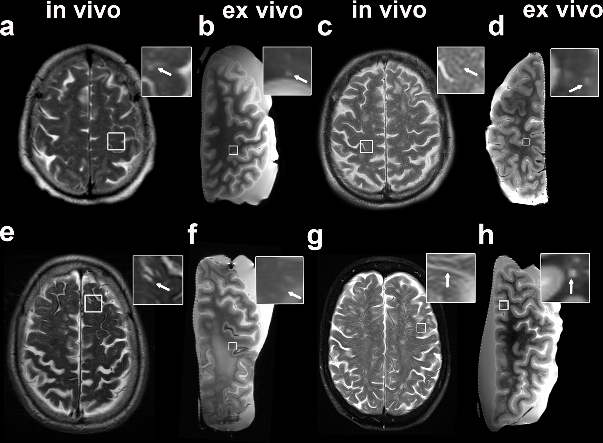
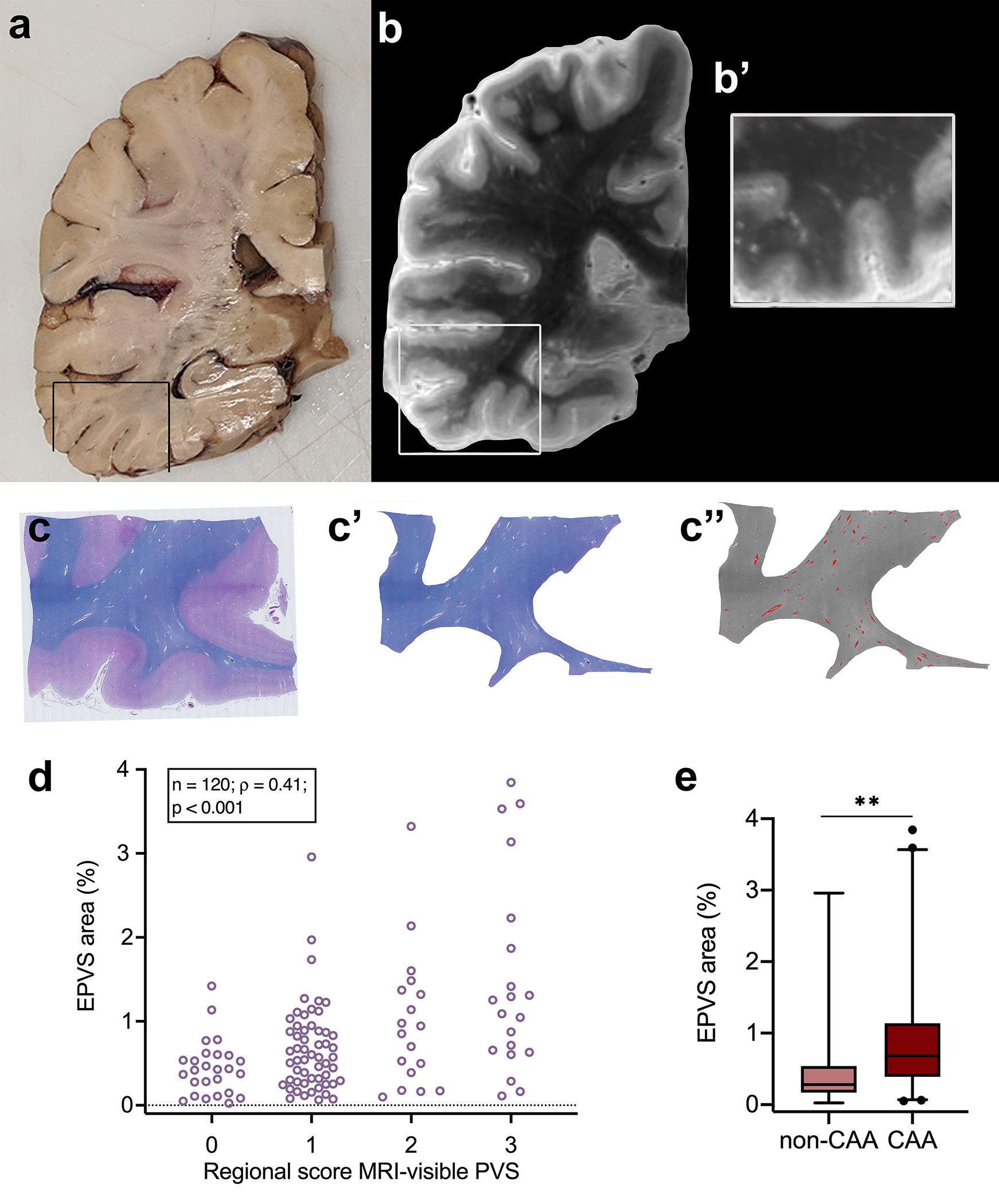
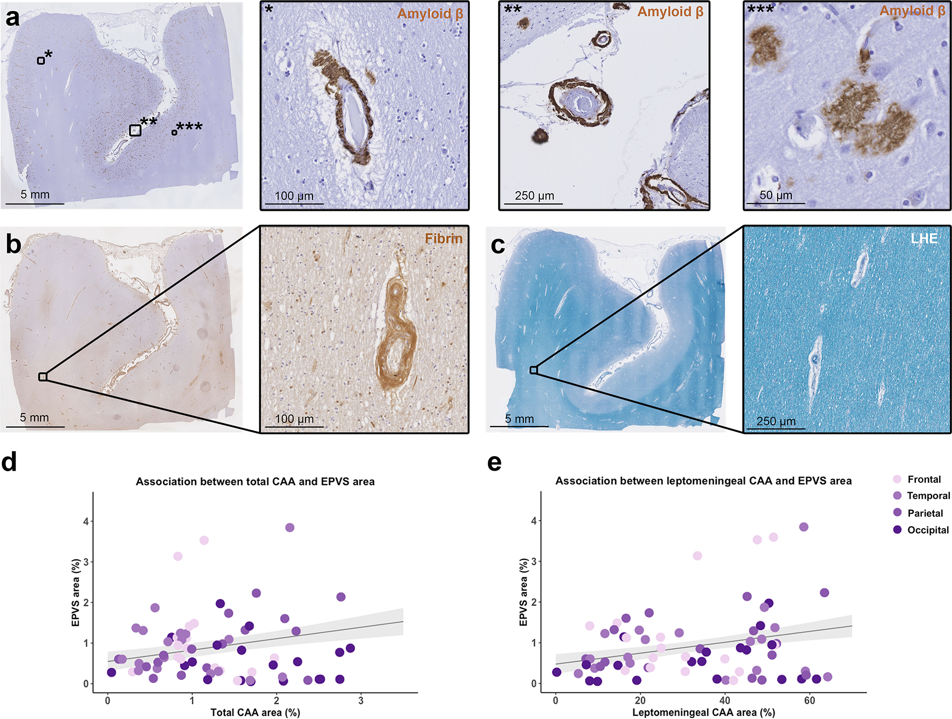
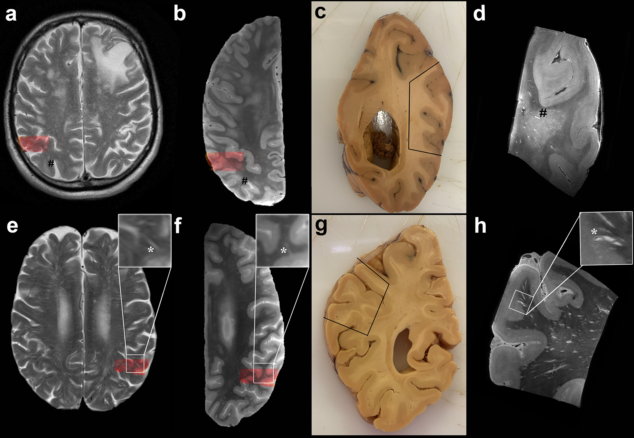
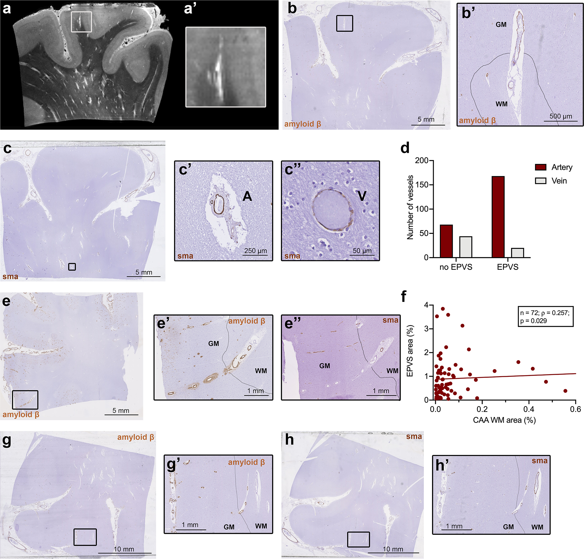
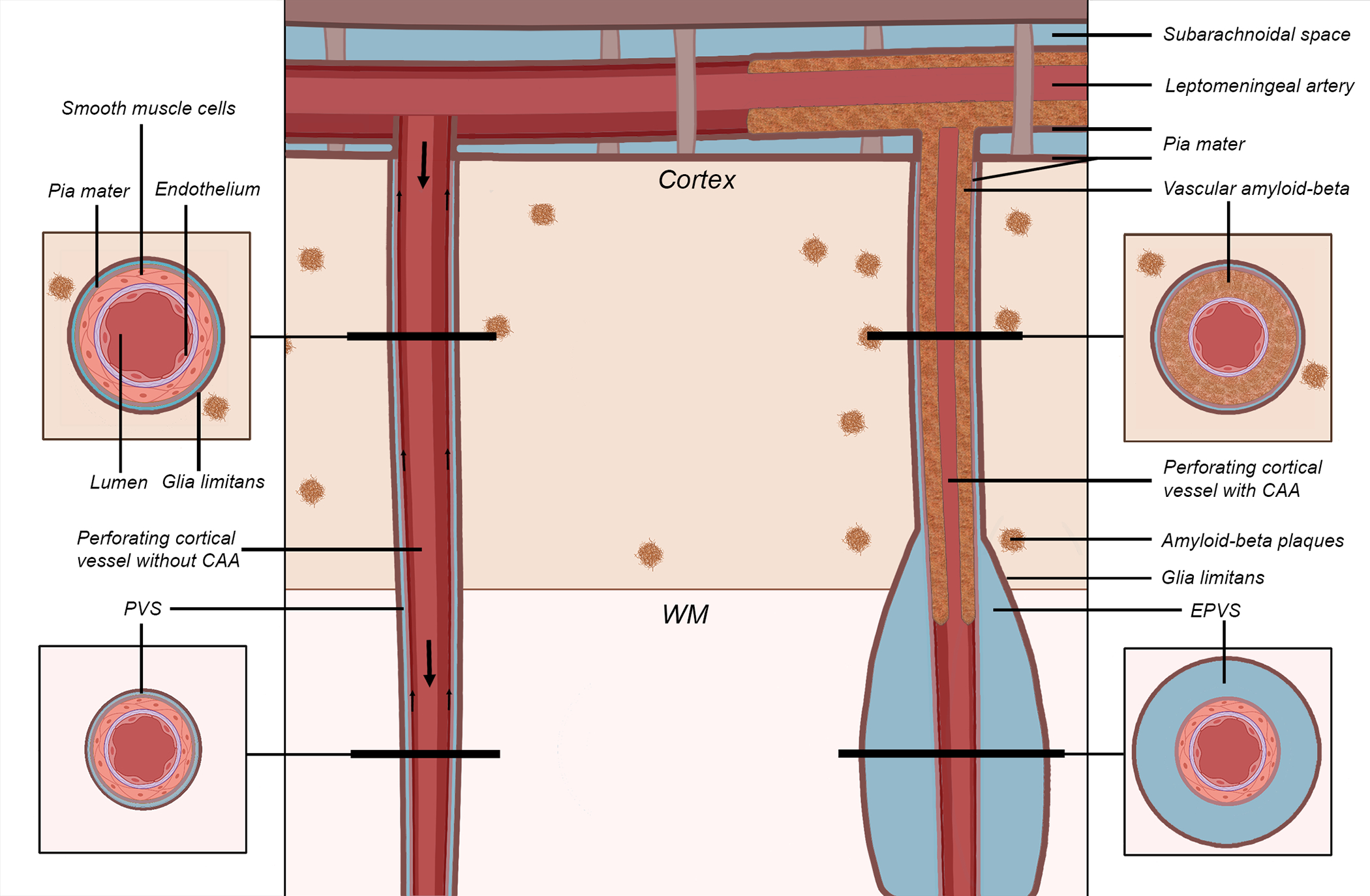
Perivascular spaces (PVS) are compartments surrounding cerebral blood vessels that become visible on MRI when enlarged. Enlarged PVS (EPVS) are commonly seen in patients with cerebral small vessel disease (CSVD) and have been suggested to reflect dysfunctional perivascular clearance of soluble waste products from the brain. In this study, we investigated histopathological correlates of EPVS and how they relate to vascular amyloid-β (Aβ) in cerebral amyloid angiopathy (CAA), a form of CSVD that commonly co-exists with Alzheimer's disease (AD) pathology. We used ex vivo MRI, semi-automatic segmentation and validated deep-learning-based models to quantify EPVS and associated histopathological abnormalities. Severity of MRI-visible PVS during life was significantly associated with severity of MRI-visible PVS on ex vivo MRI in formalin fixed intact hemispheres and corresponded with PVS enlargement on histopathology in the same areas. EPVS were located mainly around the white matter portion of perforating cortical arterioles and their burden was associated with CAA severity in the overlying cortex. Furthermore, we observed markedly reduced smooth muscle cells and increased vascular Aβ accumulation, extending into the WM, in individually affected vessels with an EPVS. Overall, these findings are consistent with the notion that EPVS reflect impaired outward flow along arterioles and have implications for our understanding of perivascular clearance mechanisms, which play an important role in the pathophysiology of CAA and AD.

Keywords: Cerebral amyloid angiopathy; Cerebral small vessel disease; Clearance; Enlarged perivascular spaces; Ex vivo MRI.


Conflict of interest statement

Competing interests

The authors declare no competing interests.

Figures

Fig. 1
Fig. 1
Examples of MRI-visible perivascular spaces (PVS) on in vivo and ex vivo MRI. The figure shows both examples of clinical in vivo (a, c, e, g) and of ex vivo (b, d, f, h) T2-weighted MRI scans of the same patient. The first is a case with mild degree of MRI-visible PVS in vivo and frequent after death (score in vivo 1 (a), ex vivo 3 (b); In vivo MRI-death interval: 7 months); the second case displays moderate MRI-visible PVS (score in vivo 2 (b), ex vivo 2 (c); In vivo MRI-death interval: 8 months). In the bottom row, a case with in vivo frequent and ex vivo severe (score in vivo 3 (e), ex vivo 4 (f); In vivo MRI-death interval: 53 months) and finally one with severe (score in vivo 4 (g), ex vivo 4 (h); In vivo MRI-death interval: 67 months) MRI-visible PVS are displayed. The insets show details of MRI-visible PVS (arrows)
Fig. 2
Fig. 2
Quantification of enlarged perivascular spaces (EPVS) on ex vivo MRI and histopathology. Example of an approximately 1 cm thick coronally cut slab (a). The box highlights the region sampled for histopathology. Coronal view of the 3 tesla ex vivo turbo spin echo sequence of the same hemisphere at the corresponding level (b). The inset shows a higher magnification of the same area at the level assessed on histopathology for EPVS severity (b’). Luxol fast blue with Hematoxylin&Eosin (LHE)-stained section from the same sample (c), with corresponding manual segmentation of the white matter (c’) and semi-automatic segmentation of the EPVS (overlaid in red) (c”). Positive correlation between the regional score of MRI-visible PVS and the percentage area of EPVS of the total white matter area, calculated on histopathological sections (one from the basal ganglia and four from cortical regions) of cases with and without CAA (d). EPVS percentage area in CAA cases was significantly higher than in non-CAA cases in the four cortical regions (frontal, temporal, parietal, and occipital). ** = p < 0.01 (e)
Fig. 3
Fig. 3
Correlation between enlarged perivascular spaces (EPVS) and cerebral amyloid angiopathy (CAA). The figure shows representative examples of the histopathological markers included in the analysis. Low magnification overview of a section that underwent immunohistochemistry against Aβ (a) and details of cortical CAA (*), leptomeningeal CAA (**) and Aβ plaques (***); an adjacent section that underwent immunohistochemistry against fibrin (b) and detail of a fibrin positive vessel in the white matter; a further adjacent section stained for Luxol fast blue with Hematoxylin&Eosin (LHE) (c) with detail of a portion of the white matter. Graphs representing the positive significant association between total CAA percentage area and EPVS percentage area in the cortical regions of CAA cases (d), and between leptomeningeal CAA percentage area and EPVS percentage area (e) (n = 19 cases; n = 72 sections; grey shadowed area shows the standard error of the model’s prediction)
Fig. 4
Fig. 4
In depth characterization of MRI-visible perivascular spaces (PVS). Top row: MRI-visible PVS (mild degree) observed in the centrum semiovale on in vivo MRI of a case with neuropathologically confirmed CAA (case no. 5) (a). Corresponding ex vivo 3 tesla T2-weighted MRI scan of the right hemisphere, where some MRI-visible PVS are also observed (b) (indicated by #). Formalin fixed tissue of the same brain, on which the parietooccipital area sampled for ultra-high resolution ex vivo 7 tesla MRI is marked by the black line (c) and co-registered to in vivo and ex vivo 3 tesla MRI (red overlaid area). The enlarged PVS are clearly visible at the 100 μm isotropic resolution T2-weighted scan (#) (d). Bottom row: Evidence of severe degree of MRI-visible PVS on the T2-weighted in vivo MRI of case no. 13 (e), with corresponding ex vivo 3 tesla MRI of the left hemisphere (f), formalin fixed tissue (g) and ultra-high resolution T2-weighted scan (h). More MRI-visible PVS are observed in case no. 13 than case 5. The MRI-visible PVS appear to be confined to the WM on in vivo and ex vivo 3 tesla MRI, whereas they can be clearly seen to continue into the cortex at ultra-high resolution in this case (insets of the bottom row, *). The EPVS-related vessels are visible at 7 tesla as hypointense structures at the center of the EPVS. They originate at the pial surface as leptomeningeal vessels, dive into the cortex as cortical perforating vessels, and continue into the white matter (h). See also Supplementary Videos 1 - 3. For the purposes of registration between in vivo and ex vivo MRI the skull and the plastic bag in which the hemispheres were placed prior to scanning, were removed from the images
Fig. 5
Fig. 5
Histopathological characterization of individual blood vessels with surrounding enlarged perivascular spaces (EPVS). Ultra-high resolution 7 tesla turbo spin echo scan of case no. 13, with an example of an EPVS extending into the cortex (a, a’). The corresponding histopathological section, on which the drawn black line represents the boundary between grey matter (indicated by GM) and white matter (indicated by WM), confirms this observation (b, b’). On serial sections immunohistochemistry for smooth muscle actin (SMA) was performed (c) and revealed, that the vast majority of vessels with an EPVS were arterioles (χ2(1) = 28.29; n = 280; p < 0.001) (d), suggesting that EPVS are mainly peri-arteriolar (c’, A = artery) and not peri-venular (c”, V = vein). On some Aβ stained cortical sections, Aβ was present not only in the wall of the cortical portion of the vessel, but to a minor extent, also in the white matter portion of the same vessel (examples are b’, e’ and g’). The same vessels showed severe loss of SMA in the cortex, but not in the WM (e”, h’). A significant positive association was found between the percentage area of EPVS and the percentage area occupied by cerebral amyloid angiopathy (CAA) in the white matter, within all CAA cases (n=72 samples in 19 cases) (f)
Fig. 6
Fig. 6
Schematic representation of the proposed mechanism of perivascular space (PVS) enlargement in cerebral amyloid angiopathy (CAA). The vessel on the left represents a healthy perforating artery (in red). The vessel on the right represents a perforating artery affected by CAA, in which Aβ accumulates within the tunica media of the wall of the cortical portion of the vessel (in brown), replacing the smooth muscle cells and leading to an enlargement of the PVS in the white matter portion of the same vessel (giving rise to the hypothesized self-reinforcing mechanism of continuing vascular Aβ accumulation). The smaller arrows on the left indicate the presumed direction of Aβ clearance along the vessel (either along the basement membranes or the perivascular compartment), while the bigger arrows represent the direction of blood flow. The results lend support to this proposed model of perivascular Aβ clearance. Key: CAA = cerebral amyloid angiopathy; EPVS = enlarged perivascular space; WM = white matter

References

    1. Abbott NJ, Pizzo ME, Preston JE, Janigro D, Thorne RG (2018) The role of brain barriers in fluid movement in the CNS: is there a ‘glymphatic’ system? Acta Neuropathol 135:387–407. doi: 10.1007/s00401-018-1812-4 - DOI - PubMed
    1. Aldea R, Weller RO, Wilcock DM, Carare RO, Richardson G (2019) Cerebrovascular smooth muscle cells as the drivers of intramural periarterial drainage of the brain. Front Aging Neurosci 11:1–17. doi: 10.3389/fnagi.2019.00001 - DOI - PMC - PubMed
    1. Atzeni A, Jansen M, Ourselin S, Iglesias JE (2018) A probabilistic model combining deep learning and multi-atlas segmentation for semi-automated labelling of histology. Lect Notes Comput Sci 11071:219–227. doi: 10.1007/978-3-030-00934-2_25 - DOI
    1. Bacyinski A, Xu M, Wang W, Hu J (2017) The Paravascular Pathway for Brain Waste Clearance: Current Understanding, Significance and Controversy. Front Neuroanat 11:1–8. doi: 10.3389/fnana.2017.00101 - DOI - PMC - PubMed
    1. Banerjee G, Kim HJ, Fox Z, Jäger HR, Wilson D, Charidimou A, Na HK, Na DL, Seo SW, Werring DJ (2017) MRI-visible perivascular space location is associated with Alzheimer’s disease independently of amyloid burden. Brain 140:1107–1116. doi: 10.1093/brain/awx003 - DOI - PubMed

Publication types

MeSH terms

Substances