. 2021 Dec 20;17(12):e1010175.
doi: 10.1371/journal.ppat.1010175. eCollection 2021 Dec.

SARS-CoV-2 receptor binding domain fusion protein efficiently neutralizes virus infection

Affiliations

SARS-CoV-2 receptor binding domain fusion protein efficiently neutralizes virus infection

Abigael Eva Chaouat et al. PLoS Pathog. .

Abstract

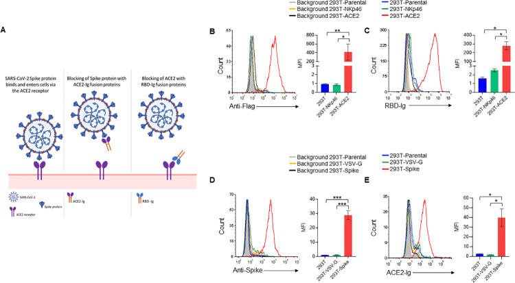
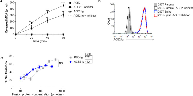
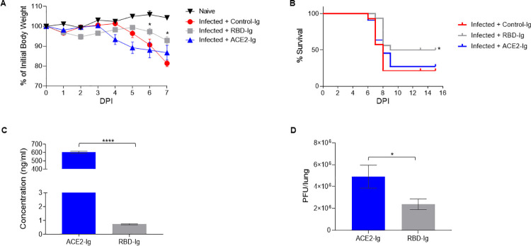
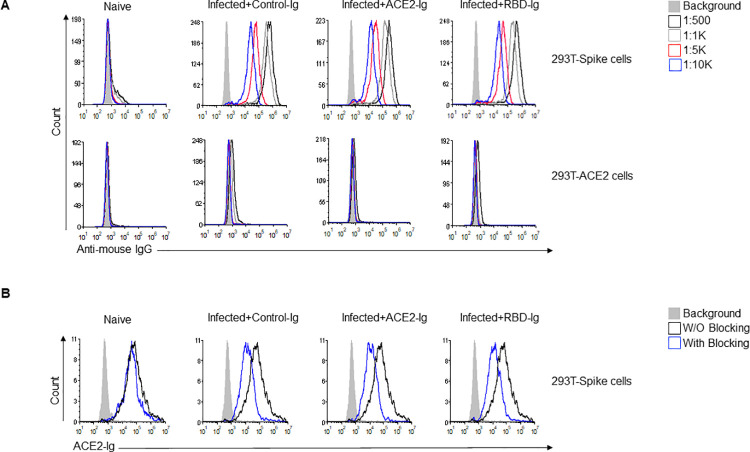
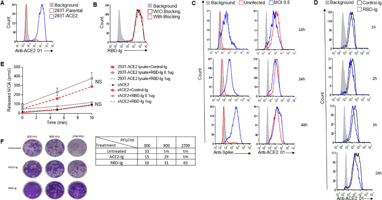
Severe acute respiratory syndrome coronavirus 2 (SARS-CoV-2) is responsible for the COVID-19 pandemic. Currently, as dangerous mutations emerge, there is an increased demand for specific treatments for SARS-CoV-2 infected patients. The spike glycoprotein on the virus envelope binds to the angiotensin converting enzyme 2 (ACE2) on host cells through its receptor binding domain (RBD) to mediate virus entry. Thus, blocking this interaction may inhibit viral entry and consequently stop infection. Here, we generated fusion proteins composed of the extracellular portions of ACE2 and RBD fused to the Fc portion of human IgG1 (ACE2-Ig and RBD-Ig, respectively). We demonstrate that ACE2-Ig is enzymatically active and that it can be recognized by the SARS-CoV-2 RBD, independently of its enzymatic activity. We further show that RBD-Ig efficiently inhibits in-vivo SARS-CoV-2 infection better than ACE2-Ig. Mechanistically, we show that anti-spike antibody generation, ACE2 enzymatic activity, and ACE2 surface expression were not affected by RBD-Ig. Finally, we show that RBD-Ig is more efficient than ACE2-Ig at neutralizing high virus titers. We thus propose that RBD-Ig physically blocks virus infection by binding to ACE2 and that RBD-Ig should be used for the treatment of SARS-CoV-2-infected patients.


Conflict of interest statement

The authors have declared that no competing interests exist.

Figures

Fig 1
Fig 1. RBD-Ig and ACE2-Ig bind their respective targets.
(A) Schematic representation of our proposed treatments. (Left panel) SARS-CoV-2 spike protein binds to the ACE2 receptor on host cells to mediate virus entry. (Middle panel) Binding of ACE2-Ig to SARS-CoV-2 Spike protein may prevent its binding to the ACE2 receptor thus blocking infection. (Right panel) Binding of RBD-Ig to the receptor ACE2 on host cells may prevent the binding of the spike protein thus blocking infection. This figure was created by us using BioRender. (B) Staining of 293T-ACE2, 293T-NKp46 and 293T parental cells using an anti-Flag antibody (left panel). Quantification of mean fluorescence intensity (MFI) of 3 repetitions (right panel). (C) Staining of 293T-ACE2, 293T-NKp46 and 293T parental cells with RBD-Ig (left panel). Quantification of MFI of 3 repetitions (right panel). (D) Spike protein surface expression on 293T cells co-transfected with either SARS-CoV-2 Spike envelope plasmid (293T-Spike cells) Vesicular stomatitis virus (VSV) G envelope plasmid (293T-VSV-G cells). 293T parental cells (untransfected) were also stained as a control (left panel). Quantification of MFI of 3 repetitions (right panel). (E) Staining of 293T-Spike/ 293T-VSV-G/ 293T parental cells with ACE2-Ig. Quantification of MFI of 3 repetitions (right panel). In all histograms except from those made for 293T parental cells, we gated on GFP positive cells. *p<0.05, **p<0.01, ***p<0.001, Student’s t-test. Data are mean ± SEM of three independent experiments.
Fig 2
Fig 2. ACE2-Ig and RBD-Ig inhibits in vitro SARS-CoV-2 infection.
(A) ACE2 enzymatic activity assay. Recombinant human ACE2 and ACE2-Ig were incubated with and without an ACE2 inhibitor, then MCA based peptide substrate was added and plate was immediately inserted in the fluorescent plate reader. *p<0.005, **p<0.0005, ***p<0.00005, Student’s t-test as compared to same treatment with inhibitor. (B) Staining of 293T-Spike cells with ACE2-Ig which was previously incubated for 15 minutes with or without an ACE2 inhibitor. (C) Plaque reduction neutralization test. Vero E6 cells were infected with SARS-CoV-2 and treated with increasing concentrations of ACE2-Ig or RBD-Ig. IC50 was calculated from the mean of the three experiments represented. Not significant (NS); Student’s t-test. Data are mean ± SEM of three independent experiments.
Fig 3
Fig 3
RBD-Ig decreases disease severity of SARS-CoV-2 infected mice. (A) Homozygous female K18-hACE2 transgenic mice were infected with SARS-CoV-2 (day 0) and treated with 75 μg/mouse of either Control-Ig, RBD-Ig or ACE2-Ig. % of initial body weight was calculated from mice which were weighed daily. *P < 0.05; Student’s t-test as compared to Infected + Control-Ig. (B) Survival percentages of SARS-CoV-2 infected mice treated as described in A. *P < 0.05; Mantel-Cox test as compared to Infected + Control-Ig. (A-B) n = 7–8, data are mean ± SEM of two independent experiments. (C-D) Homozygous female K18-hACE2 transgenic mice were infected with SARS-CoV-2 (day 0) and treated at 1, 2, 3 DPI with 2nmol/mouse of RBD-Ig (n = 10) or ACE2-Ig (n = 10), at 5 DPI mice were sacrificed, sera was collected and lung harvested. *P < 0.05; *** P < 0.0005; Student’s t-test. Data are mean ± SEM. (C) ELISA was performed from collected sera to detect fusion protein presence. (D) PFU in the harvested lungs of mice treated with ACE2-Ig or RBD-Ig.
Fig 4
Fig 4. Blocking anti-Spike antibodies are generated in all immunized mice.
(A) Anti-Spike IgG antibodies generated by mice following infection with SARS-CoV-2. At 15 DPI mice were bled through the venous tail and sera were obtained from all mice groups and from naïve mice and diluted as indicated (upper right). Sera was incubated either with 293T-Spike cells (upper histograms) or with 293T-ACE2 cells (lower histograms) as a primary antibody then cells were stained with Alexa fluor 647 anti-mouse IgG secondary antibody. (B) ACE2-Ig staining of 293T-Spike cells in the presence or absence of sera from the various groups. Sera from all indicated groups were incubated with 293T-Spike cells for 1 hour at 4°C followed by staining with ACE2-Ig. All histograms were gated on GFP positive cells. Figure shows one representative experiment out of two performed.
Fig 5
Fig 5. RBD-Ig neutralizes in-vitro infection regardless of SARS-CoV-2 titers.
(A) Staining of 293T-Parental cells and 293T-ACE2 cells with the mAb anti-ACE2 01 we generated. (B) Staining of 293T-Parental cells and 293T-ACE2 cells with RBD-Ig. Cells were incubated with or without anti-ACE2 01 for 1 hour at 4°C, washed and then staining was performed. (C) Staining of infected (MOI 0.5) and uninfected VERO E6 cells with either an anti-Spike antibody to verify infection (left panel) or with our anti-ACE2 01 antibody (right panel) at 16,24,48 hours PI. (D) Staining with anti-ACE 01 of 293T-ACE2 cells which were incubated with 1 μg of either Control-Ig or RBD-Ig for 1, 2, 6 and 24 hours. (A-D) All histograms were gated on GFP positive cells. (E) ACE2 enzymatic activity assay. Recombinant human ACE2 and 293T-ACE2 cells lysate (10 μg) were incubated with either Control-Ig (1 μg) or RBD-Ig (0.1 μg or 1 μg), then MCA based peptide substrate was added and plate was immediately read in the fluorescent plate reader. Not significant (NS), Student’s t-test as compared with Control-Ig. (F) Plaque reduction neutralization test. Vero E6 cells were infected with increasing SARS-CoV-2 titers and treated with 0.1 nmol/well of ACE2-Ig or RBD-Ig, picture of the plaques can be seen on the left panel and average number of plaques in the table on the right panel. Too many plaques to count (tm). Figures shows one representative experiment out of three performed.

References

    1. Chakraborty I, Maity P. COVID-19 outbreak: Migration, effects on society, global environment and prevention. Sci Total Environ. 2020;728. doi: 10.1016/j.scitotenv.2020.138882 - DOI - PMC - PubMed
    1. Administration D. Pfizer COVID-19 Vaccine EUA Letter of Authorization reissued 12-23-20. 2020.
    1. Administration D. Moderna COVID-19 Vaccine EUA Letter of Authorization. 2020.
    1. A Study of Ad26.COV2.S for the Prevention of SARS-CoV-2-Mediated COVID-19 in Adult Participants—Full Text View—ClinicalTrials.gov. [cited 6 Apr 2021]. Available: https://clinicaltrials.gov/ct2/show/NCT04505722#wrapper
    1. Polack FP, Thomas SJ, Kitchin N, Absalon J, Gurtman A, Lockhart S, et al.. Safety and Efficacy of the BNT162b2 mRNA Covid-19 Vaccine. N Engl J Med. 2020;383: 2603–2615. doi: 10.1056/NEJMoa2034577 - DOI - PMC - PubMed

Publication types

MeSH terms

Substances