. 2022 Jan;298(1):101500.
doi: 10.1016/j.jbc.2021.101500. Epub 2021 Dec 17.

Quantitative analysis of the formation of nucleoprotein complexes between HIV-1 Gag protein and genomic RNA using transmission electron microscopy

Affiliations

Quantitative analysis of the formation of nucleoprotein complexes between HIV-1 Gag protein and genomic RNA using transmission electron microscopy

Stéphanie Durand et al. J Biol Chem. 2022 Jan.

Abstract

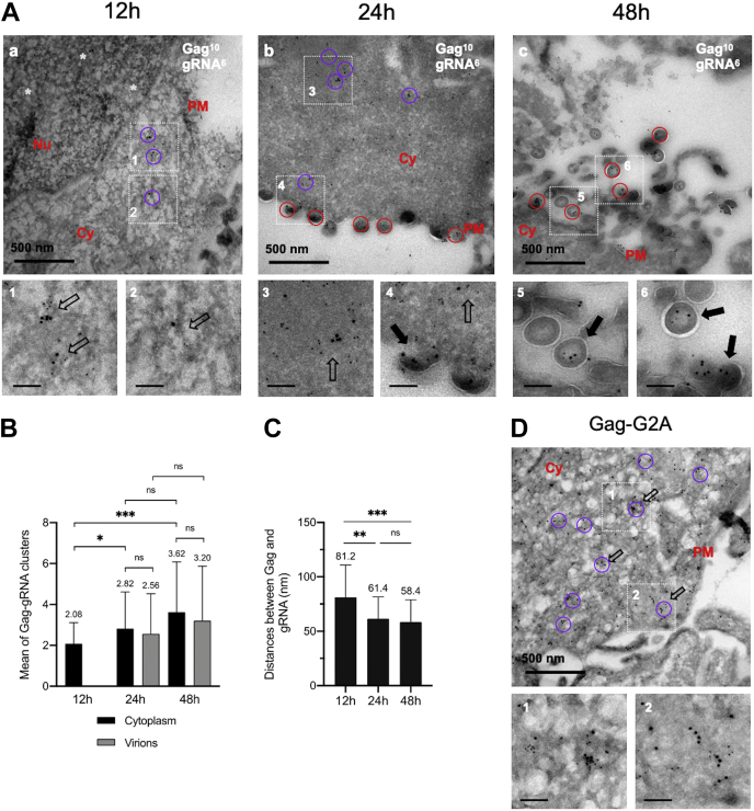
In HIV, the polyprotein precursor Gag orchestrates the formation of the viral capsid. In the current view of this viral assembly, Gag forms low-order oligomers that bind to the viral genomic RNA triggering the formation of high-ordered ribonucleoprotein complexes. However, this assembly model was established using biochemical or imaging methods that do not describe the cellular location hosting Gag-gRNA complex nor distinguish gRNA packaging in single particles. Here, we studied the intracellular localization of these complexes by electron microscopy and monitored the distances between the two partners by morphometric analysis of gold beads specifically labeling Gag and gRNA. We found that formation of these viral clusters occurred shortly after the nuclear export of the gRNA. During their transport to the plasma membrane, the distance between Gag and gRNA decreases together with an increase of gRNA packaging. Point mutations in the zinc finger patterns of the nucleocapsid domain of Gag caused an increase in the distance between Gag and gRNA as well as a sharp decrease of gRNA packaged into virions. Finally, we show that removal of stem loop 1 of the 5'-untranslated region does not interfere with gRNA packaging, whereas combined with the removal of stem loop 3 is sufficient to decrease but not abolish Gag-gRNA cluster formation and gRNA packaging. In conclusion, this morphometric analysis of Gag-gRNA cluster formation sheds new light on HIV-1 assembly that can be used to describe at nanoscale resolution other viral assembly steps involving RNA or protein-protein interactions.

Keywords: Gag; HIV-1; assembly; genomic RNA; morphometric TEM analysis; transmission electron microscopy.


Conflict of interest statement

Conflict of interest The authors declare that they have no conflicts of interest with the contents of this article.

Figures

Figure 1
Figure 1
Scheme of HIV-1 gRNA-MS2-Δenv and Gag or Gag derivatives.A, HIV-1 gRNA-MS2-Δenv is expressed from pNL4.3-MS2-Δenv plasmid and is translated under the control of HIV-1 LTR promotor in all viral genes except pol and env. The 5′ UTR contains highly structured regions important for HIV-1 replication, notably four stem-loops (SL1–SL4). This gRNA-MS2-Δenv is detected by the insertion of 24 stem-loops (orange symbols) of the MS2 bacteriophage in the pol gene interacting with MCP-eGFP-NLS proteins (green circles). B, HIV-1 Gag domains are shown as empty boxes, whereas NC is represented by its primary sequence. Deletion of each domain is symbolized by a bridge that links the flanking sequences. The substitution mutants are shown with their position and the replacing amino acid. eGFP, enhanced GFP; gRNA, genomic RNA.
Figure 2
Figure 2
Dual immunolabeling of HIV-1 Gag and gRNA as function of the time by transmission electron microscopy and morphometric analysis of Gag-gRNA clusters.A, HeLa cells were transiently transfected with a mixture of DNA plasmids expressing gRNA-MS2-Δenv and MCP-eGFP-NLS proteins (Ratio 0.6/0.4). After 12 h (a), 24 h (b), or 48 h (c) of expression, the cells were GFP-based sorted, fixed by 4% PFA, embedded in gelatin, and cryo-protected in 2.3 M sucrose. Ultrathin sections were cut and incubated first with a mixture of mouse anti-p24 and rabbit anti-eGFP antibodies, and then with anti-mouse and anti-rabbit antibodies conjugated with 10 nm and 6 nm gold particles, respectively. The circles of 100 to 130 nm in diameters correspond to Gag-gRNA clusters found in the cytoplasm (magenta) or in virions (red). Insets 1 to 6 show Gag-gRNA clusters, either free in the cytoplasm (hollow arrow) or associated with a nascent virion (full arrow). The scale is reported at the bottom left. Note that at early PT time, Gag-gRNA clusters localized in the cytoplasm, whereas at later, PT Gag-gRNA clusters are found in the virions. White stars represent 10 nm gold beads corresponding to Gag in the nucleus. B, quantification of Gag-gRNA clusters in cytoplasm and in viruses at each time PT. The data represent the mean ± SEM n = 50 cells from three independent experiments. The p values were obtained from Welsch t tests to compare the mean values (ns: nonsignificative, ∗p < 0.05, ∗∗∗p < 0.0005). C, histogram represents the mean distances between Gag and gRNA in each cluster by the measurement of the distance between each 10 nm gold bead and the surrounding 6 nm gold bead at three times PT. The beads distribution has been analyzed bioinformatically. The data shown are the mean ± the SEM from n = 90 cells from three independent experiments. The p values were obtained from Welsch t tests to compare the mean distances (ns: nonsignificative, ∗∗p < 0.0051, ∗∗∗p < 0.0001). D, HeLa cells were transiently transfected with a mixture of DNA plasmids expressing gRNA-G2A-MS2-Δenv and MCP-eGFP-NLS protein (Ratio 0.6/0.4) and analyzed, as previously described by TEM. Nu: Nucleus. Cy: Cytoplasm. PM: Plasma Membrane. The scale bar in black for zoom magnification corresponds to 100 nm. gRNA, genomic RNA; PFA, paraformaldehyde; PT, posttransfection.
Figure 3
Figure 3
Impact of NC deletions or ZF punctual mutations on HIV-1 Gag and gRNA clustering, monitored by TEM. HeLa cells were cotransfected with a mixture of modified pNL4.3-MS2-Δenv encoding for Gag (A), Gag-ΔNC (B), Gag-ΔZF1ΔZF2 (C), Gag-F16A (D), Gag-W37A (E), Gag-F16AW37A (F), Gag-H23C (G), Gag-H44C (H), or Gag-H23CH44C (I) and a plasmid expressing the MCP-eGFP-NLS protein. The cells were observed 24 h PT by TEM. The cells were prepared and stained as presented in Figure 2. Each panel shows the major observed phenotype. For each image, insets 1 to 3 show the clusters of Gag and gRNA-MS2-Δenv either free in the cytoplasm (hollow arrow) or associated with a nascent virus-like particle (full arrow). White stars represent 10 nm gold beads corresponding to Gag in the nucleus. The scale bar in black for zoom magnification corresponds to 100 nm. gRNA, genomic RNA; PT, posttransfection; TEM, transmission electronic microscopy; ZF, zinc finger.
Figure 4
Figure 4
Impact of HIV-1 Gag or gRNA mutations on Gag-gRNA clustering, monitored by TEM. HeLa cells were cotransfected with a mixture of modified pNL4.3-MS2-Δenv encoding for Gag-Δp6 (A), gRNA-ΔSL1 (B), or gRNA-ΔSL1ΔSL3 (C) and a plasmid expressing the MCP-eGFP-NLS protein. The cells were prepared and stained as presented in Figure 2. Each panel shows the major observed phenotype. For each image, insets 1 to 3 show the clusters of Gag and gRNA-MS2-Δenv either free in the cytoplasm (hollow arrow) or associated with a nascent virus (full arrow). The scale bar in black for zoom magnification corresponds to 100 nm. gRNA, genomic RNA; TEM, transmission electronic microscopy.

References

    1. Sundquist W.I., Krausslich H.-G. HIV-1 assembly, budding, and maturation. Cold Spring Harb. Perspect. Med. 2012;2 - PMC - PubMed
    1. Comas-Garcia M., Davis S., Rein A. On the selective packaging of genomic RNA by HIV-1. Viruses. 2016;8:246. - PMC - PubMed
    1. Ono A. HIV-1 assembly at the plasma membrane: Gag trafficking and localization. Future Virol. 2009;4:241–257. - PMC - PubMed
    1. Kutluay S.B., Bieniasz P.D. Analysis of the initiating events in HIV-1 particle assembly and genome packaging. PLoS Pathog. 2010;6 - PMC - PubMed
    1. Chen J., Grunwald D., Sardo L., Galli A., Plisov S., Nikolaitchik O.A., Chen D., Lockett S., Larson D.R., Pathak V.K., Hu W.-S. Cytoplasmic HIV-1 RNA is mainly transported by diffusion in the presence or absence of Gag protein. Proc. Natl. Acad. Sci. U. S. A. 2014;111:E5205–E5213. - PMC - PubMed

Publication types

LinkOut - more resources