Review
. 2021 Dec 20;16(1):84.
doi: 10.1186/s13024-021-00503-x.

TDP-43 Pathology in Alzheimer's Disease

Affiliations
Review

TDP-43 Pathology in Alzheimer's Disease

Axel Meneses et al. Mol Neurodegener. .

Abstract

Transactive response DNA binding protein of 43 kDa (TDP-43) is an intranuclear protein encoded by the TARDBP gene that is involved in RNA splicing, trafficking, stabilization, and thus, the regulation of gene expression. Cytoplasmic inclusion bodies containing phosphorylated and truncated forms of TDP-43 are hallmarks of amyotrophic lateral sclerosis (ALS) and a subset of frontotemporal lobar degeneration (FTLD). Additionally, TDP-43 inclusions have been found in up to 57% of Alzheimer's disease (AD) cases, most often in a limbic distribution, with or without hippocampal sclerosis. In some cases, TDP-43 deposits are also found in neurons with neurofibrillary tangles. AD patients with TDP-43 pathology have increased severity of cognitive impairment compared to those without TDP-43 pathology. Furthermore, the most common genetic risk factor for AD, apolipoprotein E4 (APOE4), is associated with increased frequency of TDP-43 pathology. These findings provide strong evidence that TDP-43 pathology is an integral part of multiple neurodegenerative conditions, including AD. Here, we review the biology and pathobiology of TDP-43 with a focus on its role in AD. We emphasize the need for studies on the mechanisms that lead to TDP-43 pathology, especially in the setting of age-related disorders such as AD.

Keywords: Alzheimer’s disease; TARDBP; TDP-43.


Conflict of interest statement

All the authors declare no competing interests.

Figures

Fig. 1
Fig. 1
Protein structure of transactive response DNA binding protein of 43 kDa (TDP-43). TDP-43 is a 414 amino acid protein with a nuclear localization sequence (NLS) followed by two RNA binding domains (RBD1 and RBD2), a nuclear export sequence (NES), and a glycine rich prion-like (GRD) C-terminus. The mutations reported to increase the risk of ALS (red), FTLD (blue), and AD (orange) are indicated
Fig. 2
Fig. 2
Representative images of TDP-43 pathology subtypes in FTLD-TDP brains. (A) Immunohistochemistry with an anti-phosphorylated-TDP-43 antibody (pSer409/pSer410) shows numerous neuronal cytoplasmic inclusions, short dystrophic neurites, and neuronal intranuclear inclusion (inset) in Type A; diffuse granular neuronal cytoplasmic inclusions in Type B; and numerous thick and long dystrophic neurites in Type C, in the superficial layer of the midfrontal gyrus (upper panel). In the dentate gyrus (lower panel), Type A shows compact neuronal cytoplasmic inclusions; Type B shows diffuse granular neuronal cytoplasmic inclusions; and Type C shows Pick body-like neuronal cytoplasmic inclusion. Scale bar = 50 μm. (B) A summary of clinical, pathological, and genetic features of TDP-43 pathology subtypes. NCI: Neuronal cytoplasmic inclusion; NII: Neuronal intranuclear inclusion; DN: Dystrophic neurite; GCI: Glia cytoplasmic inclusion; DG: Dentate gyrus; FTD: Frontotemporal dementia; bvFTD: Behavioral variant frontotemporal dementia; PNFA: Progressive non-fluent aphasia; MND: Motor neuron disease; and SD: Semantic dementia
Fig. 3
Fig. 3
Illustration of the involvement of TDP-43 in the progression of Alzheimer’s disease. In the brain of Alzheimer’s disease (AD), the amyloid-β (Aβ) peptide is produced through the proteolytic processing of a transmembrane protein, amyloid precursor protein (APP) by β- and γ-secretases. The accumulation of soluble Aβ monomers in the brain parenchyma leads to the formation of Aβ oligomers, fibrils, and eventually Aβ plaques, due to overproduction and/or impaired Aβ clearance pathways contributing to the development of AD. The pathological changes of tau protein decrease its microtubule binding capacity and disrupts microtubule stability causing microtubule disintegration. The intracellular aggregates of tau protein form the neurofibrillary tangles. Tau deposits are also found in neuronal cell processes (“neuropil threads”) and in dystrophic neurites within Aβ plaques. Aβ plaques are heterogeneous lesions containing not only amyloid deposits and tau-positive neurites, but also neurites with degenerating pre- and post-synaptic elements (neurite dystrophy), as well as activated microglia, reactive astrocytes, and dysfunction of oligodendrocytes causing demyelination. TDP-43 is synthesized in the cytoplasm and retains the ability to shuttle from the cytoplasm into the nucleus where it primarily resides to perform its physiological functions such as RNA splicing. During the progression of AD, the pathogenic events lead to TDP-43 depletion from the nucleus, TDP-43 mislocalization into the cytoplasm, and the formation of insoluble TDP-43 aggregates. The neurodegeneration brought about by pathological TDP-43 can be caused by a potential combination of both a loss of physiological function and a gain of toxic functions
Fig. 4
Fig. 4
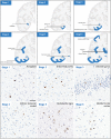
Distribution pattern of TDP-43 pathology in AD. (Upper panel) Illustration of TDP-43 stage in AD. An anterior coronal section depicts TDP-43 pathology progression from the amygdala (Stage 1), into the subiculum and entorhinal cortex (Stage 2), and then leads into the occipitotemporal cortex and dentate gyrus (Stage 3) of the hippocampus, followed by the insular cortex and the inferior temporal cortex (Stage 4). After progressing into the substantia nigra (Stage 5), the pathology reaches its final stage at the basal ganglia (putamen and globus pallidus) and middle frontal cortex (Stage 6). (Lower panel) Immunohistochemistry with an anti-phosphorylated-TDP-43 antibody shows representative images of TDP-43 pathology in different brain regions of each stage. Stage 1, amygdala; Stage 2, entorhinal cortex; Stage 3, dentate gyrus; Stage 4, inferior temporal gyrus; Stage 5, substantia nigra; Stage 6, middle frontal gyrus. Scale bar = 50 μm

References

    1. DeTure MA, Dickson DW. The neuropathological diagnosis of Alzheimer's disease. Mol Neurodegener. 2019;14:32. - PMC - PubMed
    1. Ghiso J, Frangione B. Cerebral amyloidosis, amyloid angiopathy, and their relationship to stroke and dementia. J Alzheimers Dis. 2001;3:65–73. - PubMed
    1. Guo T, Zhang D, Zeng Y, Huang TY, Xu H, Zhao Y. Molecular and cellular mechanisms underlying the pathogenesis of Alzheimer's disease. Mol Neurodegener. 2020;15:40. - PMC - PubMed
    1. Spillantini MG, Goedert M. Tau protein pathology in neurodegenerative diseases. Trends Neurosci. 1998;21:428–433. - PubMed
    1. Spillantini MG, Murrell JR, Goedert M, Farlow M, Klug A, Ghetti B. Mutations in the tau gene (MAPT) in FTDP-17: the family with Multiple System Tauopathy with Presenile Dementia (MSTD) J Alzheimers Dis. 2006;9:373–380. - PubMed

Publication types

MeSH terms