. 2021 Dec 20;13(1):11.
doi: 10.1038/s41419-021-04458-1.

TMEM165 a new player in proteoglycan synthesis: loss of TMEM165 impairs elongation of chondroitin- and heparan-sulfate glycosaminoglycan chains of proteoglycans and triggers early chondrocyte differentiation and hypertrophy

Affiliations

TMEM165 a new player in proteoglycan synthesis: loss of TMEM165 impairs elongation of chondroitin- and heparan-sulfate glycosaminoglycan chains of proteoglycans and triggers early chondrocyte differentiation and hypertrophy

Sajida Khan et al. Cell Death Dis. .

Abstract

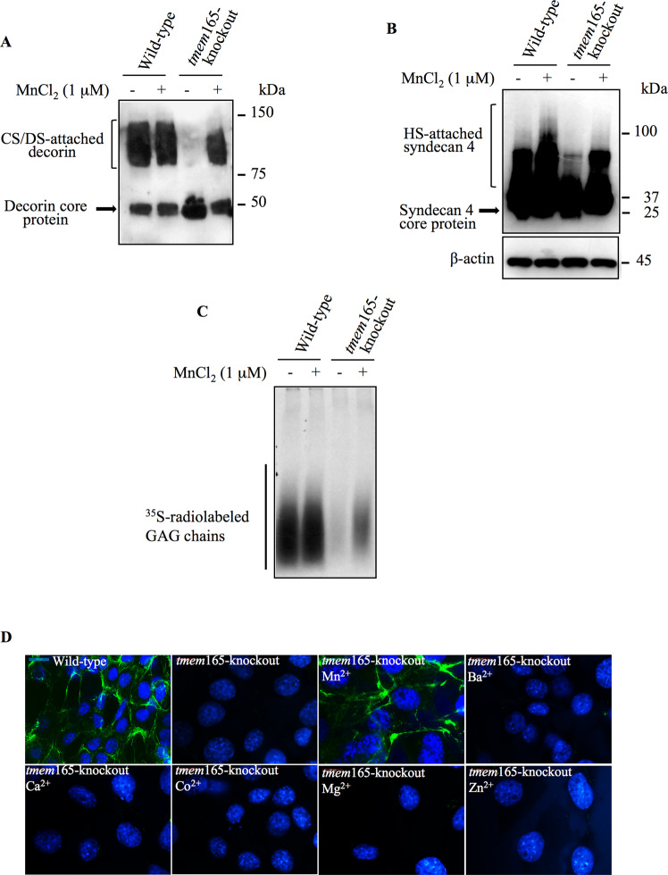
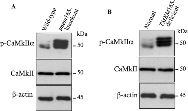
TMEM165 deficiency leads to skeletal disorder characterized by major skeletal dysplasia and pronounced dwarfism. However, the molecular mechanisms involved have not been fully understood. Here, we uncover that TMEM165 deficiency impairs the synthesis of proteoglycans by producing a blockage in the elongation of chondroitin-and heparan-sulfate glycosaminoglycan chains leading to the synthesis of proteoglycans with shorter glycosaminoglycan chains. We demonstrated that the blockage in elongation of glycosaminoglycan chains is not due to defect in the Golgi elongating enzymes but rather to availability of the co-factor Mn2+. Supplementation of cell with Mn2+ rescue the elongation process, confirming a role of TMEM165 in Mn2+ Golgi homeostasis. Additionally, we showed that TMEM165 deficiency functionally impairs TGFβ and BMP signaling pathways in chondrocytes and in fibroblast cells of TMEM165 deficient patients. Finally, we found that loss of TMEM165 impairs chondrogenic differentiation by accelerating the timing of Ihh expression and promoting early chondrocyte maturation and hypertrophy. Collectively, our results indicate that TMEM165 plays an important role in proteoglycan synthesis and underline the critical role of glycosaminoglycan chains structure in the regulation of chondrogenesis. Our data also suggest that Mn2+ supplementation may be a promising therapeutic strategy in the treatment of TMEM165 deficient patients.


Conflict of interest statement

The authors declare no competing interests.

Figures

Fig. 1
Fig. 1. CRISPR/Cas9 knockdown of TMEM165.
A Alignment of TMEM165 targeted sequence from wild-type and mutant mouse ATDC5 cells. B Detection of TMEM165 in cell lysates from wild-type and tmem165-knockout mouse ATDC5 cells (mutant 1, 2, 3 and 4) using anti-TMEM165 specific antibodies. β-actin was used as loading control (n = 3). C Immunofluorescence analysis of the expression of TMEM165 in mouse ATDC5 control and tmem165-knockout cells using antibodies against TMEM165 (green). GM130 (red) was used as a Golgi marker. The nucleus was stained with DAPI (blue) (n = 3). Digital images were captured with an inverted microscope, Leica DMI3000. Representative images from three independent experiments are shown. Scale bar: 50 μm.
Fig. 2
Fig. 2. GAG chain elongation is impaired in TMEM165-deficient cells.
A PG anabolism evaluation in wild-type and tmem165-knockout mouse ATDC5 cells by measurement of the incorporation rate of [35S]-sulfate into the GAG chains (n = 3). B SDS-PAGE and autoradiography analysis of neosynthesized radiolabelled PG-GAG chains in wild-type and tmem165-knockout mouse ATDC5 cells (n = 3). C Detection of decorin in conditioned medium of wild-type and tmem165-knockout mouse ATDC5 cells and (D) in wild-type and TMEM165-knockout HEK293 cells (n = 3). E Detection of decorin S34A mutant lacking GAG chain in conditioned medium of wild-type and mutant mouse ATDC5 cells (n = 3). F Analysis of the sensitivity to degradation by chondroitinase ABC of GAG chains of decorin in conditioned medium of wild-type and tmem165-knockout mouse ATDC5 cells (n = 3). G SDS-PAGE and autoradiography analysis of neosynthesized radiolabelled GAG chains primed with 4MU-Xyl in wild-type and tmem165-knockout mouse ATDC5 cells (n = 3). H Detection of HA-syndecan 4 in cell lysates of wild-type and tmem165-knockout mouse ATDC5 cells transfected with HA-syndecan-4 expression vector. β-actin was used as loading control. (n = 3) I Immunofluorescence analysis of cell surface HS GAG chains using anti-HS specific antibodies (green) and of the expression of TMEM165 (red) in wild-type and tmem165-knockout mouse ATDC5 cells. The nucleus was stained with DAPI (blue). Digital images were captured with an inverted microscope, Leica DM13000. Representative images from three independent experiments are shown. Scale bar: 50 μm.
Fig. 3
Fig. 3. Expression of CS polymerizing enzymes did not overcome GAG elongation defects in TMEM165-deficient cells.
A Fold changes in mRNA expression of CS polymerizing enzymes CHSY1 and CHSY2, and HS polymerizing enzymes EXT1 and EXT2 in tmem165-knockout mouse ATDC5 cells normalized to wild-type mouse ATDC5 cells and B in TMEM165-deficient fibroblasts normalized to fibroblast control cells. qPCR values were normalized for the housekeeping gene ribosomal protein S29 and are expressed as the relative expression compared with control. Data are expressed as mean ± S.D. Statistical analysis was performed with an unpaired Student’s t test (n = 3; *p < 0.05; **p < 0.01). C Detection of decorin in conditioned medium of wild-type and mutant ATDC5 cells transfected with empty vector, Myc-tagged CHSY1, HA-tagged CHSY2 or Myc-tagged CHSY1 and HA-tagged CHSY2 along with decorin expression vector. D Detection of CHSY1 and (E) of CHSY2 in wild-type and tmem165-knockout mouse ATDC5 cells. β-actin was used as loading control (n = 3). Representative images from three independent experiments are shown.
Fig. 4
Fig. 4. Manganese supplementation rescue GAG elongation in TMEM165-deficient cells.
A Detection of decorin in conditioned medium of wild-type and tmem165-knockout mouse ATDC5 cells transfected with the expression vector for decorin. (B) Detection of HA-tagged syndecan 4 in cell lysates of wild-type and tmem165-knockout mouse ATDC5 cells transfected with HA-tagged syndecan 4 expression vector and grown in the presence or absence of MnCl2 (1 µM). β-actin was used as loading control (n = 3). C SDS-PAGE and autoradiography of [35S]-sulfate radiolabelled GAG chains primed with 4MU-Xyl in wild-type and tmem165-mutant mouse ATDC5 cells grown in the presence or absence of MnCl2 (1 µM) (n = 3). One representative blot of three independent experiments is shown. D Immunofluorescence analysis of cell surface HSGAG chains using anti-HS specific antibodies (green) in wild-type and tmem165-knockout mouse ATDC5 cells cultured in the absence and presence of Mn2+, Ba2+, Ca2+, Co2+, Mg2+ and Zn2+. The nucleus was stained with DAPI (blue). Digital images were captured with an inverted microscope, Leica DM13000. Representative images from three independent experiments are shown. Scale bar: 50 μm.
Fig. 5
Fig. 5. Phospho-CaMKIIα is activated in TMEM165-deficient cells.
A Immunoblot analysis of phospho-CaMkIIα (pCaMkIIα) level in cell lysates of wild-type and tmem165-knockout mouse ATDC5 cells and (B) in cell lysates of normal fibroblasts and TMEM165-deficient fibroblasts from CDG patients, using specific antiphospho-CaMkIIα and anti-CaMkIIα antibodies. β-actin was used as loading control (n = 3). Representative images from three independent experiments are shown.
Fig. 6
Fig. 6. TGF-β signaling pathway is impaired in TMEM165-deficient cells.
A Detection of phosphorylated Smad2 (pSmad2) and total Smad2 (Smad2) in cell lysates from wild-type and tmem165-knockout mouse ATDC5 cells (n = 3). B Fold changes of serpine expression in tmem165-knockout cells normalized to wild-type mouse ATDC5 cells. C Fold changes of TGF-β reporter activity in tmem165-knockout cells normalized to wild-type mouse ATDC5. D Detection of phosphorylated Smad2 (pSmad2) and total Smad2 in cell lysates from control and TMEM165-deficient CDG patient fibroblast cells (n = 3). E Fold changes of serpine expression in TMEM165-deficient fibroblasts normalized to normal fibroblast cells. F Detection of p-Smad2 in cell lysates from wild-type and tmem165-knockout mouse ATDC5 cells and (G) from normal and TMEM165-deficient CDG patient fibroblast cells treated or not with TGFβ1 (1 ng/ml) for 1 hour (n = 3). H Fold changes of TGFβR1 and TGFβR2 expression in tmem165-knockout mouse ATDC5 cells normalized to wild-type mouse ATDC5 cells. I Detection of TGFβR2 in cell lysates from wild-type and tmem165-mutant mouse ATDC5 (n = 3). J Fold changes of TGFβR1 and TGFβR2 expression in TMEM165-deficient fibroblasts from CDG patient normalized to normal fibroblast cells. K Detection of TGFβR2 in cell lysates from normal fibroblasts and TMEM165-deficient CDG patient fibroblast cells (n = 3). L Fold changes of asporin expression in tmem165-knockout cells normalized to wild-type mouse ATDC5 cells and in TMEM165-deficient fibroblast cells normalized to normal fibroblast cells. M Detection of asporin in conditioned medium of wild-type and tmem165-knockout mouse ATDC5 cells and of normal fibroblasts and TMEM165-deficient CDG patient fibrobast cells. β-actin was used as loading control (n = 3). qPCR values were normalized for the housekeeping gene ribosomal protein S29 and are expressed as the relative expression compared with control. Data are expressed as mean ± S.D. Statistical analysis was performed with an unpaired Student’s t test (n = 3; *p < 0.05; **p < 0.01). Representative images from three independent experiments are shown.
Fig. 7
Fig. 7. BMP signaling is activated in TMEM165-deficient cells.
A Detection of phosphorylated Smad1, 5, 9 (pSmad1,5,9) and total Smad in cell lysates from wild-type and tmem165-mutant mouse ATDC5 cells and (B) from normal fibroblasts and TMEM165-deficient CDG patient fibroblast cells (n = 3). C Fold changes of Id1 expression in tmem165-knockout cells normalized to wild-type mouse ATDC5 cells. D Fold changes of BMP reporter activity in tmem165-knockout cells normalized to wild-type mouse ATDC5 cells. E Fold changes of BMPR1A, BMPR1B and BMPR2 expression in tmem165-knockout cells normalized to wild-type mouse ATDC5 cells. F Detection of BMPR2 in cell lysates of wild-type and tmem165-knockout mouse ATDC5 cells. β-actin was used as loading control (n = 3). G Fold changes of BMPR1A, BMPR1B and BMPR2 expression in TMEM165-deficient fibroblasts normalized to normal fibroblast cells. H Detection of BMPR2 in cell lysates of normal fibroblasts and TMEM165-deficient CDG patient fibroblast cells. β-actin was used as loading control (n = 3). I Fold changes of Noggin expression in tmem165-knockout mouse ATDC5 cells normalized to wild-type ATDC5 cells and (J) in TMEM165-deficient CDG patient fibroblasts normalized to normal fibroblast cells. qPCR values were normalized for the housekeeping gene ribosomal protein S29 and are expressed as the relative expression compared with control. Data are expressed as mean ± S.D. Statistical analysis was performed with an unpaired Student’s t test (n = 3; *p < 0.05; **p < 0.01). K Detection of phosphorylated Smad2 (pSmad2) and Smad1, 5, 9 (pSmad1,5,9) and of total Smad in cell lysates from wild-type and tmem165-mutant mouse ATDC5 cells cultured in medium with or without Mn2+ supplementation. β-actin was used as loading control (n = 3). Representative images from three independent experiments are shown.
Fig. 8
Fig. 8. Early hypertrophic differentiation of tmem165-deficient mouse ATDC5 cells.
A Fold changes of chondrogenic markers expression in wild-type mouse ATDC5 cells and tmem165-knockout cells. RT-qPCR analysis of the mRNA levels of chondrogenic markers SOX9, Col2A, and Aggrecan, and (B) of hypertrophic markers Ihh and OCN. qPCR values were normalized for the housekeeping gene ribosomal protein S29 and are expressed as the relative expression compared with control. Data are expressed as mean ± S.D. Statistical analysis was performed with an unpaired Student’s t test (n = 3; *p < 0.05; **p < 0.01). C Mineralization of wild-type and tmem165-deficient mouse ATDC5 cells analyzed by Alizarin red staining at Days 0, 14, 21, and 28. Representative images from three independent experiments are shown. D Loss of TMEM165 function impairs Golgi Mn2+ homeostasis necessary for glycosyltransferase polymerization activities leading to blockage in the elongation of GAG chains of proteoglycans. Blockage in the elongation of GAG chains by loss of TMEM165 may account for dysregulation of TGF/BMP and Ihh signaling, and therefore in defects in chondrocyte differentiation and maturation. However, other mechanisms can’t be ruled-out.

References

    1. Varki A. Biological roles of oligosaccharides: all of the theories are correct. Glycobiology. 1993;3:97–130. doi: 10.1093/glycob/3.2.97. - DOI - PMC - PubMed
    1. Sharon N, Lis H. Lectins as cell recognition molecules. Science. 1989;246:227–34. doi: 10.1126/science.2552581. - DOI - PubMed
    1. Demaegd D, Foulquier F, Colinet AS, Gremillon L, Legrand D, Mariot P, et al. Newly characterized Golgi-localized family of proteins is involved in calcium and pH homeostasis in yeast and human cells. Proc Natl Acad Sci USA. 2013;110:6859–64. doi: 10.1073/pnas.1219871110. - DOI - PMC - PubMed
    1. Foulquier F, Amyere M, Jaeken J, Zeevaert R, Schollen E, Race V, et al. TMEM165 deficiency causes a congenital disorder of glycosylation. Am J Hum Genet. 2012;91:15–26. doi: 10.1016/j.ajhg.2012.05.002. - DOI - PMC - PubMed
    1. Morelle W, Potelle S, Witters P, Wong S, Climer L, Lupashin V, et al. Galactose supplementation in patients with TMEM165-CDG rescues the glycosylation defects. J Clin Endocrinol Metab. 2017;102:1375–86.. doi: 10.1210/jc.2016-3443. - DOI - PMC - PubMed

Publication types

MeSH terms