. 2021 Dec 20;6(1):85.
doi: 10.1038/s41536-021-00195-3.

A bioinspired gelatin-hyaluronic acid-based hybrid interpenetrating network for the enhancement of retinal ganglion cells replacement therapy

Affiliations

A bioinspired gelatin-hyaluronic acid-based hybrid interpenetrating network for the enhancement of retinal ganglion cells replacement therapy

Pierre C Dromel et al. NPJ Regen Med. .

Abstract

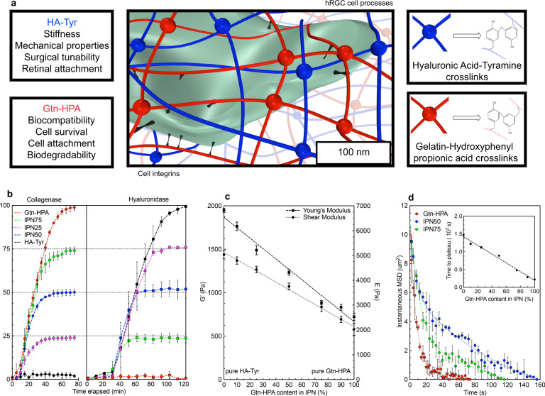
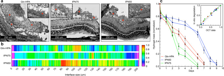
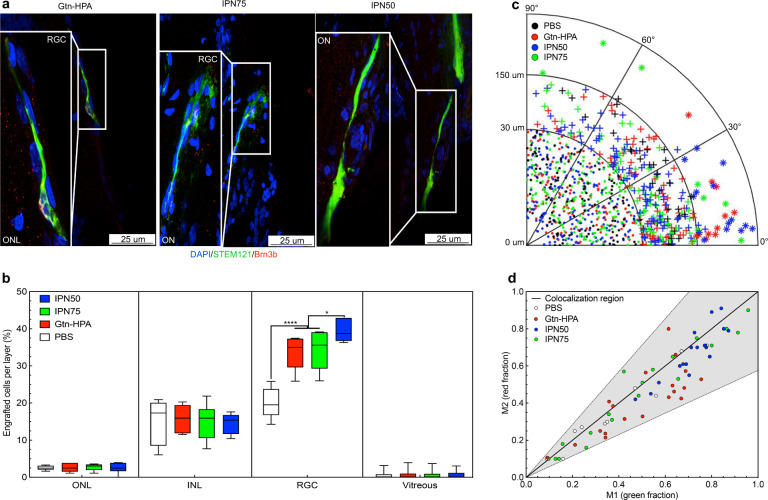
Biomaterial-based cell replacement approaches to regenerative medicine are emerging as promising treatments for a wide array of profound clinical problems. Here we report an interpenetrating polymer network (IPN) composed of gelatin-hydroxyphenyl propionic acid and hyaluronic acid tyramine that is able to enhance intravitreal retinal cell therapy. By tuning our bioinspired hydrogel to mimic the vitreous chemical composition and mechanical characteristics we were able to improve in vitro and in vivo viability of human retinal ganglion cells (hRGC) incorporated into the IPN. In vivo vitreal injections of cell-bearing IPN in rats showed extensive attachment to the inner limiting membrane of the retina, improving with hydrogels stiffness. Engrafted hRGC displayed signs of regenerating processes along the optic nerve. Of note was the decrease in the immune cell response to hRGC delivered in the gel. The findings compel further translation of the gelatin-hyaluronic acid IPN for intravitreal cell therapy.


Conflict of interest statement

The authors declare no competing interests.

Figures

Fig. 1
Fig. 1. The Gelatin and Hyaluronic Acid interpenetrating network system and its mechanical and chemical characterization.
a Schematic and 3D model of a human cell encapsulated in the IPN made of Gelatin-HPA (red) and HA-Tyr (blue) with integrins bonding to Gtn-HPA only (black). Legends shows the two different crosslinks with their respective chemical structures. b Hydrogels’ degradation assays performed with Collagenase and Hyaluronidase treatment for Gtn-HPA (red), IPN75 (green), IPN50 (blue), IPN25 (pink) and HA-Tyr (black). Degradation assay, comprising n = 15 replicates, was measured every 5 min with collagenase and every 10 min with Hyaluronidase. c Shear (dot line) and Young’s (dash line) moduli measurements for multiple IPN with different Gtn-HPA content (ranging from 10% to 90%). Linear regression was applied both set of values, with n = 15. d Mean square displacement measurements of PLGA microbeads encapsulated in materials during microrheology assay for Gtn-HPA (red), IPN75 (green) and IPN50 (blue). N = 15 microbeads were tracked for each sample and the data were fitted with a double exponential decay regression. Inset (d) shows the time to reach stability of the different IPN during the gelation process. Linear regression was applied to the data confirming the increase of gelation time with the addition of HA-Tyr in the mix. All data is shown as mean ± SEM.
Fig. 2
Fig. 2. Optimal interpenetrating network candidates for human retinal ganglion cells viability, encapsulation, and in vivo release.
a Viability assay at day 3 of encapsulated human retinal ganglion cells in IPN with different Gtn-HPA content and a range of crosslinker concentration compared with PBS. Data were measured for 10 field of views of live/dead staining. Data shown as mean ± SEM and one-way ANOVA followed by student t test was performed showing a statistically high significant difference between H2O2-1mM and all others for more than 50% Gtn-HPA in the IPN (****p < 0.0001). b Viability assay through the time of encapsulated hRGC in hydrogel candidates (Gtn-HPA, IPN75, and IPN50) with media or PBS. Plain bars show cells deprived from nutrients while hashed bars represent cells in their defined medium. Data shown as mean ± SEM of triplicate wells with 15 different fields. Two-way ANOVA, followed by Tukey’s test, was performed and shows a statistically high significant difference between samples including media and all other groups (**p = 0.005 and ***p = 0.0001) at all time points; and between hydrogels groups deprived from nutrients compared to PBS (*p = 0.01 and ****p < 0.0001). c Relative fluorescence intensity of hRGC stained with live/dead assay, imaged with confocal microscopy for Gtn-HPA, IPN75, IPN50, and PBS, on 150 um thick slides, in the function of their z position. d. 3D fluorescence image of live (CalceinAM-green) and dead (Ethidium Bromide-red) hRGC encapsulated in the hydrogel candidates, imaged with confocal microscopy on 1.2×1.2×0.3 mm samples. All images were taken at 10X magnification under fluorescence microscopy.
Fig. 3
Fig. 3. In vivo proof of attachment of hydrogel candidates to the inner limiting membrane (ILM) of the retina.
a Hematoxylin and Eosin (H&E) staining of rat’s retina 3 days post hydrogel injections for Gtn-HPA, IPN75, and IPN50 groups. Images were taken under bright-field microscopy at 10X and 63X magnification for respectively large image and inset. Hydrogel presence is observed and labelled with red stars. Inset shows higher magnification and interface of gel with ILM to prove attachment. The scale bar is 250 um. b. Analysis of hydrogel-ILM interface from H&E staining with heat map. 200 um length interfaces were analyzed for all three samples. Heat map ranges from 0 with no attachment at the interface to 1 with attachment seen on all different slides. c. Gtn-HPA, IPN75, and IPN50 hydrogels mass degraded through time during in vivo injection compared with in vitro degradation. In vitro degradation (plain lines) was performed using concentrations of collagenase and hyaluronidase found in the eye,, while in vivo degradation was measured using SD-OCT and image processing (for more information see Method and Supplementary Fig. 10). Normalized hydrogel mass was measured for N = 15 replicates every 1–2 days. The correlation coefficient between in vitro and in vivo was measured and are shown in inset. R2 = 0.97 was found for all samples with no statistical difference found between in vivo and in vitro data. Data are shown as mean ± SEM.
Fig. 4
Fig. 4. In vivo vitreal injection of encapsulated human retinal ganglion cells in Long Evans rats.
a Fluorescence microscopy images of retina sections 1-month post transplantation. All images were taken at 63X magnification with confocal microscopy (scale bar – 25 um). Slides were stained with DAPI-VioBlue (nuclei staining), STEM121-FITC (human marker), and Brn3b-TdTomato (RGC marker present in all injected cells and some host RGC). Immunohistochemistry shows cell bodies expressing human marker extending processes inside the retina towards the optic nerve, suggesting engraftment, for all samples injected in hydrogels. No such findings were seen cells injected in PBS. b. Statistical analysis, using one-way ANOVA followed by Tukey’s test, of the percentage of engrafted hRGC per layer after 1-month. N = 60 field of views were analyzed to localize injected cells in their retinal layer and extrapolate the percentage of engrafted cells. hRGC engraftment percentage was significantly higher in hydrogels groups compared to PBS (****p < 0.0001) in the target layer (RGC): IPN50 showing a higher engraftment (high average showed with center line) and narrower distribution (smaller box corresponding to 75% interval and smaller whiskers corresponding to SEM) compared to other hydrogels (*p = 0.01). Cell size and orientation were measured for more than 1×10³ cells and is shown as a polar plot for all groups in c Cells were divided into three categories: undifferentiated round cells (r < 30 um), medium-size processes extension (30 < r < 150 um) and long processes extension (r > 150 um). All long cells were observed only in the hydrogel groups, being parallel to the retina. d Colocalization analysis of cells extended processes. All events were located in the co-localization region, confirming the presence of both STEM121 and Brn3b markers in injected cells.

References

    1. Kim SU, De Vellis J. Stem cell‐based cell therapy in neurological diseases: a review. J. Neurosci. Res. 2009;87:2183–2200. - PubMed
    1. Daley GQ, Scadden DT. Prospects for stem cell-based therapy. Cell. 2008;132:544–548. - PubMed
    1. Singh R, et al. Pluripotent Stem Cells for Retinal Tissue Engineering: Current Status and Future Prospects. Stem Cell Rev. Rep. 2018;14:463–483. - PMC - PubMed
    1. Murikipudi S, Methe H, Edelman ER. The effect of substrate modulus on the growth and function of matrix-embedded endothelial cells. Biomaterials. 2013;34:677–684. - PMC - PubMed
    1. Parhi R. Cross-linked hydrogel for pharmaceutical applications: a review. Adv. Pharm. Bull. 2017;7:515–530. - PMC - PubMed