. 2022 Apr;126(6):927-936.
doi: 10.1038/s41416-021-01652-y. Epub 2021 Dec 20.

Intratumoural haematopoietic stem and progenitor cell differentiation into M2 macrophages facilitates the regrowth of solid tumours after radiation therapy

Affiliations

Intratumoural haematopoietic stem and progenitor cell differentiation into M2 macrophages facilitates the regrowth of solid tumours after radiation therapy

Tyler M Parsons et al. Br J Cancer. 2022 Apr.

Abstract

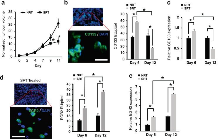
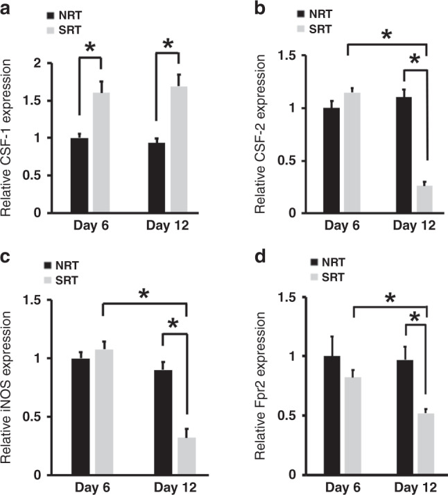
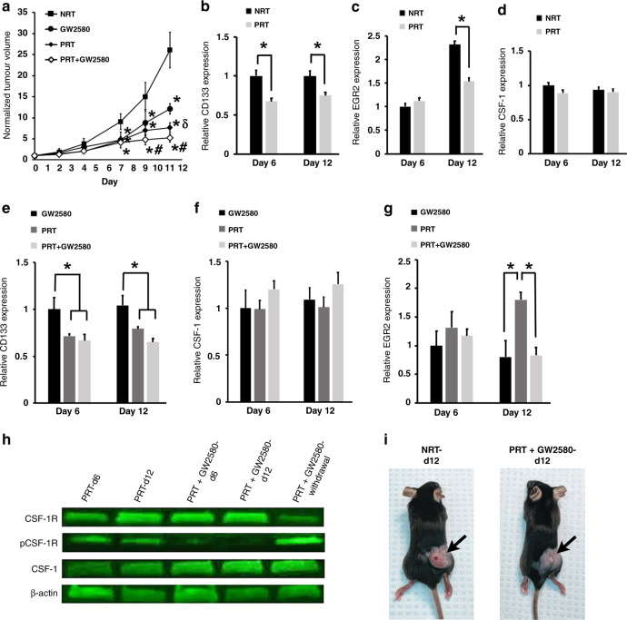
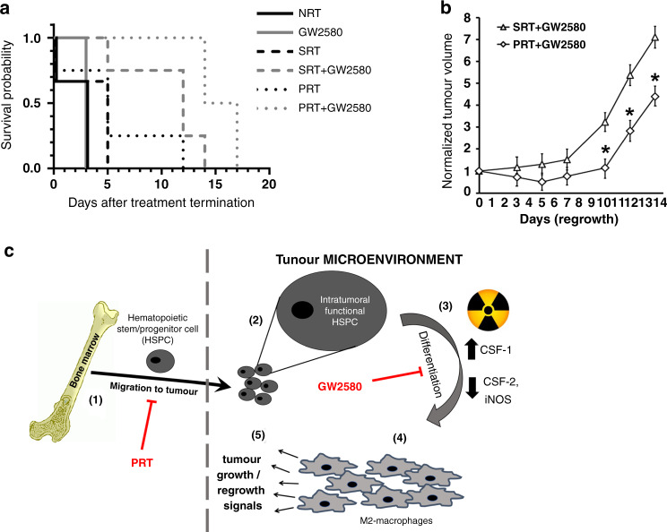
Background: Bone-marrow-derived haematopoietic stem and progenitor cells (HSPCs) are a prominent part of the highly complex tumour microenvironment (TME) where they localise within tumours and maintain haematopoietic potency. Understanding the role HSPCs play in tumour growth and response to radiation therapy (RT) may lead to improved patient treatments and outcomes.

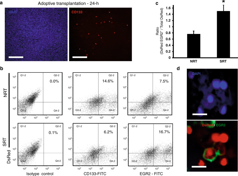
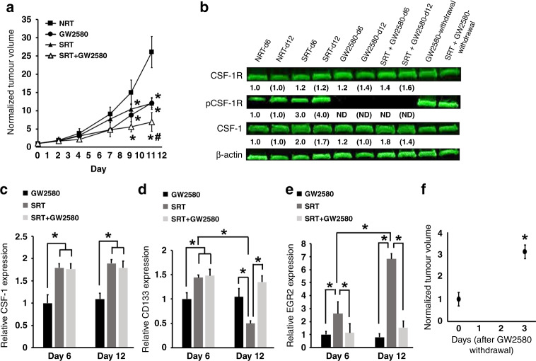
Methods: We used a mouse model of non-small cell lung carcinoma where tumours were exposed to RT regimens alone or in combination with GW2580, a pharmacological inhibitor of colony stimulating factor (CSF)-1 receptor. RT-PCR, western blotting and immunohistochemistry were used to quantify expression levels of factors that affect HSPC differentiation. DsRed+ HSPC intratumoural activity was tracked using flow cytometry and confocal microscopy.

Results: We demonstrated that CSF-1 is enhanced in the TME following exposure to RT. CSF-1 signaling induced intratumoural HSPC differentiation into M2 polarised tumour-associated macrophages (TAMs), aiding in post-RT tumour survival and regrowth. In contrast, hyperfractionated/pulsed radiation therapy (PRT) and GW2580 ablated this process resulting in improved tumour killing and mouse survival.

Conclusions: Tumours coopt intratumoural HSPC fate determination via CSF-1 signaling to overcome the effects of RT. Thus, limiting intratumoural HSPC activity represents an attractive strategy for improving the clinical treatment of solid tumours.


Conflict of interest statement

The authors declare no competing interests.

Figures

Fig. 1
Fig. 1. SRT modulates tumour growth and levels of intratumoural HSPCs and M2 macrophages.
a Tumour bearing mice treated with SRT produced significantly smaller tumours compared to non-treated controls (n = 15). b, c Analysis of tumours by IHC and image analysis (b) as well as RT-PCR (c) showed an initial increase in HSPC numbers during SRT (n = 9) followed by a decrease at later times (n = 9). A representative image identifies CD133+ cells used for analysis. Scale bar: 15 μm. d Image analysis showed a significant increase in EGR2+ M2 macrophages in tumours during treatment (n = 9). An image is shown to demonstrate EGR2 cell surface staining. Scale bar: 30 μm. e RT-PCR confirmed the increased presence of EGR2 expressing M2 macrophages (n = 9). *p < 0.05 for indicated comparisons, no animals were excluded from analysis, relative gene expression values were normalised to both TBP (housekeeping gene) and NRT day 6 values.
Fig. 2
Fig. 2. SRT induces TME alterations that favour HSPC differentiation into M2 macrophages.
a RT-PCR showed that SRT promoted an increase in the expression of CSF-1 a factor known to promote HSPCs differentiation into M2 polarised macrophages (n = 9). b–d The mRNA levels of CSF-2 (b), iNOS (c) and Fpr2 (d), factors associated with M1 macrophage phenotypes were significantly lower in the TME during treatment (n = 9). *p < 0.05 for indicated comparisons, relative gene expression values were normalised to both TBP (housekeeping gene) and NRT day 6 values.
Fig. 3
Fig. 3. HSPCs can directly differentiate into M2 macrophages in response to SRT.
a CD133+ HSPCs were isolated from DsRed transgenic mice and transplanted intravenously into tumour bearing mice. Twenty four hours later tumours were harvested and showed that the CD133+DsRed HSPCs had migrated to tumours and taken up residence. Scale bar: 100 μm. b, c Representative flow plots (b) and analysis of dissociated tumours showing a decrease in CD133+ HSPCs and an increase in EGR2+ M2 macrophages with SRT (n = 6) (c). Isotype control plots are shown and depict gating strategy. Gating was established to capture changes in DsRed CD133+ HSPCs and DsRed EGR2+ M2 macrophages. Since tumours were initially transplanted with DsRed cells from transgenic mice, a DsRed population was present in isotype controls. To account for this, horizontal quadrant lines were set to differentiate between DsRed+ and DsRed cells present in tumours and to minimise false DsRed+ cells from the DsRed population. Vertical quadrant lines were set to ensure 0.1% or less of events appeared in the upper/lower right quadrants. d An image showing an EGR2+ cell that was derived from the originally transplanted DsRed CD133+ HSPCs. Scale bar: 15 μm. *p < 0.05 for indicated comparison, no animals were excluded from analysis.
Fig. 4
Fig. 4. Use of the CSF-1R active inhibitor GW2580 prevents differentiation of HSPCs into M2 macrophages after RT reducing tumour regrowth.
a The effects of treating tumour bearing mice with SRT and GW2580 show that each treatment alone or in combination resulted in smaller tumour volumes in comparison to non-treated controls. *p < 0.05 in comparison to NRT-treated tumours; #p < 0.05 in comparison to GW2580 and SRT alone (n = 7). b Western blot analysis demonstrates both the ability of GW2580 to inhibit CSF-1R phosphorylation during dose delivery and the loss of inhibition after dose withdrawal. The fold changes for the densitometry measurements, normalised to β-actin and then compared to NRT controls, are indicated below the corresponding lane. Numbers without brackets compare day 6 changes (n = 3); numbers with brackets compare day 12 changes (n = 6); ND denotes “not detected”. c–e RT-PCR analysis shows that use of GW2580 has no effect on CSF-1 production (c), results in tumour maintenance of CD133+ HSPCs (d), and decreased levels of M2 macrophages (e) (day 6 n = 3, day 12 n = 6). f Tumours significantly regrow after withdrawal of GW2580 from tumour bearing mice (n = 4). *p < 0.05 for indicated comparisons, no animals were excluded from analysis, relative gene expression values were normalised to both TBP (housekeeping gene) and NRT day 6 values.
Fig. 5
Fig. 5. Use of fractionated RT decreases HSPC migration and differentiation into M2 macrophages reducing tumour growth.
a The effects of treating tumour bearing mice with combinations of PRT and GW2580 were tested and show that each treatment alone (n = 19) or in combination (n = 10) resulted in smaller tumour volume in comparison to non-treated controls. *p < 0.05 in comparison to NRT-treated tumours (n = 18); δp < 0.05 in comparison to GW2580 alone (n = 13); #p < 0.05 in comparison to GW2580 and PRT alone. b–d Following PRT treatment, lower levels of CD133+ HSPCs (b) and M2 macrophages (c) was observed in tumours in comparison to controls, with no effects on CSF-1 levels (d) (n = 9). e–g RT-PCR analysis shows that use of GW2580 is able to augment PRT effects resulting in lower HSPCs (e), no change in CSF-1 production compared to NRT (f) and decreased levels of M2 macrophages (g) (GW2580: day 6 n = 3, day 12 n = 6; PRT n = 9; PRT + GW2580 n = 3). h Western blot demonstrates the ability of GW2580 to prevent CSF-1R phosphorylation (n = 3). *p < 0.05 for indicated comparisons. i Images demonstrating the drastic effects of treatment on tumour size. No animals were excluded from analysis, relative gene expression values were normalised to both TBP (housekeeping gene) and NRT day 6 values.
Fig. 6
Fig. 6. Augmentative treatment with GW2580 enhances RT effects and abrogates HSPC activity in the TME.
a The survival of tumour bearing mice treated with SRT, PRT and GW2580 alone or in combination was assessed (n = 4). Kaplan–Meier analysis demonstrated that the combination of PRT + GW2580 provided the best survival (p = 0.0002). b Tumour volumes were tracked upon termination of both the PRT + GW2580 and SRT + GW2580 combination therapies (n = 4). The results show that tumour regrowth is significantly prevented in the PRT + GW2580 cohort. *p < 0.05 for indicated comparisons, no animals were excluded from analysis. c Schematic of the proposed activity of HSPCs in solid tumour growth and regrowth post-RT. HSPCs normally migrate to growing tumours (step 1) where they are maintained in an undifferentiated state (step 2). When tumours are exposed to radiation, there is an increased production of factors such as CSF-1 (step 3), which create a TME that induces HSPC differentiation into M2 macrophages (step 4). These newly formed TAMs aid in tumour regrowth (step 5). Therapies aimed at preventing these processes such as PRT and GW2580 are able to abrogate this supportive mechanism resulting in smaller tumours and enhanced mouse survival.

References

    1. Kane J, Krueger SA, Dilworth JT, Torma JT, Wilson GD, Marples B, et al. Hematopoietic stem and progenitor cell migration after hypofractionated radiation therapy in a murine model. Int J Radiat Oncol Biol Phys. 2013;87:1162–70. doi: 10.1016/j.ijrobp.2013.08.044. - DOI - PubMed
    1. Bissell MJ, Hines WC. Why don’t we get more cancer? A proposed role of the microenvironment in restraining cancer progression. Nat Med. 2011;17:320–9. doi: 10.1038/nm.2328. - DOI - PMC - PubMed
    1. Liu Y, Cao X. The origin and function of tumor-associated macrophages. Cell Mol Immunol. 2015;12:1–4. doi: 10.1038/cmi.2014.83. - DOI - PMC - PubMed
    1. Kim J, Bae JS. Tumor-associated macrophages and neutrophils in tumor microenvironment. Mediators Inflamm. 2016;2016:6058147. - PMC - PubMed
    1. Bingle L, Brown NJ, Lewis CE. The role of tumour-associated macrophages in tumour progression: implications for new anticancer therapies. J Pathol. 2002;196:254–65. doi: 10.1002/path.1027. - DOI - PubMed

Publication types