

Schwannoma of the Appendix Orifice
- PMID: 34931151
- PMCID: PMC8684518
- DOI: 10.1155/2021/7250145
Schwannoma of the Appendix Orifice
Abstract
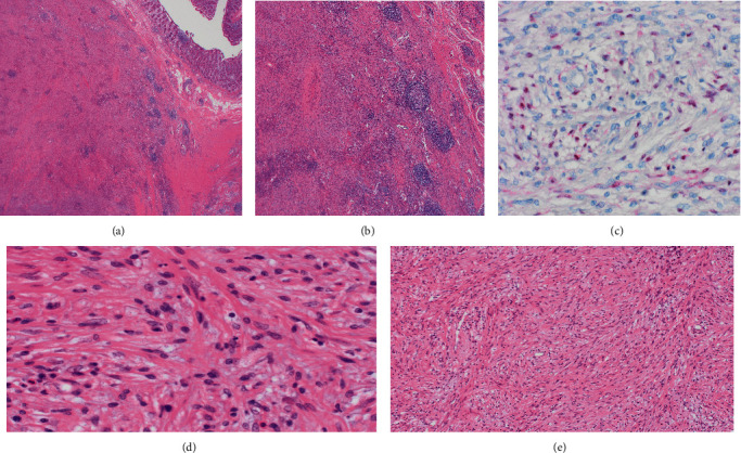
Schwannomas are rare mesenchymal tumors. They are usually diagnosed incidentally during endoscopic or diagnostic imaging for another reason. Malignant transformation is rare. In this case report, we present an incidental schwannoma protruding through the appendiceal orifice diagnosed during endoscopy. A healthy 56-year-old female underwent a surveillance colonoscopy for family history of colorectal cancer. A prominent and edematous appendiceal orifice was noted, and the area was aggressively biopsied. Histopathological assessment revealed a benign schwannoma. Computerized topography was unremarkable. Subsequently, the patient underwent a right hemicolectomy. Patient is scheduled to undergo routine surveillance in three years. Grossly, schwannomas are white, encapsulated, and well-circumscribed lesions that stain strongly positive for S100, GFAP, and CD57. Histologically, schwannomas demonstrate spindle cell proliferation. Several imaging modalities have been utilized in the diagnosis and management of mesenchymal neoplasms. Despite the benign nature of the diagnosis, complete surgical resection with clear margins remains the gold standard management strategy. Our case highlights the presence of a relatively uncommon tumor in an unusual anatomical location.
Copyright © 2021 Maha Alkhattab et al.
Conflict of interest statement
The authors declare that they have no conflicts of interest.
Figures

References
-
- Maciejewski A., Lange D., Wloch J. Case report of schwannoma of the rectum--clinical and pathological contribution. Medical Science Monitor . 2000;6(4) - PubMed
Publication types
LinkOut - more resources
Full Text Sources
Miscellaneous
